Abstract
Objective
To assess the relationship of spinal cord injury level and duration to peak aerobic capacities during arms-only (AO) rowing compared to hybrid Functional Electrical Stimulation (FES) rowing.
Design
Comparison of peak aerobic capacity (VO2peak), peak ventilation (VEpeak), peak respiratory exchange ratio (RERpeak), and peak heart rate (HRpeak) were measured during AO-rowing and FES-rowing obtained from graded exercise tests.
Results
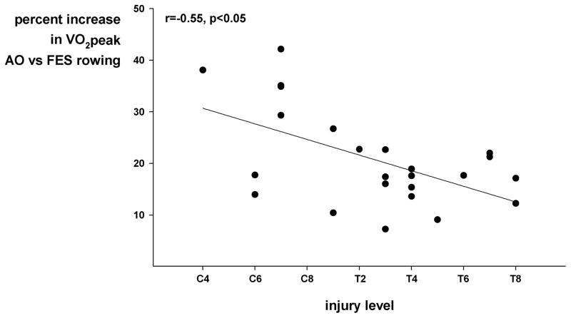
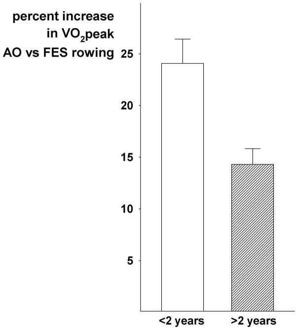
Peak aerobic values were strongly related to injury level and injury duration for both AO-rowing (r=0.67, p<0.05) and FES-rowing (r=0.61, p<0.05). Peak aerobic capacities were greater across all injury levels and durations with FES-rowing compared to AO-rowing. Differences in VO2peak were inversely related to injury level (r=0.55, p<0.05) with greater increases in VO2 in higher level injuries. Injury durations <2 years had greater percent increases in VO2 with FES-rowing.
Conclusions
FES-rowing acutely post injury may have the greatest effect to maintain function and improve peak aerobic capacity. This impact appears to be greatest in those with higher level injuries.
Keywords: spinal cord injury, exercise, functional electrical stimulation, peak aerobic capacity
INTRODUCTION
The amount of muscle mass engaged in dynamic exercise directly determines the magnitude of aerobic demand. After a spinal cord injury (SCI), both loss of innervation and progressive atrophy of muscle lead to lesser muscle mass engaged with exercise, with greater improvement as injury level moves higher. Previous work has clearly demonstrated that Functional Electrical Stimulation (FES) of the paralyzed lower limbs (specifically, the quadriceps and hamstrings) in conjunction with voluntary upper body exercise engages greater muscle mass and elicits a greater metabolic demand. FES-cycling and FES-rowing produce peak aerobic capacities (VO2peak) greater than arms-only (AO)1 or FES alone.2,3 However, the use of arms in conjunction with FES of the legs, specifically to produce the rowing motion, seems to elicit the greatest aerobic response.1–6 This may derive from the coordinated momentum of the leg extension followed by the arm pull to more efficiently utilize a majority of muscle mass for a well-coordinated movement.
We and others before us have found a strong inverse relation of exercise capacity to injury level during arm crank ergometry.7–9 In fact, a relationship remains even with the addition of more muscle mass via hybrid FES exercise. However, the relationship is weaker in those with lower level injuries. Forty percent of the variance in aerobic capacity during hybrid exercise (FES-rowing) is predicted by injury level in those with lesions T3 and below,5 whereas 70% is predicted in high level injuries (T3 and above).4 Hence, the difference between AO exercise and FES-rowing may be lesser at higher level of injury. On the other hand, the addition of FES to the paralyzed legs in higher level spinal cord injuries may produce greater increases in peak aerobic capacity due to the relatively greater amount of muscle mass engaged relative to AO exercise. However, it should be noted that the data suggest those with higher level injuries consistently have lower metabolic responses to exercise, even with the addition of FES.4,5
One might also expect that duration of injury negatively impacts exercise capacity. Skeletal muscle atrophy is significant within one year post injury across all levels, with ~15% of leg lean mass being lost.10 Furthermore, longer duration of injury directly relates to greater loss of muscle mass,11 with the largest atrophy occurring within two years post injury.11,12 In addition, those with higher level injuries have a significantly lower amount of arm lean mass compared to those with lower level injuries.11 Hence, those with higher level injuries have significant loss of whole body muscle mass over time, not only loss of leg lean mass. Indeed, those with higher injury levels may have similar leg lean mass as those with lower level injuries at a similar injury duration, but have lower arm and whole body lean mass.11
We set out to compare the impact of FES-rowing on aerobic capacity as compared to AO-rowing with respect to injury level and duration. Data suggest that those with higher level injuries consistently have lower metabolic responses to exercise, even with the addition of FES. Hence, it is unknown to what extent FES might increase capacity as it relates to injury. Moreover, there are very little data accounting for duration as well as level of injury. We hypothesized that those with high level injuries would have greater increases in VO2peak with FES-rowing due to the relatively greater muscle mass engaged as compared with AO-rowing. Furthermore, we hypothesized that those with lesser time since injury would have greater increases in VO2peak with FES-rowing due to lesser leg muscle atrophy. To test these hypotheses, we assessed peak aerobic capacity during AO- and FES-rowing in individuals with SCI across a range of injury levels and durations.
METHODS
Subjects
Twenty-four individuals with motor injury levels C4-T8 (2 female) and American Spinal Injury Association (AIS) levels A, B, or C were recruited to participate in an FES-row training study at Spaulding Hospital Cambridge. We restricted the subject age to below 40 years to avoid the confound of the effect of aging on aerobic capacity. In addition, we only studied those with adult onset injuries to avoid the potential effect of lack of skeletal muscle development during maturation. Therefore, subjects were aged 22–35 years and were 0.3 months to 8.0 years post injury. A complete health history and physical evaluation were obtained prior to any activity. Individuals were excluded if they were on cardioactive drugs or had any previous cardiovascular or pulmonary disease, diabetes, neurological disorders (other than SCI), pressure induced injuries, or any other contra-indications to vigorous exercise. Written informed consents were provided and all procedures were approved by the Spaulding Hospital Cambridge Institutional Review Board.
FES-Rowing Protocol
A period of pre-training is necessary for individuals to become familiarized with the rowing stroke and to have sufficient leg strength to perform a peak FES-rowing test. This involved 1–2 sessions of FES leg strengthening via electrical stimulation (Odstock, Salisbury, UK; note this is not FDA approved) followed by sessions consisting of short intervals (2–5 minutes) of FES-rowing interspersed with rest for 2–3 sessions per week over ~5 weeks prior to baseline testing. FES-rowing has been described previously,1 but in brief, the Concept-2 adapted rowing machine (Morrisville, VT, USA) provides trunk stability and leg control so that leg contractions can initiate and contribute to the rowing stroke. Individuals synchronize the upper body elbow flexion with leg extension by timing the electrical stimulation of the affected limbs. Subjects were instructed on proper rowing technique, form, and timing of stimulation. Sessions were 2–3 days per week with increasing duration of rowing. On average, individuals required 5.2 ± 0.7 weeks (8.5 ± 1.3 training sessions averaging 170.0 ± 31.2 total minutes) before being able to complete a graded FES-rowing exercise test up to maximal effort. Subjects had to be able to row continuously for a minimum of six minutes with increasing intensities to be considered ready for testing. Subjects were familiarized to arms only (AO) rowing over 2–3 sessions before a graded AO-rowing exercise test to maximal effort. After complete familiarization to both FES- and AO-rowing, peak aerobic capacity tests for each were performed.
Aerobic Capacity Testing
Prior to FES and AO metabolic testing, individuals refrained from high intensity activity for 24 hours. All studies were performed in the morning at the same time with subjects well-rested and hydrated. Testing protocols were graded with target wattage increases every one to two minutes with a goal of reaching maximal effort in six to twelve minutes. Work rates were tailored to each subject based on their pre-test training and were most commonly 5- or 10-watt increments. Minute ventilation and expired O2 and CO2 concentrations were measured (ParvoMedics, Sandy, UT, USA) to determine oxygen consumption (VO2), CO2 production, respiratory exchange ratio (RER), and ventilation (VE). Heart rate was recorded via a Garmin strap and a 5-lead electrocardiogram. To ensure a maximal effort was achieved, we followed ACSM Guidelines for Exercise Testing and Prescription 10th edition, and at least three of the following criteria were met: 1) RER >1.10, 2) lactate via finger prick >8.0 mmol/L, 3) plateau in oxygen consumption (failure to increase VO2 by 150 mL/min with increased workload), 4) a rating of perceived exertion >17 on the Borg scale, and 5) for FES-rowing, a precipitous decline in power >20 watts despite maximal leg stimulation. For lactate, blood samples were taken directly from the finger using a lancet approximately 150 seconds after the cessation of exercise and was measured using a Lactate Plus analyzer (Nova Biomedical, USA). A criterion for 85% of peak heart (220-age) was only applied in individuals with injury level T1 and below, because higher level injuries reduce cardiac sympathetic drive in response to exercise.
Data and Statistical Analysis
Injury scores from neurological examination were transformed into ordinal number, with C1 corresponding to 1 and T1 corresponding to 9, etc. Peak aerobic capacity (VO2peak), peak minute ventilation (VEpeak), peak respiratory exchange ratio (RERpeak), and peak heart rate (HRpeak) were derived from 30-second averages. Differences were calculated via a paired t-test; relations between aerobic capacity and injury characteristics were assessed with linear regression. Data are presented as means with standard errors. A P level of less than 0.05 was considered significant.
RESULTS
Seven subjects had tetraplegia with levels C4-C7 (17 had paraplegia ranging T1-T8). Two-thirds of the subjects were <2 years post injury. Age did not differ between those who were <2 years and those who were >2 years post injury (28.9 ± 1.3 vs. 29.6 ± 1.8 yrs). Injuries were complete (AIS scale A) in 16 individuals, and incomplete in eight (AIS B N=6, AIS C N=2). It should be noted that two subjects used compression socks and one also used an abdominal binder. Both were <2 years since injury, one was higher level AIS C injury and the other was lower level AIS A injury. Their VO2 data were no different from the group means.
For AO-rowing, VO2peak averaged 16.4 ± 1.1 ml/kg/min with a VEpeak of 49.5 ± 3.6 L/min and a RERpeak of 1.26 ± 0.02. Peak heart rate averaged 160 ± 6 bpm. There was a strong relationship between neurological level of injury and VO2peak (r=0.67, p<0.05), VEpeak (r=0.60, p<0.05), and HRpeak (r=0.75, p<0.05) with greater aerobic values at lower injury levels. There was a modest relationship of injury duration to VO2peak (r=0.52, p<0.05).
VO2peak for FES-rowing averaged 20.4 ± 1.1 ml/kg/min with a VEpeak of 50.0 ± 2.9 L/min and RERpeak of 1.17 ± 0.01. Peak heart rate averaged 163 ± 5 bpm. Injury level was strongly related to VO2peak (r=0.61, p<0.05), VEpeak (r=0.66, p<0.05), and HRpeak (r=0.63, p<0.05). Those with lower-level injuries had higher VO2peak, VEpeak, and HRpeak. There was a modest relationship of VO2peak to duration of injury (r=0.40, p<0.05).
VO2peak with FES-rowing was higher than with AO-rowing in all subjects, ranging from 7.3% to 42.2% and averaging 20.8 ± 1.9% higher. However, RERpeak was greater with AO-rowing, whereas VEpeak was similar. Peak heart rate was 2–36 bpm higher with FES-rowing in 15 of 24 individuals, but this did not achieve statistical significance (p=0.10). The difference in VO2peak was inversely related to injury level (r=0.55, p<0.05) with greater improvements in VO2 with FES-rowing in those with higher injury levels (Figure 1). Additionally, a similar relationship was seen in heart rate differences between the two modes of exercise; subjects with higher injury levels showed larger differences in heart rate (r=0.43, p<0.05). In addition, injury duration had an impact on the difference in VO2peak with AO-rowing versus FES-rowing; those with injury duration <2 years demonstrated a greater percent increase in VO2peak with FES-rowing compared to those >2 years (24 ± 2 % vs. 14 ± 2%, p<0.05; Figure 2).
Figure 1.
Relations among injury level and VO2peak percent changes with FES-rowing.
Figure 2.
Relations among injury duration and VO2peak percent changes with FES-rowing.
DISCUSSION
Prior work has shown hybrid FES-rowing elicits a greater aerobic response in comparison to AO-rowing.1–6 The addition of FES to the paralyzed lower limbs increases the amount of muscle mass engaged in exercise and consequently increases aerobic capacity. Our previous work found a 25% greater peak aerobic capacity with the addition of FES in comparison to AO-rowing,1 however peak aerobic capacity with FES-rowing tends to be lower in those with higher level injuries.4 Our current work showed that the addition of FES had a greater relative improvement on VO2peak in those with higher level injuries and even more so in those with acute injuries. This is some of the first work to consider if injury level and duration impact the aerobic capacity improvements with hybrid FES exercise.
As injury level increases, progressively more muscle mass is paralyzed leading to lesser aerobic capacity. This relationship remains even with the addition of FES. This may be due to a similar amount of lower limb muscle mass being engaged by FES in both high and low level injuries and so the amount of upper body mass engaged still determines the relationship. However, the addition of FES for those with higher level injuries affected a marked increase in VO2peak due to the relatively greater muscle mass engaged. For example, one subject who was ~1 year from a complete C5 injury nearly doubled his peak aerobic capacity with the addition of FES. In contrast, another subject who was ~1 year from a complete T4 injury demonstrated only a ~20% increase in VO2peak from AO- to FES-rowing. Hence, the addition of FES in higher level injuries provides a markedly more beneficial exercise stimulus which would otherwise not exist without the electrical stimulation.
Our current work showed that injury duration had a modest relationship to VO2peak during both AO- and FES-rowing. This likely is due to greater muscle atrophy as time since injury progresses. Data suggest that within two years post injury there are significant declines in total lean muscle mass11,12 which then plateaus as duration progresses up to ten years and beyond.13 Hence, it may follow that our data showed that those <2 years post injury had nearly a 40% average increase in peak aerobic capacity with the addition of FES compared to a less than 20% average increase in those >2 years post injury. This suggests that the optimal time to introduce hybrid FES exercise to augment VO2peak is likely within the first two years post injury.
We did not consider any relationship to completeness of injury as there were only two subjects with an AIS C classification (motor incomplete). We would hypothesize that there might be a greater effect on VO2peak with motor incomplete injuries due to the preserved motor function and relatively greater muscle mass. However, we did not have a large enough sample size to determine any relationship. (Parenthetically, including the two individuals with an AIS C classification did not have a significant effect on the outcome of our results.) Further work examining a broader range of injury completeness may provide clearer information, however when motor function is partially preserved there may be minimal increases in VO2peak with the addition of FES.
Our data shows that those with higher level injuries have a greater relative increase in VO2peak with the addition of FES, and this is even more pronounced within 2 years post injury. The effects of hybrid FES-rowing in those with spinal cord injuries can have immense benefits including increases in muscle mass and improvements in aerobic capacity and metabolic demand.1 Therefore, FES-rowing may be crucial to maintaining aerobic fitness levels specifically for those with higher level injuries and acutely post injury.
Acknowledgments
We thank Colleen Caty, Kailyn Burke, and Kristianna Landry for their assistance with the training in this study, as well as our subjects for their participation. We thank the Exercise for Persons with Disabilities program at Spaulding Hospital Cambridge as well.
Footnotes
Disclosures:
We have no conflicts of interests, financial benefits to authors or any prior presentation of this manuscript. Disclosure of funding: National Institutes of Health (R01HL117037).
References
- 1.Taylor JA, Picard G, Widrick JJ. Aerobic capacity with hybrid FES rowing in spinal cord injury: comparison with arms-only exercise and preliminary findings with regular training. PM R. 2011;3:817–24. doi: 10.1016/j.pmrj.2011.03.020. [DOI] [PubMed] [Google Scholar]
- 2.Brurok B, Helgerud J, Karlsen T, Leivseth G, Hoff J. Effect of aerobic high-intensity hybrid training on stroke volume and peak oxygen consumption in men with spinal cord injury. Am J Phys Med Rehabil. 2011;90:407–14. doi: 10.1097/PHM.0b013e31820f960f. [DOI] [PubMed] [Google Scholar]
- 3.Verellen J, Vanlandewijck Y, Andrews B, Wheeler GD. Cardiorespiratory responses during arm ergometry, functional electrical stimulation cycling, and two hybrid exercise conditions in spinal cord injured. Disabil Rehabil Assist Technol. 2007;2:127–32. doi: 10.1080/09638280600765712. [DOI] [PubMed] [Google Scholar]
- 4.Qiu S, Alzhab S, Picard G, Taylor JA. Ventilation Limits Aerobic Capacity after Functional Electrical Stimulation Row Training in High Spinal Cord Injury. Med Sci Sports Exerc. 2016;48:1111–8. doi: 10.1249/MSS.0000000000000880. [DOI] [PMC free article] [PubMed] [Google Scholar]
- 5.Taylor JA, Picard G, Porter A, Morse LR, Pronovost MF, Deley G. Hybrid functional electrical stimulation exercise training alters the relationship between spinal cord injury level and aerobic capacity. Arch Phys Med Rehabil. 2014;95:2172–9. doi: 10.1016/j.apmr.2014.07.412. [DOI] [PMC free article] [PubMed] [Google Scholar]
- 6.Phillips WT, Burkett LN. Augmented upper body contribution to oxygen uptake during upper body exercise with concurrent leg functional electrical stimulation in persons with spinal cord injury. Spinal Cord. 1998;36:750–5. doi: 10.1038/sj.sc.3100692. [DOI] [PubMed] [Google Scholar]
- 7.Battikha M, Sa L, Porter A, Taylor JA. Relationship between pulmonary function and exercise capacity in individuals with spinal cord injury. Am J Phys Med Rehabil. 2014;93:413–21. doi: 10.1097/PHM.0000000000000046. [DOI] [PubMed] [Google Scholar]
- 8.Lassau-Wray ER, Ward GR. Varying physiological response to arm-crank exercise in specific spinal injuries. J Physiol Anthropol Appl Human Sci. 2000;19:5–12. doi: 10.2114/jpa.19.5. [DOI] [PubMed] [Google Scholar]
- 9.Burkett LN, Chisum J, Stone W, Fernhall B. Exercise capacity of untrained spinal cord injured individuals and the relationship of peak oxygen uptake to level of injury. Paraplegia. 1990;28:512–21. doi: 10.1038/sc.1990.68. [DOI] [PubMed] [Google Scholar]
- 10.Wilmet E, Ismail AA, Heilporn A, Welraeds D, Bergmann P. Longitudinal study of the bone mineral content and of soft tissue composition after spinal cord section. Paraplegia. 1995;33:674–7. doi: 10.1038/sc.1995.141. [DOI] [PubMed] [Google Scholar]
- 11.Spungen AM, Adkins RH, Stewart CA, Wang J, Pierson RN, Jr, Waters RL, Bauman WA. Factors influencing body composition in persons with spinal cord injury: a cross-sectional study. J Appl Physiol. 2003;95:2398–407. doi: 10.1152/japplphysiol.00729.2002. [DOI] [PubMed] [Google Scholar]
- 12.Giangregorio L, McCartney N. Bone loss and muscle atrophy in spinal cord injury: epidemiology, fracture prediction, and rehabilitation strategies. J Spinal Cord Med. 2006;29:489–500. doi: 10.1080/10790268.2006.11753898. [DOI] [PMC free article] [PubMed] [Google Scholar]
- 13.Kern H, Hofer C, Modlin M, Mayr W, Vindigni V, Zampieri S, Boncompagni S, Protasi F, Carraro U. Stable muscle atrophy in long-term paraplegics with complete upper motor neuron lesion from 3- to 20-year SCI. Spinal Cord. 2008;46:293–304. doi: 10.1038/sj.sc.3102131. [DOI] [PubMed] [Google Scholar]


