Abstract
Background
The reliability of MUC2 as a prognostic marker in colorectal cancer (CRC) is controversial. This study evaluated the association between MUC2 expression levels in CRC tissues and prognosis.
Methods
The PubMed, Web of Science, Embase, Cochrane Library, China Biology Medicine disc (CBMdisc), Wanfang Database, and China National Knowledge Infrastructure (CNKI) databases were searched to identify studies exploring the relationship between MUC2 expression in CRC tissues and overall survival (OS). Pooled hazard ratios (HRs) and risk ratios (RRs) with 95% confidence intervals (CIs) were used to evaluate the associations between MUC2 expression levels and prognosis and MUC2 expression levels and CRC clinicopathological characteristics, respectively.
Results
The meta-analysis included 11 studies (2619 patients). Low MUC2 expression level was significantly associated with poor OS (HR, 1.67; 95% CI, 1.43–1.94; P < 0.00001) and disease-free survival (DFS)/recurrence-free survival (RFS) (HR, 1.60; 95% CI, 1.21–2.12; P = 0.001) in patients with CRC. Low MUC2 expression level was associated with advanced TNM stage (RR, 1.42; 95% CI, 1.26–1.60; P < 0.00001), lymph node metastasis (RR, 1.41; 95% CI, 1.25–1.60; P < 0.00001), lymphatic invasion (RR,1.64; 95% CI, 1.26–2.12; P = 0.0002), rectal tumor site (RR, 1.26; 95% CI, 1.09–1.46; P = 0.001), and large tumor size (RR,1.32; 95% CI, 1.02–1.70; P = 0.03). There were no associations between low MUC2 expression level and gender, histological grade, depth of invasion, and distant metastasis.
Conclusion
The low levels of MUC2 in CRC tissues are poor prognostic factor independent of stage or other well-recognized markers of later-stage disease. Large well-designed cohort studies are required to validate MUC2 as a biomarker for poor prognosis in CRC.
1. Introduction
Colorectal cancer (CRC) is associated with substantial morbidity and is ranked the third leading cause of cancer-related mortality in the world [1, 2]. The 5-year and 10-year survival rates for CRC are 65% and 58%, respectively [3]. Recurrence is very common in CRC [4, 5], and there is a high risk of subsequent primary cancers in the colon, rectum, and other parts of the digestive system [6]. CRC incidence and mortality rates are rising rapidly in many low- and middle-income countries. The incidence is highest in highly developed countries, but the rates are stabilizing or decreasing in these regions. A 60% increase in the global burden of CRC with more than 2.2 million new cases and 1.1 million deaths is predicted by 2030.
The American Joint Committee on Cancer/Union for International Cancer Control tumor-node-metastasis (TNM) system provides the strongest prognostic parameters for CRC and serves as the basis for treatment decisions [7]. However, the TNM system is less able to predict outcomes in patients with intermediate levels of CRC [8], and there are no definitive biomarkers for monitoring the efficacy of CRC therapies [9]. Therefore, it is necessary to identify new molecular markers that have the potential to predict therapeutic outcomes, serve as therapeutic targets, and improve clinical management in CRC.
Mucins are a family of high molecular weight glycosylated proteins [10] that protect epithelial cells and form the ductal surfaces of several organs [11–13]. To date, approximately 20 mucins have been identified, which can be divided into two subfamilies based on their structure and function, the secreted gel-forming mucins and the transmembrane mucins [14]. Among these, MUC2 is a secreted gel-forming mucin that is encoded within a cluster of genes at the chromosomal locus 11p15 and is thought to share a common ancestor with von Willebrand factor (VWF) [15, 16].
Secreted MUC2 mucin constitutes the major structural component of the mucus in the colon. Colonic mucus has a stratified appearance; the inner mucus layer is attached to the epithelium, is compact, and is devoid of bacteria, while the outer mucus layer is not attached to the epithelium and has an expanded volume due to the action of endogenous proteases, which allows it to be colonized by intestinal bacteria [17]. The inner mucus layer is impervious to bacteria and provides a protective barrier for the colon epithelium. The downregulation of MUC2 expression eliminates this protective mucus barrier, creating a microenvironment in which bacteria can contact the epithelial surface and activate an inflammatory response. Chronic inflammation leads to cellular damage and molecular changes that transform the inflamed epithelium to low-grade dysplasia (LGD), high-grade dysplasia (HGD), and finally CRC [18]. Functionally, MUC2 inhibits the intestinal inflammatory response, thus suppressing the development of intestinal tumors [19, 20]. Conversely, decreased MUC2 expression contributes to CRC by promoting interleukin-6-induced epithelial to mesenchymal transition (EMT), thereby influencing the invasiveness of cancer cells [21, 22]. These mechanisms suggest that MUC2 is an attractive biomarker for diagnosis, immunotherapy, and prognosis in CRC.
Evidence suggests that MUC2 expression is associated with invasion and metastasis in various malignant tumors, including pancreatic cancer [23], gastric carcinoma [24], gallbladder carcinoma [25], extrahepatic bile duct carcinoma [26], breast cancer [27], ovarian cancer [28], ampullary cancer [29], prostate cancer [30], laryngeal cancer [31], and lung cancer [32]. However, the association between MUC2 expression and prognosis in CRC remains to be elucidated. Some studies showed that a low level of MUC2 expression in CRC tissues is associated with poor prognosis [33], while other studies report no obvious correlation [34–37]. Therefore, the objective of the current meta-analysis was to determine the prognostic value of MUC2 in CRC by assessing the association between MUC2 expression levels in CRC tissues and survival. The associations between MUC2 expression levels and several CRC clinicopathological characteristics were also investigated.
2. Materials and Methods
2.1. Search Strategy
Two reviewers (Chao Li, Didi Zuo) independently searched the PubMed, Web of Science, Embase, Cochrane Library, China Biology Medicine disc (CBMdisc), Wanfang Database, and China National Knowledge Infrastructure (CNKI) databases from inception through November 9, 2017, using the following MeSH terms and free-text words: “colorectal neoplasms”/“colorectal cancer”/“colon cancer”/“rectal cancer” and “mucin 2”/“MUC2” and “survival”/“outcome”/“prognosis”/“mortality”. A manual search of the reference lists of relevant articles was conducted to identify additional relevant studies. The search was limited to articles published in the English or Chinese language.
2.2. Inclusion and Exclusion Criteria
Inclusion criteria were as follows: (1) study design: cohort, (2) population: patients with CRC, (3) parameter: MUC2 expression levels in CRC tissue samples, and (4) outcomes: associations between MUC2 expression levels in CRC tissues and overall survival (OS). Exclusion criteria were as follows: (1) duplicate publications; (2) in vitro or animal studies; (3) conference reports, reviews, books, case reports, or letters; or (4) insufficient data. When articles reported data from the same study, data from the most recent article was included.
2.3. Study Selection and Data Extraction
Two reviewers (Chao Li, Didi Zuo) independently examined titles and abstracts to select eligible studies. The full text of potentially relevant studies was retrieved and examined to determine which studies met the inclusion criteria.
Two reviewers (Chao Li, Didi Zuo) independently extracted data from eligible studies including first author's last name, year of publication, country, number of patients, mean age of patients, time of follow-up, MUC2 detection method, MUC2 antibody, cutoff values used to assess MUC2 expression levels, and clinical outcomes. Disagreements about study selection and data extraction were resolved by discussion with a third reviewer (Libin Yin) until consensus was reached.
2.4. Methodological Quality
Two reviewers (Chao Li, Didi Zuo) independently assessed the methodological quality of the included studies using the modified Newcastle-Ottawa Scale (NOS) [38], which allocates a maximum of 9 points according to the quality of the selection, comparability, and outcomes of the study populations. Study quality was defined as poor (0–3), fair (4–6), or good (7–9). Publication bias was assessed using Begg's rank correlation test and Egger's linear regression [39].
Disagreements about the assessment of methodological quality were resolved by discussion with a third reviewer (Libin Yin) until consensus was reached.
2.5. Statistical Analysis
Statistical analyses were performed using Review Manager, version 5.3 (Cochrane Collaboration, Copenhagen, Denmark), and STATA, version 12.0 (Stata Corporation, College Station, TX, USA). Hazard ratios (HRs) with 95% confidence intervals (CIs) were used to evaluate the association between MUC2 expression levels (low versus high) in CRC tissues and OS. HR data were obtained directly from studies or were calculated from Kaplan-Meier curves using Engauge Digitizer, version 4.1 (http://markummitchell.github.io/engauge-digitizer/) [40]. An HR > 1 suggested a worse prognosis in CRC patients with a low level of MUC2 expression, and an HR < 1 indicated a better prognosis. Risk ratios (RRs) with 95% CIs were used to evaluate the associations between MUC2 expression levels (low versus high) in CRC tissues and CRC clinicopathological characteristics, including TNM stage, lymph node metastasis, lymphatic invasion, tumor site, tumor size, gender, histological grade, depth of invasion, and distant metastasis. An RR > 1 suggested that a clinicopathological characteristic was associated with a low level of MUC2 expression, and an RR < 1 indicated a characteristic was associated with a high level of MUC2 expression. A random-effects model was used to pool studies with significant heterogeneity, as determined by the chi-squared test (P ≤ 0.10) and the inconsistency index (I 2 ≥ 50%) [41, 42]. Sources of heterogeneity were explored using metaregression. Sensitivity analysis omitting one study at a time was conducted to investigate the robustness of the findings. P < 0.05 was considered statistically significant.
3. Results
3.1. Search Results
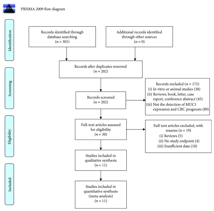
The searches identified 301 articles. Titles and abstracts were screened, and 99 duplicates and 172 studies that did not meet the inclusion criteria were excluded. The full text of 30 articles was retrieved for further review. Of these, 5 review articles, 4 studies that did not report an endpoint, and 10 studies with insufficient data were excluded. Finally, 11 studies [37, 43–52] were found eligible for inclusion in our review (Figure 1).
Figure 1.
Flow chart of literature search. From: Moher D, Libertati A, Tetzlaff J, Altman DG, The PRISMA Group (2009). Preferred Reporting Items for Systematic Reviews and Meta-Analyses: The PRISMA Statement. PloS Med 6(7): e1000097. doi:10.1371/journal.pmed1000097. For more information, visit http://www.prisma-statement.org.
3.2. Characteristics of the Included Studies
The characteristics of the included studies are shown in Table 1. The 11 eligible studies [37, 43–52] were published between 2007 and 2017. Overall, the studies included 2619 patients (range, 35–938 patients). The mean age of patients ranged from 52.9 to 70.5 years, and the median follow-up ranged from 36 to 128 months. Various anti-MUC2 monoclonal antibodies were utilized, including Ccp-58 MRQ-18, NCL-MUC2, and H300. All studies quantified MUC2 expression levels in CRC tissues by immunohistochemistry (IHC). The measurements in all of the included studies were of overall intensity of staining or of percentage cells that were stained. However, each study used a different cutoff point.
Table 1.
Main characteristics of the included publications.
| Publication | Year | Country | Patient number | Gender | Antibody | Cutoff (low/high level) | Method | Outcome | TNM stage | Mean age (years) | Median follow-up (months) | NOS score |
|---|---|---|---|---|---|---|---|---|---|---|---|---|
| Adams et al. | 2009 | Switzerland | 938 | 422/510 | NR | High (>5%) | IHC | OS | I–IV | 70.5 | 128 | 7 |
| Betge et al. | 2016 | Germany | 381 | 215/166 | Ccp-58 | High (>0%) | IHC | OS/DFS | I–IV | 68.5 | NR | 8 |
| Elzagheid et al. | 2013 | Libya | 141 | 55/86 | MRQ-18 | High (>0%) | IHC | OS/DFS | I–IV | NR | 77 | 8 |
| Imai et al. | 2013 | Japan | 250 | 136/114 | Ccp-58 | High (≥25%) | IHC | OS/RFS | I–IV | NR | NR | 8 |
| Kang et al. | 2011 | Korea | 229 | NR | NR | High (staining score ≥ 6) | IHC | OS | II-III | NR | 108 | 7 |
| Khanh et al. | 2013 | Japan | 206 | 114/92 | Ccp-58 | High (≥5%) | IHC | OS/RFS | I–IV | NR | NR | 8 |
| Lu et al. | 2014 | China | 60 | 33/27 | Ccp-58 | High (>5%) | IHC | OS | I–IV | 52.9 | NR | 8 |
| Perez et al. | 2008 | Brazil | 35 | 20/15 | Ccp-58 | High (>10%) | IHC | OS/DFS | I–IV | 62.2 | NR | 7 |
| Wang et al. | 2017 | China | 139 | 76/63 | NCL-MUC2 | High (>20%) | IHC | OS | II–IV | NR | NR | 8 |
| Yu et al. | 2007 | China | 150 | 95/55 | Ccp-58 | High (staining score ≥ 2) | IHC | OS | I–IV | 57.5 | NR | 8 |
| Zwenger et al. | 2014 | Argentina | 90 | 52/38 | H300 | High (staining score > 0) | IHC | OS | I–IV | NR | NR | 8 |
IHC: immunohistochemistry; OS: overall survival; DFS: disease-free survival; RFS: recurrence-free survival; NR: not reported; NOS score: Newcastle-Ottawa Scale score.
3.3. Methodological Quality
The methodological quality of all included studies was good (NOS score > 7) (Table 2). Begg's rank correlation test and Egger's linear regression revealed no publication bias (Begg's test: OS, P = 0.152; DFS/RFS, P = 0.806; TNM stage, P = 0.711; lymph node metastasis, P = 0.536; lymphatic invasion, P = 1.000; tumor site, P = 1.000; tumor size, P = 1.000; gender, P = 0.060, histological grade, P = 0.707; depth of invasion, P = 0.707; and distant metastasis, P = 1.000) (Supplementary File 1).
Table 2.
Quality assessment of the included studies.
| First author, year | Selection1 | Comparability2 | Outcome3 | ||||||
|---|---|---|---|---|---|---|---|---|---|
| Representativeness of exposed cohort ★ | Selection of nonexposed cohort ★ | Ascertainment of exposure ★ | No primary outcome was present at the start of study ★ | Comparable on confounder ★★ | Outcome assessment ★ | Adequate follow-up ★ | Loss to follow-up ★ | Total score | |
| Adams et al., 2009 | ★ | ★ | ★ | ★★ | ★ | ★ | 7 | ||
| Betge et al., 2016 | ★ | ★ | ★ | ★ | ★ | ★ | ★ | ★ | 8 |
| Elzagheid et al., 2013 | ★ | ★ | ★ | ★★ | ★ | ★ | ★ | 8 | |
| Imai et al., 2013 | ★ | ★ | ★ | ★★ | ★ | ★ | ★ | 8 | |
| Kang et al., 2011 | ★ | ★ | ★ | ★ | ★ | ★ | ★ | 7 | |
| Khanh et al., 2013 | ★ | ★ | ★ | ★★ | ★ | ★ | ★ | 8 | |
| Lu et al., 2014 | ★ | ★ | ★ | ★★ | ★ | ★ | ★ | 8 | |
| Perez et al., 2008 | ★ | ★ | ★ | ★ | ★ | ★ | ★ | 7 | |
| Wang et al., 2017 | ★ | ★ | ★ | ★★ | ★ | ★ | ★ | 8 | |
| Yu et al., 2007 | ★ | ★ | ★ | ★★ | ★ | ★ | ★ | 8 | |
| Zwenger et al., 2014 | ★ | ★ | ★ | ★★ | ★ | ★ | ★ | 8 | |
1“Selection” part includes representativeness of cases, selection of controls, exposure ascertainment, and no death when investigation begins. 2“Comparability” part includes comparable on confounders. 3“Outcome” part includes outcome assessment, adequate follow-up, and loss to follow-up rate. ★ represents score of 1. ★★ represents score of 2.
3.4. Outcomes
3.4.1. MUC2 Expression and Overall Survival in CRC
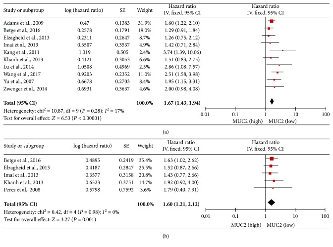
The association between the MUC2 expression level in CRC tissues and OS was investigated in 10 studies. The meta-analysis demonstrated that a low level of MUC2 expression was associated with poor OS in patients with CRC (HR, 1.67; 95% CI, 1.43–1.94; P < 0.00001; Figure 2(a)). There was no evidence of significant heterogeneity between the studies (P = 0.28, I 2 = 17%).
Figure 2.
Associations between the MUC2 expression level and OS (a) and DFS/RFS (b) in CRC.
3.4.2. MUC2 Expression and Disease-Free Survival/Recurrence-Free Survival
The association between the MUC2 expression level in CRC tissues and DFS/RFS was investigated in 5 studies. The meta-analysis demonstrated that a low level of MUC2 expression was associated with shorter DFS/RFS in patients with CRC (HR, 1.60; 95% CI, 1.21–2.12; P = 0.001; Figure 2(b)). There was no evidence of significant heterogeneity between the studies (P = 0.98, I 2 = 0%).
3.4.3. MUC2 Expression and TNM Stage
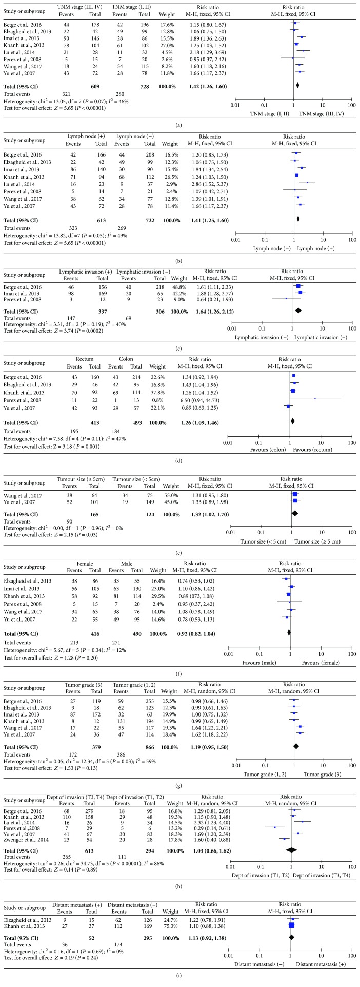
The association between the MUC2 expression level in CRC tissues and TNM stage was investigated in 8 studies. The meta-analysis demonstrated that a low level of MUC2 expression was associated with CRC in the advanced stages (TNM stage III/IV) compared to the localized stages (TNM stage I/II) (RR, 1.42; 95% CI, 1.26–1.60; P < 0.00001; Figure 3(a)). There was no evidence of significant heterogeneity between the studies (P = 0.007, I 2 = 46%).
Figure 3.
Associations between the MUC2 expression level and CRC clinicopathological characteristics. (a) TNM stage, (b) lymph node metastasis, (c) lymphatic invasion, (d) tumor site, (e) tumor size, (f) gender, (g) histological grade, (h) depth of invasion, (i) distant metastasis.
3.4.4. MUC2 Expression and Lymph Node Metastasis
The association between the MUC2 expression level in CRC tissues and lymph node metastasis was investigated in 8 studies. The meta-analysis demonstrated that a low level of MUC2 expression was associated with lymph node metastasis in patients with CRC (RR, 1.41; 95% CI, 1.25–1.60; P < 0.00001; Figure 3(b)). There was no evidence of significant heterogeneity between the studies (P < 0.00001, I 2 = 49%).
3.4.5. MUC2 Expression and Lymphatic Invasion
The association between the MUC2 expression level in CRC tissues and lymphatic invasion was investigated in 3 studies. The meta-analysis demonstrated that a low level of MUC2 expression was associated with lymphatic invasion in patients with CRC (RR, 1.64; 95% CI, 1.26–2.12; P = 0.0002; Figure 3(c)). There was no evidence of significant heterogeneity between the studies (P = 0.19, I 2 = 40%).
3.4.6. MUC2 Expression and Tumor Site
The association between the MUC2 expression level in CRC tissues and tumor site was investigated in 5 studies. The meta-analysis demonstrated that a low level of MUC2 expression was associated with CRC in the rectum compared to the colon (RR, 1.26; 95% CI, 1.09–1.46; P = 0.001; Figure 3(d)). There was no evidence of significant heterogeneity between the studies (P = 0.11, I 2 = 47%).
3.4.7. MUC2 Expression and Tumor Size
The association between the MUC2 expression level in CRC tissues and tumor size was investigated in 2 studies. The meta-analysis demonstrated that a low level of MUC2 expression was associated with large tumors compared to small tumors in patients with CRC (RR, 1.32; 95% CI, 1.02–1.70; P = 0.03; Figure 3(e)). There was no evidence of heterogeneity between the studies (P = 0.96, I 2 = 0%).
3.4.8. MUC2 Expression and Other Clinical Features
The associations between the MUC2 expression level in CRC tissues and other clinicopathological characteristics were investigated The meta-analysis demonstrated that a low level of MUC2 expression did not show an association with gender (female versus male: RR, 0.92; 95% CI, 0.82–1.04; P = 0.20; Figure 3(f)), histological grade (RR 1.19; 95% CI, 0.95–1.50; P = 0.13; Figure 3(g)), depth of invasion (T3, T4 versus T1, T2: RR, 1.03; 95% CI, 0.66–1.62; P = 0.89; Figure 3(h)), and distant metastasis (positive versus negative: RR, 1.13;95% CI, 0.92–1.38; P = 0.24; Figure 3(i)).
3.5. Sensitivity Analysis
Sensitivity analysis omitting one study at a time indicated that the findings of this meta-analysis were robust (Supplementary File 2).
3.6. Metaregression
The metaregression of factors influencing the association of MUC2 expression with OS and DFS/RFS in CRC was performed. None of the covariates analyzed, including year, country, antibody, or cutoff values, influenced the association (Supplementary File 3).
4. Discussion
Evidence suggests that CRC tissues express low levels of MUC2 and that MUC2 plays a role in the development and progression of CRC. However, the prognostic value of MUC2 in CRC remains to be elucidated. Although a previous meta-analysis [53] investigated the association between MUC2 expression and CRC clinicopathological characteristics, to the authors' knowledge, the current study is the first meta-analysis to evaluate the prognostic value of MUC2 expression in CRC. The results showed that a low level of MUC2 expression in CRC tissues was associated with poor OS and DFS/RFS. These findings suggest that MUC2 has a protective role in CRC, which may be explained by several mechanisms. MUC2 silencing may promote CRC metastasis by interleukin-6-induced EMT, which contributes to the invasiveness of cancer cells [21, 54]. MUC2 downregulation may contribute to chronic inflammation [55], generating a microenvironment that results in genomic instability [56]. In addition, MUC2 downregulation has been associated with increased expression of tumor-associated antigen carcinoembryonic antigen-related cell adhesion molecules 5 and 6 (CEACAM5 and CEACAM6), which are involved in cell adhesion, migration, tumor invasion, and metastasis [57, 58]. Taken together, these data indicate that MUC2 may serve as a therapeutic target with potential to improve clinical management in CRC and suggest that randomized controlled clinical trials investigating the role of MUC2 in CRC therapy are warranted.
In accordance with our findings, previous studies indicated that a low level of MUC2 expression in CRC tissues is an indicator of poor prognosis. Betge et al. [48], showed that loss of MUC2 expression in CRC tissues was a predictor of adverse outcome. Wang et al. [45] reported that low MUC2 expression in CRC tissues was significantly associated with lymph node metastasis, poor cellular differentiation, and an advanced tumor stage in CRC, and patients with high MUC2 expression in CRC tissues had higher 5-year survival than patients with low MUC2 expression. Lugli et al. [59] found that the loss of MUC2 in CRC tissues was an adverse prognostic factor for survival in mismatch-repair- (MMR-) proficient and MLH1-negative CRC. In contrast, other studies showed a lower 3-year survival rate in patients with high MUC2 expression in CRC tissues compared to low MUC2 expression (0% versus 60%, resp.) [60]. Espinoza et al. [34] reported that MUC2 expression levels in CRC tissues did not significantly correlate with DFS among African Americans and Caucasian Americans.
As CRC clinicopathological characteristics are often used in clinical practice to predict prognosis, the current study comprehensively explored the association between MUC2 expression levels in CRC tissues and CRC clinicopathological characteristics. The results showed that a low level of MUC2 expression was associated with advanced TNM stage, lymph node metastasis, lymphatic invasion, tumor in the rectum versus the colon, and large tumor size. However, there were no associations between MUC2 expression level and gender, histological grade, depth of invasion, and distant metastasis. Previous reports have demonstrated that advanced TNM stage, lymph node metastasis, lymphatic invasion, rectal tumor site, and large tumor size are predictors of poor prognosis in CRC [44, 46, 61–63]. These data, together with the findings from the current study, imply that the low levels of MUC2 expression in CRC tissues may be used as a biomarker for poor prognosis.
As MUC2 expression levels in CRC tissues are important for diagnosis and prognosis in CRC, MUC2 levels in CRC tissues may be used to guide clinical decision-making. MUC2 may be detected by immunohistochemistry, which is a relatively simple and cost-effective method that could gain widespread acceptance. However, as MUC2 is determined postoperatively in CRC tissue samples, continuous monitoring of MUC2 expression levels throughout the course of disease and with treatment will be challenging.
This study was associated with some limitations. First, some of the included studies did not directly report HRs; instead, they had to be extracted from Kaplan-Meier curves, which may have affected the robustness of our results. Second, the potential sources of heterogeneity between the studies included publication year, country, MUC2 antibody, and cutoff values for MUC2 expression; however, the metaregression analysis revealed that none of these factors were significant sources of heterogeneity. Last, the sample size in the study was small; therefore, the findings should be considered preliminary.
In conclusion, the current study suggests that a low level of MUC2 expression is an independent factor of poor prognosis in colorectal cancer and also associated with later TNM stage, presence of lymph node metastasis, rectal tumor site, and large tumor size. However, the clinical relevance of MUC2 downregulation in CRC tissues remains to be elucidated. Large well-designed cohort studies are required to validate MUC2 as a biomarker for poor prognosis in CRC.
Conflicts of Interest
The authors declare no competing financial interests.
Authors' Contributions
Chao Li helped in the data curation, investigation, methodology, resources, validation, and writing of the original draft. Didi Zuo helped in the data curation, formal analysis, and investigation. Libin Yin helped in the analysis, investigation, and validation. Yuyang Lin helped in doing formal analysis and designed a software. Chenguang Li designed a software. Tao Liu also designed a software. Lei Wang helped in the conceptualization, funding acquisition, project administration, supervision, visualization, and writing—review and editing.
Supplementary Materials
Supplementary File 1: publication bias. (a) OS, (b) DFS/RFS, (c) TNM stage, (d) lymph node metastasis, (e) lymphatic invasion, (f) tumor site, (g) tumor size, (h) gender, (i) tumor grade, (j) depth of invasion, (k) distant.
Supplementary File 2: sensitivity analysis. (a) OS, (b) DFS/RFS, (c) TNM stage, (d) lymph node metastasis, (e) lymphatic invasion, (f) tumor site, (g) tumor size, (h) gender, (i) tumor grade, (j) depth of invasion, (k) distant.
Supplementary File 3: results of metaregression analysis exploring the source of heterogeneity with OS and DFS.
References
- 1.Henley S. J., Singh S. D., King J., Wilson R. J., O’Neil M. E., Ryerson A. B. Invasive cancer incidence and survival — United States, 2012. MMWR. Morbidity and Mortality Weekly Report. 2015;64(49):1353–1358. doi: 10.15585/mmwr.mm6449a1. [DOI] [PubMed] [Google Scholar]
- 2.Siegel R. L., Miller K. D., Jemal A. Cancer statistics, 2015. CA: A Cancer Journal for Clinicians. 2015;65(1):5–29. doi: 10.3322/caac.21254. [DOI] [PubMed] [Google Scholar]
- 3.Miller K. D., Siegel R. L., Lin C. C., et al. Cancer treatment and survivorship statistics, 2016. CA: A Cancer Journal for Clinicians. 2016;66(4):271–289. doi: 10.3322/caac.21349. [DOI] [PubMed] [Google Scholar]
- 4.Primrose J. N., Perera R., Gray A., et al. Effect of 3 to 5 years of scheduled CEA and CT follow-up to detect recurrence of colorectal cancer: the FACS randomized clinical trial. JAMA. 2014;311(3):263–270. doi: 10.1001/jama.2013.285718. [DOI] [PubMed] [Google Scholar]
- 5.Tjandra J. J., Chan M. K. Y. Follow-up after curative resection of colorectal cancer: a meta-analysis. Diseases of the Colon & Rectum. 2007;50(11):1783–1799. doi: 10.1007/s10350-007-9030-5. [DOI] [PubMed] [Google Scholar]
- 6.Mariotto A. B., Rowland J. H., Ries L. A. G., Scoppa S., Feuer E. J. Multiple cancer prevalence: a growing challenge in long-term survivorship. Cancer Epidemiology, Biomarkers & Prevention. 2007;16(3):566–571. doi: 10.1158/1055-9965.EPI-06-0782. [DOI] [PubMed] [Google Scholar]
- 7.Compton C. C. Optimal pathologic staging: defining stage II disease. Clinical Cancer Research. 2007;13(22):6862s–6870s. doi: 10.1158/1078-0432.CCR-07-1398. [DOI] [PubMed] [Google Scholar]
- 8.Ueno H., Mochizuki H., Akagi Y., et al. Optimal colorectal cancer staging criteria in TNM classification. Journal of Clinical Oncology. 2012;30(13):1519–1526. doi: 10.1200/JCO.2011.39.4692. [DOI] [PubMed] [Google Scholar]
- 9.Paul D., Kumar A., Gajbhiye A., Santra M. K., Srikanth R. Mass spectrometry-based proteomics in molecular diagnostics: discovery of cancer biomarkers using tissue culture. BioMed Research International. 2013;2013:16. doi: 10.1155/2013/783131.783131 [DOI] [PMC free article] [PubMed] [Google Scholar]
- 10.Andrianifahanana M., Moniaux N., Batra S. K. Regulation of mucin expression: mechanistic aspects and implications for cancer and inflammatory diseases. Biochimica et Biophysica Acta (BBA) - Reviews on Cancer. 2006;1765(2):189–222. doi: 10.1016/j.bbcan.2006.01.002. [DOI] [PubMed] [Google Scholar]
- 11.Hollingsworth M. A., Swanson B. J. Mucins in cancer: protection and control of the cell surface. Nature Reviews Cancer. 2004;4(1):45–60. doi: 10.1038/nrc1251. [DOI] [PubMed] [Google Scholar]
- 12.Vandenhaute B., Buisine M. P., Debailleul V., et al. Mucin gene expression in biliary epithelial cells. Journal of Hepatology. 1997;27(6):1057–1066. doi: 10.1016/S0168-8278(97)80150-X. [DOI] [PubMed] [Google Scholar]
- 13.Lakshmanan I., Ponnusamy M. P., Macha M. A., et al. Mucins in lung cancer: diagnostic, prognostic, and therapeutic implications. Journal of Thoracic Oncology. 2015;10(1):19–27. doi: 10.1097/JTO.0000000000000404. [DOI] [PubMed] [Google Scholar]
- 14.Itoh Y., Kamata-Sakurai M., Denda-Nagai K., et al. Identification and expression of human epiglycanin/MUC21: a novel transmembrane mucin. Glycobiology. 2008;18(1):74–83. doi: 10.1093/glycob/cwm118. [DOI] [PubMed] [Google Scholar]
- 15.Desseyn J. L., Buisine M. P., Porchet N., Aubert J. P., Degand P., Laine A. Evolutionary history of the 11p15 human mucin gene family. Journal of Molecular Evolution. 1998;46(1):102–106. doi: 10.1007/PL00006276. [DOI] [PubMed] [Google Scholar]
- 16.Desseyn J. L., Aubert J. P., Porchet N., Laine A. Evolution of the large secreted gel-forming mucins. Molecular Biology and Evolution. 2000;17(8):1175–1184. doi: 10.1093/oxfordjournals.molbev.a026400. [DOI] [PubMed] [Google Scholar]
- 17.Johansson M. E. V., Phillipson M., Petersson J., Velcich A., Holm L., Hansson G. C. The inner of the two Muc2 mucin-dependent mucus layers in colon is devoid of bacteria. Proceedings of the National Academy of Sciences of the United States of America. 2008;105(39):15064–15069. doi: 10.1073/pnas.0803124105. [DOI] [PMC free article] [PubMed] [Google Scholar]
- 18.Herszényi L., Barabás L., Miheller P., Tulassay Z. Colorectal cancer in patients with inflammatory bowel disease: the true impact of the risk. Digestive Diseases. 2015;33(1):52–57. doi: 10.1159/000368447. [DOI] [PubMed] [Google Scholar]
- 19.Velcich A., Yang W., Heyer J., et al. Colorectal cancer in mice genetically deficient in the mucin Muc2. Science. 2002;295(5560):1726–1729. doi: 10.1126/science.1069094. [DOI] [PubMed] [Google Scholar]
- 20.Van der Sluis M., De Koning B. A. E., De Bruijn A. C. J. M., et al. Muc2-deficient mice spontaneously develop colitis, indicating that MUC2 is critical for colonic protection. Gastroenterology. 2006;131(1):117–129. doi: 10.1053/j.gastro.2006.04.020. [DOI] [PubMed] [Google Scholar]
- 21.Neurath M. F., Finotto S. IL-6 signaling in autoimmunity, chronic inflammation and inflammation-associated cancer. Cytokine & Growth Factor Reviews. 2011;22(2):83–89. doi: 10.1016/j.cytogfr.2011.02.003. [DOI] [PubMed] [Google Scholar]
- 22.Shan Y.-S., Hsu H.-P., Lai M.-D., et al. Suppression of mucin 2 promotes interleukin-6 secretion and tumor growth in an orthotopic immune-competent colon cancer animal model. Oncology Reports. 2014;32(6):2335–2342. doi: 10.3892/or.2014.3544. [DOI] [PMC free article] [PubMed] [Google Scholar]
- 23.Yang H. S., Tamayo R., Almonte M., et al. Clinical significance of MUC1, MUC2 and CK17 expression patterns for diagnosis of pancreatobiliary arcinoma. Biotechnic & Histochemistry. 2012;87(2):126–132. doi: 10.3109/10520295.2011.570276. [DOI] [PubMed] [Google Scholar]
- 24.Xiao L. J., Zhao S., Zhao E. H., et al. Clinicopathological and prognostic significance of MUC-2, MUC-4 and MUC-5AC expression in Japanese gastric carcinomas. Asian Pacific Journal of Cancer Prevention. 2012;13(12):6447–6453. doi: 10.7314/APJCP.2012.13.12.6447. [DOI] [PubMed] [Google Scholar]
- 25.Hiraki T., Yamada S., Higashi M., et al. Immunohistochemical expression of mucin antigens in gallbladder adenocarcinoma: MUC1-positive and MUC2-negative expression is associated with vessel invasion and shortened survival. Histology and Histopathology. 2017;32(6):585–596. doi: 10.14670/HH-11-824. [DOI] [PubMed] [Google Scholar]
- 26.Hong S. M., Cho H., Moskaluk C. A., Frierson H. F., Jr, Yu E., Ro J. Y. CDX2 and MUC2 protein expression in extrahepatic bile duct carcinoma. American Journal of Clinical Pathology. 2005;124(3):361–370. doi: 10.1309/GTU1Y77MVR4DX5A2. [DOI] [PubMed] [Google Scholar]
- 27.Patel D. S., Khandeparkar S. G. S., Joshi A. R., et al. Immunohistochemical study of MUC1, MUC2 and MUC5AC expression in primary breast carcinoma. Journal of Clinical and Diagnostic Research. 2017;11(4):EC30–EC34. doi: 10.7860/jcdr/2017/26533.9707. [DOI] [PMC free article] [PubMed] [Google Scholar]
- 28.He Y. F., Zhang M. Y., Wu X., et al. High MUC2 expression in ovarian cancer is inversely associated with the M1/M2 ratio of tumor-associated macrophages and patient survival time. PLoS One. 2013;8(12, article e79769) doi: 10.1371/journal.pone.0079769. [DOI] [PMC free article] [PubMed] [Google Scholar]
- 29.Moriya T., Kimura W., Hirai I., Takasu N., Mizutani M. Expression of MUC1 and MUC2 in ampullary cancer. International Journal of Surgical Pathology. 2011;19(4):441–447. doi: 10.1177/1066896911405654. [DOI] [PubMed] [Google Scholar]
- 30.Cozzi P. J., Wang J., Delprado W., et al. MUC1, MUC2, MUC4, MUC5AC and MUC6 expression in the progression of prostate cancer. Clinical & Experimental Metastasis. 2005;22(7):565–573. doi: 10.1007/s10585-005-5376-z. [DOI] [PubMed] [Google Scholar]
- 31.Jeannon J. P., Aston V., Stafford F. W., Soames J. V., Wilson J. A. Expression of MUC1 and MUC2 glycoproteins in laryngeal cancer. Clinical Otolaryngology and Allied Sciences. 2001;26(2):109–112. doi: 10.1046/j.1365-2273.2001.00437.x. [DOI] [PubMed] [Google Scholar]
- 32.Awaya H., Takeshima Y., Yamasaki M., Inai K. Expression of MUC1, MUC2, MUC5AC, and MUC6 in atypical adenomatous hyperplasia, bronchioloalveolar carcinoma, adenocarcinoma with mixed subtypes, and mucinous bronchioloalveolar carcinoma of the lung. American Journal of Clinical Pathology. 2004;121(5):644–653. doi: 10.1309/U4WGE9EBFJN6CM8R. [DOI] [PubMed] [Google Scholar]
- 33.Betge J., Schneider N. I., Harbaum L., et al. Expression profiles and clinical impact of MUC1, MUC2, MUC5AC and MUC6 in colorectal cancer. Virchows Archiv. 2016;469:S156–S157. doi: 10.1007/s00428-016-1970-5. [DOI] [PMC free article] [PubMed] [Google Scholar]
- 34.Espinoza I. C., Reddy A., Zhang X., et al. Abstract 4716: stemness markers in colorectal cancer: analysis in a racially-diverse population. Cancer Research. 2017;77, article 4716(Supplement 13) doi: 10.1158/1538-7445.AM2017-4716. [DOI] [Google Scholar]
- 35.Baldus S. E., Monig S. P., Hanisch F. G., et al. Comparative evaluation of the prognostic value of MUC1, MUC2, sialyl-Lewisa and sialyl-Lewisx antigens in colorectal adenocarcinoma. Histopathology. 2002;40(5):440–449. doi: 10.1046/j.1365-2559.2002.01389.x. [DOI] [PubMed] [Google Scholar]
- 36.Manne U., Weiss H. L., Grizzle W. E. Racial differences in the prognostic usefulness of MUC1 and MUC2 in colorectal adenocarcinomas. Clinical Cancer Research. 2000;6(10):4017–4025. [PubMed] [Google Scholar]
- 37.Zwenger A., Rabassa M., Demichelis S., Grossman G., Segal-Eiras A., Croce M. V. High expression of sLex associated with poor survival in Argentinian colorectal cancer patients. The International Journal of Biological Markers. 2014;29(1):e30–e39. doi: 10.5301/jbm.5000060. [DOI] [PubMed] [Google Scholar]
- 38.Stang A. Critical evaluation of the Newcastle-Ottawa Scale for the assessment of the quality of nonrandomized studies in meta-analyses. European Journal of Epidemiology. 2010;25(9):603–605. doi: 10.1007/s10654-010-9491-z. [DOI] [PubMed] [Google Scholar]
- 39.Begg C. B., Mazumdar M. Operating characteristics of a rank correlation test for publication bias. Biometrics. 1994;50(4):1088–1101. doi: 10.2307/2533446. [DOI] [PubMed] [Google Scholar]
- 40.Tierney J. F., Stewart L. A., Ghersi D., Burdett S., Sydes M. R. Practical methods for incorporating summary time-to-event data into meta-analysis. Trials. 2007;8(1):p. 16. doi: 10.1186/1745-6215-8-16. [DOI] [PMC free article] [PubMed] [Google Scholar]
- 41.Higgins J. P. T., Thompson S. G., Deeks J. J., Altman D. G. Measuring inconsistency in meta-analyses. BMJ. 2003;327(7414):557–560. doi: 10.1136/bmj.327.7414.557. [DOI] [PMC free article] [PubMed] [Google Scholar]
- 42.DerSimonian R., Laird N. Meta-analysis in clinical trials revisited. Contemporary Clinical Trials. 2015;45(Part A):139–145. doi: 10.1016/j.cct.2015.09.002. [DOI] [PMC free article] [PubMed] [Google Scholar]
- 43.Yu X. W., Rong W., Xu F. L., Xu G. Y., Sun Y. R., Feng M. Y. Expression and clinical significance of mucin and E-cadherin in colorectal tumors. Ai zheng= Aizheng= Chinese journal of cancer. 2007;26(11):1204–1210. [PubMed] [Google Scholar]
- 44.Imai Y., Yamagishi H., Fukuda K., Ono Y., Inoue T., Ueda Y. Differential mucin phenotypes and their significance in a variation of colorectal carcinoma. World Journal of Gastroenterology. 2013;19(25):3957–3968. doi: 10.3748/wjg.v19.i25.3957. [DOI] [PMC free article] [PubMed] [Google Scholar]
- 45.Wang H., Jin S., Lu H., et al. Expression of survivin, MUC2 and MUC5 in colorectal cancer and their association with clinicopathological characteristics. Oncology Letters. 2017;14(1):1011–1016. doi: 10.3892/ol.2017.6218. [DOI] [PMC free article] [PubMed] [Google Scholar]
- 46.Kang H., Min B. S., Lee K. Y., et al. Loss of E-cadherin and MUC2 expressions correlated with poor survival in patients with stages II and III colorectal carcinoma. Annals of Surgical Oncology. 2011;18(3):711–719. doi: 10.1245/s10434-010-1338-z. [DOI] [PubMed] [Google Scholar]
- 47.Elzagheid A., Emaetig F., Buhmeida A., et al. Loss of MUC2 expression predicts disease recurrence and poor outcome in colorectal carcinoma. Tumor Biology. 2013;34(2):621–628. doi: 10.1007/s13277-012-0588-8. [DOI] [PubMed] [Google Scholar]
- 48.Betge J., Schneider N. I., Harbaum L., et al. MUC1, MUC2, MUC5AC, and MUC6 in colorectal cancer: expression profiles and clinical significance. Virchows Archiv. 2016;469(3):255–265. doi: 10.1007/s00428-016-1970-5. [DOI] [PMC free article] [PubMed] [Google Scholar]
- 49.Perez R. O., Bresciani B. H., Bresciani C., et al. Mucinous colorectal adenocarcinoma: influence of mucin expression (Muc1, 2 and 5) on clinico-pathological features and prognosis. International Journal of Colorectal Disease. 2008;23(8):757–765. doi: 10.1007/s00384-008-0486-0. [DOI] [PubMed] [Google Scholar]
- 50.Adams H., Tzankov A., Lugli A., Zlobec I. New time-dependent approach to analyse the prognostic significance of immunohistochemical biomarkers in colon cancer and diffuse large B-cell lymphoma. Journal of Clinical Pathology. 2009;62(11):986–997. doi: 10.1136/jcp.2008.059063. [DOI] [PubMed] [Google Scholar]
- 51.Khanh D. T., Mekata E., Mukaisho K. I., et al. Transmembrane mucin MUC1 overexpression and its association with CD10+ myeloid cells, transforming growth factor-β1 expression, and tumor budding grade in colorectal cancer. Cancer Science. 2013;104(7):958–964. doi: 10.1111/cas.12170. [DOI] [PMC free article] [PubMed] [Google Scholar]
- 52.Lu M., He J., Ye W. Combined expression and clinicopathological correlation study of MUC1 and MUC2 in colorectal adenomas and colorectal carcinoma. Journal of Colorectal & Anal Surgery. 2014;03:158–163. [Google Scholar]
- 53.Li L., Huang P. L., Yu X. J., Bu X. D. Clinicopathological significance of mucin 2 immunohistochemical expression in colorectal cancer: a meta-analysis. Chinese Journal of Cancer Research. 2012;24(3):190–195. doi: 10.1007/s11670-012-0190-z. [DOI] [PMC free article] [PubMed] [Google Scholar]
- 54.Hsu H. P., Lai M. D., Lee J. C., et al. Mucin 2 silencing promotes colon cancer metastasis through interleukin-6 signaling. Scientific Reports. 2017;7(1):p. 5823. doi: 10.1038/s41598-017-04952-7. [DOI] [PMC free article] [PubMed] [Google Scholar]
- 55.Kufe D. W. Mucins in cancer: function, prognosis and therapy. Nature Reviews Cancer. 2009;9(12):874–885. doi: 10.1038/nrc2761. [DOI] [PMC free article] [PubMed] [Google Scholar]
- 56.Feagins L. A., Souza R. F., Spechler S. J. Carcinogenesis in IBD: potential targets for the prevention of colorectal cancer. Nature Reviews Gastroenterology & Hepatology. 2009;6(5):297–305. doi: 10.1038/nrgastro.2009.44. [DOI] [PubMed] [Google Scholar]
- 57.Zheng C., Feng J., Lu D., et al. A novel anti-CEACAM5 monoclonal antibody, CC4, suppresses colorectal tumor growth and enhances NK cells-mediated tumor immunity. PLoS One. 2011;6(6, article e21146) doi: 10.1371/journal.pone.0021146. [DOI] [PMC free article] [PubMed] [Google Scholar]
- 58.Beauchemin N., Arabzadeh A. Carcinoembryonic antigen-related cell adhesion molecules (CEACAMs) in cancer progression and metastasis. Cancer Metastasis Reviews. 2013;32(3-4):643–671. doi: 10.1007/s10555-013-9444-6. [DOI] [PubMed] [Google Scholar]
- 59.Lugli A., Zlobec I., Baker K., et al. Prognostic significance of mucins in colorectal cancer with different DNA mismatch-repair status. Journal of Clinical Pathology. 2007;60(5):534–539. doi: 10.1136/jcp.2006.039552. [DOI] [PMC free article] [PubMed] [Google Scholar]
- 60.Fujishima Y., Goi T., Kimura Y., Hirono Y., Katayama K., Yamaguchi A. MUC2 protein expression status is useful in assessing the effects of hyperthermic intraperitoneal chemotherapy for peritoneal dissemination of colon cancer. International Journal of Oncology. 2012;40(4):960–964. doi: 10.3892/ijo.2012.1334. [DOI] [PMC free article] [PubMed] [Google Scholar]
- 61.Yuan H., Dong Q., Zheng B., Hu X., Xu J. B., Tu S. Lymphovascular invasion is a high risk factor for stage I/II colorectal cancer: a systematic review and meta-analysis. Oncotarget. 2017;8(28):46565–46579. doi: 10.18632/oncotarget.15425. [DOI] [PMC free article] [PubMed] [Google Scholar]
- 62.Hassan M. R., Suan M. A., Soelar S. A., Mohammed N. S., Ismail I., Ahmad F. Survival analysis and prognostic factors for colorectal cancer patients in Malaysia. Asian Pacific Journal of Cancer Prevention. 2016;17(7):3575–3581. [PubMed] [Google Scholar]
- 63.Siegel R. L., Miller K. D., Fedewa S. A., et al. Colorectal cancer statistics, 2017. CA: A Cancer Journal for Clinicians. 2017;67(3):177–193. doi: 10.3322/caac.21395. [DOI] [PubMed] [Google Scholar]
Associated Data
This section collects any data citations, data availability statements, or supplementary materials included in this article.
Supplementary Materials
Supplementary File 1: publication bias. (a) OS, (b) DFS/RFS, (c) TNM stage, (d) lymph node metastasis, (e) lymphatic invasion, (f) tumor site, (g) tumor size, (h) gender, (i) tumor grade, (j) depth of invasion, (k) distant.
Supplementary File 2: sensitivity analysis. (a) OS, (b) DFS/RFS, (c) TNM stage, (d) lymph node metastasis, (e) lymphatic invasion, (f) tumor site, (g) tumor size, (h) gender, (i) tumor grade, (j) depth of invasion, (k) distant.
Supplementary File 3: results of metaregression analysis exploring the source of heterogeneity with OS and DFS.


