Abstract
Primary Sjögren's syndrome (pSS) is a rheumatic disease characterized by the destruction of salivary and lacrimal glands, and its pathogenesis mechanism remains unclear. Gαq is the α-subunit of the heterotrimeric Gq protein. Researches demonstrated that Gαq was involved in the pathogenesis regulation of several rheumatic diseases. This study explored the role of Gαq in pSS. Gαq mRNA levels in peripheral blood mononuclear cells (PBMCs) from 39 patients and 26 healthy controls (HCs) were investigated using real-time PCR. IL-17A serum concentrations in 22 pSS patients and 23 HCs were tested by ELISA, and the clinical significance of Gαq was analyzed. The association of Gαq with interleukin-17A (IL-17A) expression was also analyzed in patients with pSS. We showed that Gαq expression in PBMCs from patients with pSS was significantly lower than that in PBMCs from HCs. Gαq expression level was closely associated with pSS disease activity. Furthermore, a negative association was also found in IL-17A and Gαq expression level. These data suggest that Gαq is involved in pSS pathogenesis regulation, possibly due to its regulation of Th17. These results provide new insights into the pSS pathogenesis mechanism involving abnormal Th17 regulation.
1. Introduction
Primary Sjögren's syndrome (pSS), one of the most common rheumatic diseases, is characterized by dry eyes and dry mouth, due to lymphoplasmocytic infiltration and destruction of lachrymal and salivary glands. In addition to the hypofunction of the salivary and lacrimal glands, other vital organs such as the lungs, kidneys, and liver can also be damaged in patients with pSS [1].
Specific details regarding the etiology of pSS remain unknown. The diagnosis of pSS is always at a relatively late stage when irreversible damages of the glands already existed, and the effective treatment strategies are limited compared with other rheumatic diseases. Intensive studies are trying to unravel the molecular, genetic, and immunological mechanisms for this disease and provide a better understanding about the pathogenesis mechanism of pSS [2]. T helper type 17 (Th17), characterized by the production of IL-17, is a subset of effector T helper cells that are distinct from Th1 and Th2 cells [3]. They have been proven to be the main pathogenic cells in inflammation and autoimmunity [4]. Several studies have indicated that Th17 cells are increased in patients with pSS and involved in the glandular tissue damage of SS [5–7]. However, the presence of Th17 is always found to be associated with the onset of gland destruction; how Th17 is regulated in pSS remains unclear. A study about the mechanism of Th17 regulation in pSS may help us have a better understanding of pSS at an early stage before the onset of gland destruction and may help us explore more treatment targets in pSS.
Gαq is encoded by gene GNAQ; it is the α-subunit of the heterotrimeric Gq protein. The Gq protein is a member of the subfamilies of the heterotrimeric G proteins. There are three subunits in the heterotrimeric G proteins, namely, α, β, and γ. According to the difference of the α-subunits, the G proteins can be classified into four subfamilies including Gαs, Gαi, Gαq/11, and G12/13. Gq is a member of the Gαq/11 subfamily [8]. Gαq is widely expressed in several kinds of cells, including lymphocytes. Gαq couples with a wide variety of membrane receptors to effector molecules inside cells. Important roles of Gαq in the immune system have been revealed in recent years, giving us new understanding about the pathogenesis mechanism of rheumatic diseases [9, 10]. Our previous researches demonstrated that Gαq is involved in the pathogenesis regulation of several rheumatic diseases, including systemic lupus erythematosus (SLE) and rheumatoid arthritis (RA), which may be related to its regulation in Th17 differentiation [11, 12]. pSS shares some similarities with the pathogenesis mechanisms of SLE or RA; however, whether Gαq is also involved in pSS pathogenesis remains unknown.
In this study, we investigated the expression of Gαq in patients with pSS and analyzed the association of Gαq expression and the clinical characteristics of patients with pSS. We then studied the level of IL-17A in the serum of patients with pSS and the association between IL-17A and Gαq expression. We found the expression of Gαq was significantly lower in patients with pSS and that the expression of Gαq was associated with the disease activity of pSS, presence of arthritis, and IgG level. The concentration of IL-17A was significantly higher in patients with pSS, and the expression of Gαq was negatively related to IL-17A. Our data suggest that Gαq is involved in pSS pathogenesis regulation, indicating that Gαq can be used as a potential target in pSS research and treatment.
2. Materials and Methods
2.1. Patients and Controls
39 patients diagnosed with pSS (22 pSS patients with both PBMCs and sera) and 40 healthy controls (HCs, 17 HCs with samples of PBMCs, 14 HCs with samples of sera, and 9 HCs with samples of both PBMCs and sera) matched for sex and age were included in this study. Patients with pSS were all from the outpatient and inpatient departments of Rheumatology and Clinical Immunology, the First Affiliated Hospital of Xiamen University. The patients with pSS were diagnosed based on the criteria of the American College of Rheumatology [13]. Data such as sex, age, history, clinical manifestations, laboratory findings, and treatment strategy on patients with pSS were collected from the patients' medical records. This study was approved by the medical ethics committee of the First Affiliated Hospital of Xiamen University. The basic clinical characteristics of the pSS patients and HCs are summarized in Table 1.
Table 1.
Demographic and clinical characteristics of pSS patients.
| pSS | Health controls | P | |
|---|---|---|---|
| Number, N | 39 | 40 | NS |
| Age (years), mean ± SD | 46.1 ± 8 | 48 ± 9 | NS |
| Sex, M/F | 3/36 | 4/36 | NS |
| Disease duration (years), mean ± SD | 2.7 ± 1.2 | ||
| Clinical manifestations, N (%) | |||
| Oral dry | 27 (69) | 0 (0) | |
| Eye dry | 24 (62) | 0 (0) | |
| Arthritis | 14 (36) | 0 (0) | |
| Pulmonary domain | 12 (31) | 0 (0) | |
| Autoimmune hemolytic anemia | 13 (43) | 0 (0) | |
| Lymphopenia | 5 (13) | 0 (0) | |
| Renal involvement | 12 (31) | 0 (0) | |
| Nervous system involvement | 2 (5) | 0 (0) | |
| ESSDAI score, mean (range) | 3 (0–10) | ||
| Active disease (ESSDAI ≥ 4) | 9 | ||
| Serological features, N (%) | |||
| ANA | 31 (79) | 0 (0) | |
| Anti-SSA/Ro | 33 (85) | 0 (0) | |
| Anti-SSB/La | 21 (54) | 0 (0) | |
| Anti-SSA/Ro and anti-SSB/La | 20 (51) | 0 (0) | |
| Low serum C3 | 19 (49) | 0 (0) | |
| High serum IgG | 16 (41) | 0 (0) | |
| CRP | 22 (56) | 0 (0) | |
| ESR | 27 (69) | 0 (0) |
2.2. Peripheral Blood Mononuclear Cell Isolation
Peripheral blood mononuclear cells (PBMCs) were isolated from blood samples of HCs and pSS patients using standard density gradient centrifugation. The heparinized blood samples were centrifuged with the addition of Ficoll-Paque Plus (Eppendorf, GER). Total RNA of PBMCs was isolated using TRIzol™ Reagent (Invitrogen, USA). RNA was then reverse transcribed following the manufacturer's instructions. The concentration of RNA was determined with a Nanodrop ND1000 spectrophotometer.
2.3. RNA Extraction and Quantification of Transcripts Using Real-Time Polymerase Chain Reaction
Total RNA of PBMCs of patients and HCs was extracted by TRIzol (Invitrogen, USA) according to the manufacturer's instructions. Briefly, total RNA was reverse-transcribed first to cDNA using reverse transcription reagent kits (Roche, CH) following the manufacturer's instructions. The reaction conditions were as follows: 50°C for 60 mins, then 85°C for 5 min, and finally 4°C for 5 min. The mRNA expression levels of GAPDH and Gαq were investigated by real-time quantitative PCR (RT-PCR) using SYBR Green (Roche, CH). A 10 μL SYBR Green RT-PCR reaction mixture containing 2 μL of cDNA, 0.2 μL of sense or antisense primer, and 7.6 μL ddH2O was used. Quantitative PCR was performed according to the manufacturer's instructions (ABI7500, USA). The PCR reactions were amplified as follows: 50°C for 2 min, 95°C for 10 min, 40 cycles at 95°C for 15 s and 60°C for 1 min, and 95°C for 1 min to denature. GAPDH expression level was used to normalize the mRNA expression level of Gαq; the 2−ΔΔCt method was used to determine the relative expression. The primers used were as follows: GAPDH, sense primer: 5′-GTGAACCATGAGAAGTATGACAAC-3′ and antisense primer: 5′-CATGAGTCCTTCCACGATACC-3′ and Gαq, sense primer: 5′- GTTGATGTGGAGAAGGTGTCTG-3′ and anti-sense primer: 5′-GTAGGCAGGTAGGCAGGGT-3′. To ensure the amplification specificity, melting curve analysis was used in the PCR products from each primer pair and then agarose gel electrophoresis was used.
2.4. Enzyme-Linked Immunosorbent Assay (ELISA)
The concentration of L-17A in the serum of HCs and pSS patients was determined by an ELISA kit (PeproTech, USA) following the manufacturer's protocols. An ELISA microplate reader was used to measure the absorbance at 450 nm.
2.5. Statistical Analysis
The results were analyzed using the Mann–Whitney U test with PRISM software (GraphPad Software, San Diego, CA, USA). Spearmen's rank test was used to investigate the correlations between the clinical parameters of patients with pSS and Gαq expression level. P values < 0.05 were considered significant.
3. Results
3.1. Expression of Gαq Was Decreased in Patients with pSS
To explore the role of Gαq in pSS, we first investigated the Gαq expression level in patients with pSS. As an imbalance in lymphocytes is a main factor in pSS pathogenesis, we collected PBMCs from patients with SS and HCs and analyzed the mRNA expression of Gαq in PBMCs. As shown in Figure 1, we found that the mRNA expression of Gαq in PBMCs was significantly lower in patients with SS compared with that in HCs. These data suggest Gαq might have a potential role in the regulation of SS pathogenesis (Figure 1).
Figure 1.
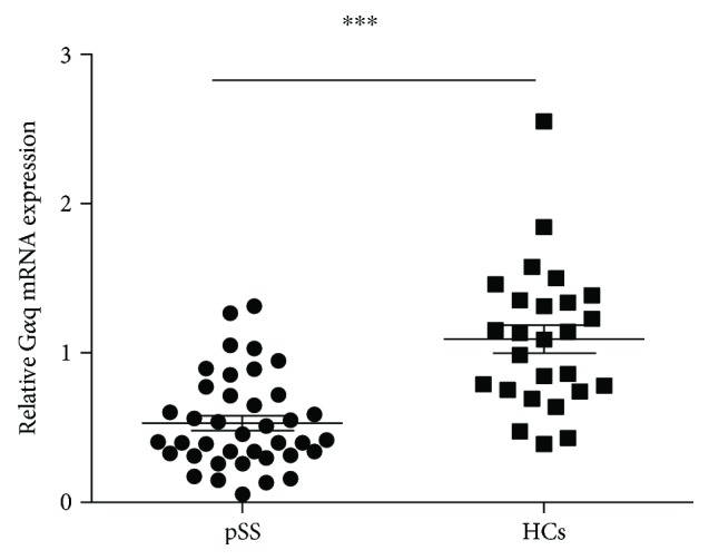
Expression of Gαq was decreased in lymphocytes of pSS patients. Peripheral blood mononuclear cells (PBMCs) from pSS (n = 39) and HC (n = 26) were collected. Gαq mRNA expression in PBMCs was analyzed by RT-PCR. The mRNA expression level of Gαq was significantly higher in pSS patients compared with HCs. ∗∗∗P < 0.001.
3.2. Expression of Gαq Was Negatively Associated with Disease Activity in Patients with pSS
To further investigate the specific role of Gαq in pSS pathogenesis regulation, we then analyzed the disease activity of patients with pSS with different expression levels of Gαq. The EULAR Sjögren's syndrome disease activity index (ESSDAI) is now widely used to evaluate pSS disease activity [14]. We analyzed the relation of Gαq mRNA expression in PBMCs with the ESSDAI in patients with pSS. As shown in Figure 2, we found the expression level of Gαq mRNA was negatively related to the ESSDAI in patients with pSS. This suggested that Gαq negatively regulates the pathogenesis of pSS and that a low level of Gαq might contribute to the disease onset of pSS.
Figure 2.
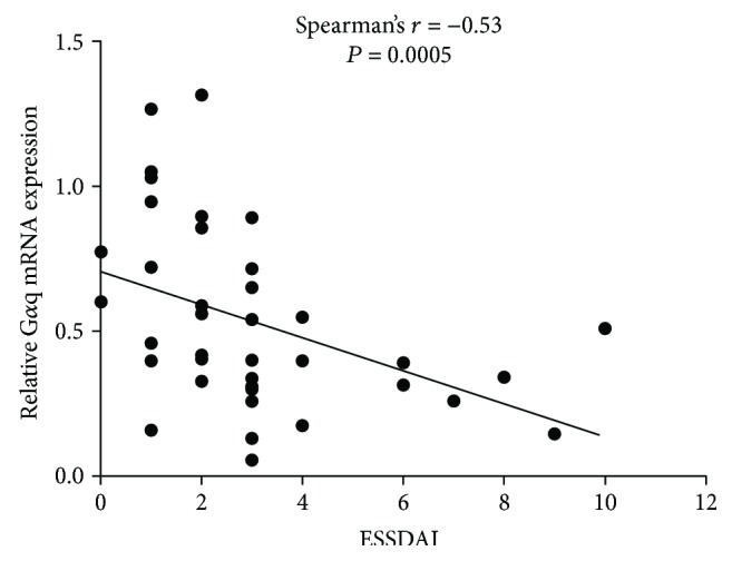
Expression of Gαq was negatively associated with the disease activity of pSS patients. Peripheral blood mononuclear cells (PBMCs) from pSS (n = 39) patients were collected. Gαq mRNA expression in PBMCs was analyzed by RT-PCR. The correlation between Gαq mRNA expression level and ESSDAI in pSS patients was determined by using Spearmen's rank test.
3.3. Clinical Significance of Gαq in Patients with pSS
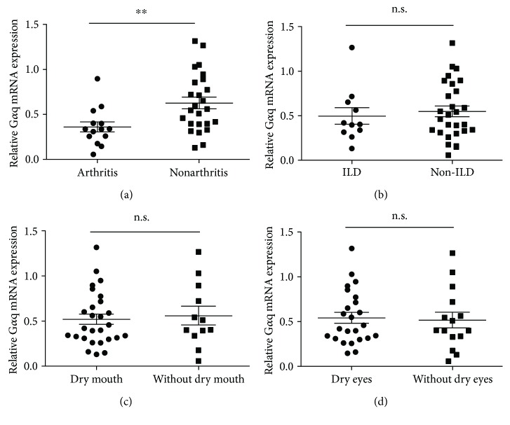
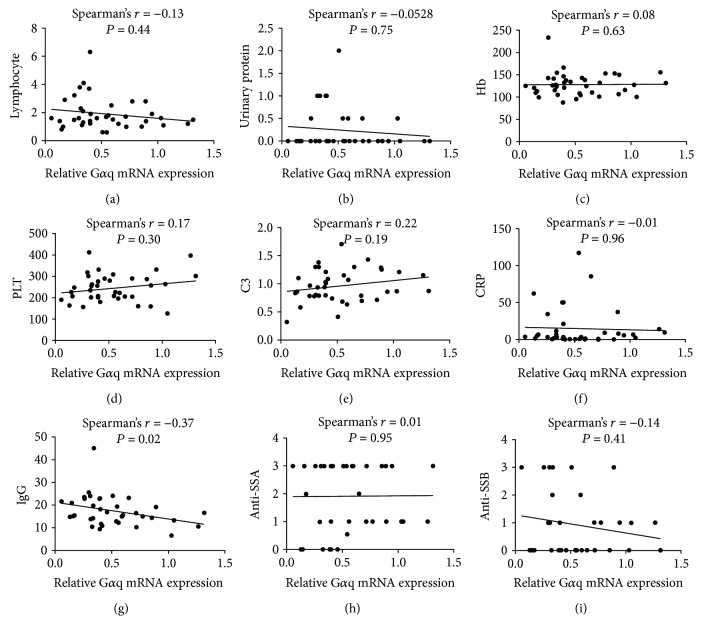
We then analyzed the association of the expression of Gαq with the clinical characteristics of patients with pSS to explore the clinical significance of Gαq in pSS. The expression of Gαq was significantly lower in pSS patients with arthritis than that in those without arthritis (Figure 3(a)), suggesting that Gαq might be involved in the pathogenesis of arthritis. However, no differences in Gαq expression between pSS patients with and without interstitial lung disease (ILD), dry eyes, and dry mouth were found (Figures 3(b)–3(d)). We further analyzed the association of Gαq mRNA expression with other clinical characteristics, including lymphocyte count in peripheral blood, urinary protein, hemoglobin (HB) level, platelet (PLT) level, complement 3 (C3) level, C-reactive protein (CRP) level, immunoglobulin G (IgG) level, anti-SSA titer, and anti-SSB titer (Figure 4). A significant negative association was found between Gαq mRNA expression and IgG level, whereas no association was found for the others. These data suggest that Gαq plays a role in the negative regulation of the immune reaction.
Figure 3.
Expression level of Gαq was much more lower in pSS patients with arthritis. Gαq mRNA expression in PBMCs of pSS (n = 39) patients was analyzed by RT-PCR. (a) Gαq mRNA expression in pSS patients with and without arthritis. (b) Gαq mRNA expression in pSS patients with and without interstitial lung diseases (ILD). (c) Gαq mRNA expression in pSS patients with and without dry mouth. (d) Gαq mRNA expression in pSS patients with and without dry eyes. ∗∗P < 0.01.
Figure 4.
Expression level of Gαq was negatively associated with IgG level. Association of Gαq mRNA expression with lymphocyte count in peripheral blood (a), urinary protein (b), level of hemoglobin (HB) (c), platelet (PLT) (d), complement 3 (C3) (e), C-reactive protein (CRP) (f), immunoglobulin G (IgG), titer of anti-SSA (h), and titer of anti-SSB (i). Significant negative association was found between Gαq mRNA expression and IgG level.
3.4. Gαq Expression Was Negatively Related to IL-17A Concentration, Which May Contribute to the Mechanism by Which Gαq Is Involved in pSS Pathogenesis Regulation
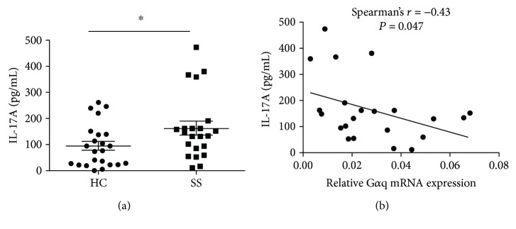
Our data suggest that Gαq is involved in pSS pathogenesis regulation, but the mechanism remains unclear. Th17 is a key factor in pSS pathogenesis, and our previous researches demonstrated that Gαq inhibited the differentiation of Th17 [11]. We further investigated the correlation between Gαq and Th17 in patients with pSS. We first investigated the level of IL-17A in the serum of patients with pSS and HCs. We found that the concentration of IL-17A was significantly higher in patients with pSS than in HCs (Figure 5(a)). We then analyzed the correlation of Gαq mRNA expression in PBMCs from patients with pSS with IL-17A. A negative correlation was found between Gαq mRNA expression and IL-17A expression (Figure 5(b)). These data suggested that Gαq might be a factor involved in Th17 regulation in patients with pSS.
Figure 5.
Level of IL-17A was increased in pSS patients and negatively correlated with Gαq mRNA expression. (a) Level of IL-17A in serum of pSS (n = 22) patients and HCs (n = 23). Level IL-17A was significantly higher in pSS patients than in HCs. (b) The correlation between Gαq mRNA expression level and IL-17A in pSS patients (n = 22) was determined by using Spearmen's rank test. ∗P < 0.05.
4. Discussion
In this study, we report for the first time that Gαq mRNA expression in lymphocytes was decreased in patients with pSS and that the expression of Gαq was closely related to disease activity. Gαq mRNA expression was negatively associated with IL-17A levels in the serum of patients with pSS. Our study revealed that Gαq plays a role in pSS pathogenesis regulation, providing a new mechanism for how Th17 cells are regulated in pSS.
pSS is a disorder in which dry eyes and dry mouth occur as a manifestation of immune dysregulation [15, 16]. Dry eyes and mouth are often the first symptoms of SS and may indicate the involvement of other organs, including the salivary glands, lungs, and kidneys. Roughly 5–10% of patients with SS develop lymphoma [17]. Treatment strategies for pSS are relatively limited because the irreversible destruction of the salivary glands is always present when pSS is diagnosed. Studies on the pathogenesis mechanism of pSS are needed to clarify the early stages of pSS. Th17 cells have been shown to be a factor in pSS pathogenesis. IL-17 KO mice were shown to be completely resistant to SS induction, and the adoptive transfer of Th17 cells induced the presence of SS symptoms in immunized IL-17 KO mice rapidly, proving the crucial role of Th17 in pSS pathogenesis [18]. Increased levels of IL-17 were also found in the serum as well as the salivary glands of patients with pSS [6, 19]. Consistent with previous studies, we also found that IL-17A was increased in the serum of patients with pSS, confirming the crucial role of Th17 cells in pSS pathogenesis. However, how the abnormal Th17 cells are regulated in pSS remains unclear. Studies on the regulation mechanism of Th17 cells in pSS can help to better understand the stage before the onset of Th17 cell upregulation and the destruction of salivary glands and help to develop better management strategies for pSS in its early stages.
Gαq is the α-subunit of the Gq protein encoded by gene GNAQ [8]. Our previous studies confirmed the vital role of Gαq in several aspects of immune regulation such as dendritic cell trafficking, B cell selection, and T cell activation [9, 10, 20]. By using Gnaq−/− chimeric mice by reconstituting lethally irradiated C57BL/6J recipient mice with Gnaq−/− bone marrow, we also revealed the role of Gαq in the pathogenesis of autoimmune disease. Autoimmunity with multiorgan involvement and arthritis can spontaneously develop in Gnaq−/− chimeric mice [10]. In previous studies, the expression of Gαq was shown to decrease in lymphocytes from patients with RA and SLE and closely related to disease activity, indicating the role of Gαq in RA and SLE pathogenesis regulation [11, 12, 21, 22]. Thus, these studies revealed a new mechanism for autoimmune disease pathogenesis regulation.
This study demonstrated the role of Gαq in pSS. We found that the expression of Gαq was also decreased in lymphocytes from patients with pSS and that the expression level of Gαq was closely associated with the disease activity of pSS, presence of arthritis, and high level of IgG. Our previous studies demonstrated that Gnaq−/− chimeric mice spontaneously developed inflammatory arthritis [10], and the expression of Gαq was shown to be decreased in lymphocytes from RA patients, suggesting that Gαq is involved in the regulation of inflammatory arthritis development. pSS shares some similarities with RA regarding the pathogenesis mechanism and is often coexisted with RA [23]. Our studies have shown that Gαq is decreased in both patients with pSS and patients with RA, suggesting that Gαq might contribute to the overlap in the pathogenesis mechanisms of pSS and RA. Furthermore, we found that Gαq expression was lower in patients with pSS with arthritis compared with that in those without arthritis, suggesting that a low expression level of Gαq may be used as a predictor for the presence of arthritis in pSS. However, prospective studies are needed to confirm the ability of the Gαq level to predict arthritis in pSS.
It was demonstrated that Gαq could negatively regulate Th17 differentiation in our previous studies, and a negative association was found between the expression of Gαq and IL-17A in patients with RA [11]. In this study, we also found a negative association between Gαq and IL-17A, revealing a novel regulation mechanism for the abnormal Th17 levels in patients with pSS. This represents a potential new research target for the early stages of pSS.
In conclusion, this study showed that Gαq expression is involved in pSS pathogenesis regulation, possibly because of its regulation of Th17 cells. These results provide a new mechanism for pSS pathogenesis regarding abnormal Th17 cell regulation.
Acknowledgments
The work was supported by NSFC (Natural Science Foundation of China) Grant U1605223 to Dr. Guixiu Shi, NSFC grant 81501407 to Dr. Yuan Liu; NSFC grant 81501369 to Dr. Shiju Chen; and the Leadership Program of the Technology Department of Fujian Province (Grant no. 2015D011) to Dr. Guixiu Shi.
Contributor Information
Yuan Liu, Email: liuyuancuto@163.com.
Guixiu Shi, Email: guixiu.shi@gmail.com.
Data Availability
The data used and/or analyzed in the current study are available from the corresponding author on reasonable request.
Conflicts of Interest
The authors declare that they have no conflict of interests.
Authors' Contributions
Yuechi Sun, Ying Wang, and Shiju Chen contributed equally to this study.
References
- 1.Tzioufas A. G., Kapsogeorgou E. K., Moutsopoulos H. M. Pathogenesis of Sjögren’s syndrome: what we know and what we should learn. Journal of Autoimmunity. 2012;39(1-2):4–8. doi: 10.1016/j.jaut.2012.01.002. [DOI] [PubMed] [Google Scholar]
- 2.Nguyen C. Q., Peck A. B. Unraveling the pathophysiology of Sjogren syndrome-associated dry eye disease. The Ocular Surface. 2009;7(1):11–27. doi: 10.1016/S1542-0124(12)70289-6. [DOI] [PMC free article] [PubMed] [Google Scholar]
- 3.Miossec P., Korn T., Kuchroo V. K. Interleukin-17 and type 17 helper T cells. The New England Journal of Medicine. 2009;361(9):888–898. doi: 10.1056/NEJMra0707449. [DOI] [PubMed] [Google Scholar]
- 4.Cua D. J., Sherlock J., Chen Y., et al. Interleukin-23 rather than interleukin-12 is the critical cytokine for autoimmune inflammation of the brain. Nature. 2003;421(6924):744–748. doi: 10.1038/nature01355. [DOI] [PubMed] [Google Scholar]
- 5.Sakai A., Sugawara Y., Kuroishi T., Sasano T., Sugawara S. Identification of IL-18 and Th17 cells in salivary glands of patients with Sjögren's syndrome, and amplification of IL-17-mediated secretion of inflammatory cytokines from salivary gland cells by IL-18. The Journal of Immunology. 2008;181(4):2898–2906. doi: 10.4049/jimmunol.181.4.2898. [DOI] [PubMed] [Google Scholar]
- 6.Katsifis G. E., Rekka S., Moutsopoulos N. M., Pillemer S., Wahl S. M. Systemic and local interleukin-17 and linked cytokines associated with Sjögren’s syndrome immunopathogenesis. The American Journal of Pathology. 2009;175(3):1167–1177. doi: 10.2353/ajpath.2009.090319. [DOI] [PMC free article] [PubMed] [Google Scholar]
- 7.Nguyen C. Q., Yin H., Lee B. H., Carcamo W. C., Chiorini J. A., Peck A. B. Pathogenic effect of interleukin-17A in induction of Sjögren’s syndrome-like disease using adenovirus-mediated gene transfer. Arthritis Research & Therapy. 2010;12(6, article R220) doi: 10.1186/ar3207. [DOI] [PMC free article] [PubMed] [Google Scholar]
- 8.Oldham W. M., Hamm H. E. Heterotrimeric G protein activation by G-protein-coupled receptors. Nature Reviews Molecular Cell Biology. 2008;9(1):60–71. doi: 10.1038/nrm2299. [DOI] [PubMed] [Google Scholar]
- 9.Shi G., Partida-Sánchez S., Misra R. S., et al. Identification of an alternative Gαq-dependent chemokine receptor signal transduction pathway in dendritic cells and granulocytes. Journal of Experimental Medicine. 2007;204(11):2705–2718. doi: 10.1084/jem.20071267. [DOI] [PMC free article] [PubMed] [Google Scholar]
- 10.Misra R. S., Shi G., Moreno-Garcia M. E., et al. Gαq-containing G proteins regulate B cell selection and survival and are required to prevent B cell–dependent autoimmunity. Journal of Experimental Medicine. 2010;207(8):1775–1789. doi: 10.1084/jem.20092735. [DOI] [PMC free article] [PubMed] [Google Scholar]
- 11.Liu Y., Wang D., Li F., Shi G. Gαq controls rheumatoid arthritis via regulation of Th17 differentiation. Immunology & Cell Biology. 2015;93(7):616–624. doi: 10.1038/icb.2015.13. [DOI] [PubMed] [Google Scholar]
- 12.He Y., Huang Y., Tu L., et al. Decreased Gαq expression in T cells correlates with enhanced cytokine production and disease activity in systemic lupus erythematosus. Oncotarget. 2016;7(52):85741–85749. doi: 10.18632/oncotarget.13903. [DOI] [PMC free article] [PubMed] [Google Scholar]
- 13.Shiboski S. C., Shiboski C. H., Criswell L. A., et al. American College of Rheumatology classification criteria for Sjögren’s syndrome: a data-driven, expert consensus approach in the Sjögren’s International Collaborative Clinical Alliance cohort. Arthritis Care & Research. 2012;64(4):475–487. doi: 10.1002/acr.21591. [DOI] [PMC free article] [PubMed] [Google Scholar]
- 14.Seror R., Ravaud P., Bowman S. J., et al. EULAR Sjögren’s syndrome disease activity index: development of a consensus systemic disease activity index for primary Sjögren’s syndrome. Annals of the Rheumatic Diseases. 2010;69(6):1103–1109. doi: 10.1136/ard.2009.110619. [DOI] [PMC free article] [PubMed] [Google Scholar]
- 15.Thanou-Stavraki A., James J. A. Primary Sjögren’s syndrome: current and prospective therapies. Seminars in Arthritis & Rheumatism. 2008;37(5):273–292. doi: 10.1016/j.semarthrit.2007.06.002. [DOI] [PubMed] [Google Scholar]
- 16.Latkany R. Dry eyes: etiology and management. Current Opinion in Ophthalmology. 2008;19(4):287–291. doi: 10.1097/ICU.0b013e3283023d4c. [DOI] [PubMed] [Google Scholar]
- 17.Theander E., Henriksson G., Ljungberg O., Mandl T., Manthorpe R., Jacobsson L. T. Lymphoma and other malignancies in primary Sjögren’s syndrome: a cohort study on cancer incidence and lymphoma predictors. Annals of the Rheumatic Diseases. 2006;65(6):796–803. doi: 10.1136/ard.2005.041186. [DOI] [PMC free article] [PubMed] [Google Scholar]
- 18.Lin X., Rui K., Deng J., et al. Th17 cells play a critical role in the development of experimental Sjögren’s syndrome. Annals of the Rheumatic Diseases. 2015;74(6):1302–1310. doi: 10.1136/annrheumdis-2013-204584. [DOI] [PubMed] [Google Scholar]
- 19.Nguyen C. Q., Hu M. H., Li Y., Stewart C., Peck A. B. Salivary gland tissue expression of interleukin-23 and interleukin-17 in Sjögren’s syndrome: findings in humans and mice. Arthritis & Rheumatism. 2008;58(3):734–743. doi: 10.1002/art.23214. [DOI] [PMC free article] [PubMed] [Google Scholar]
- 20.Wang D., Zhang Y., He Y., Li Y., Lund F. E., Shi G. The deficiency of Gαq leads to enhanced T-cell survival. Immunology & Cell Biology. 2014;92(9):781–790. doi: 10.1038/icb.2014.53. [DOI] [PubMed] [Google Scholar]
- 21.Wang D., Liu Y., Li Y., He Y., Zhang J., Shi G. Gαq regulates the development of rheumatoid arthritis by modulating Th1 differentiation. Mediators of Inflammation. 2017;2017:9. doi: 10.1155/2017/4639081.4639081 [DOI] [PMC free article] [PubMed] [Google Scholar]
- 22.Wang Y., Li Y., He Y., et al. Expression of G protein αq subunit is decreased in lymphocytes from patients with rheumatoid arthritis and is correlated with disease activity. Scandinavian Journal of Immunology. 2012;75(2):203–209. doi: 10.1111/j.1365-3083.2011.02635.x. [DOI] [PubMed] [Google Scholar]
- 23.He J., Ding Y., Feng M., et al. Characteristics of Sjögren’s syndrome in rheumatoid arthritis. Rheumatology. 2013;52(6):1084–1089. doi: 10.1093/rheumatology/kes374. [DOI] [PubMed] [Google Scholar]
Associated Data
This section collects any data citations, data availability statements, or supplementary materials included in this article.
Data Availability Statement
The data used and/or analyzed in the current study are available from the corresponding author on reasonable request.


