Abstract
Manifestation of appropriate behavior in adult animals requires developmental mechanisms that help in the formation of correctly wired neural circuits. Flight circuit development in Drosophila requires store-operated calcium entry (SOCE) through the STIM/Orai pathway. SOCE-associated flight deficits in adult Drosophila derive extensively from regulation of gene expression in pupal neurons, and one such SOCE-regulated gene encodes the small GTPase Ral. The cellular mechanism by which Ral helps in maturation of the flight circuit was not understood. Here, we show that knockdown of components of a Ral effector, the exocyst complex, in pupal neurons also leads to reduced flight bout durations, and this phenotype derives primarily from dopaminergic neurons. Importantly, synaptic release from pupal dopaminergic neurons is abrogated upon knockdown of dSTIM, Ral, or exocyst components. Ral overexpression restores the diminished synaptic release of dStim knockdown neurons as well as flight deficits associated with dSTIM knockdown in dopaminergic neurons. These results identify Ral-mediated vesicular release as an effector mechanism of neuronal SOCE in pupal dopaminergic neurons with functional consequences on flight behavior.
Keywords: Exo84, Neural Circuit, SOCE, Synaptic Maturation
Significance Statement
Appropriate wiring of neuronal circuits during development is essential for adult behavior, and cellular mechanisms such as calcium signaling orchestrate the pattern and strength of such neuronal connections to a significant extent. In Drosophila, flight behavior impacts multiple aspects of life. Calcium, especially through the store-operated calcium entry (SOCE) pathway, regulates gene expression during flight circuit maturation. Ral encodes a small GTPase and is one such SOCE-regulated gene required for Drosophila flight. In this paper, we show that SOCE-related loss of flight is determined to a significant extent by Ral and the exocyst complex–driven synaptic vesicle release in pupal dopaminergic neurons. Thus, SOCE-regulated release of dopamine ensures correct wiring of the flight circuit in Drosophila.
Introduction
The coordinated development and maturation of neuronal circuits is essential for ensuring appropriate circuit function underlying adult behavior. Neural circuits develop and reach functional maturity through a complex process that begins with genetically encoded programs for neural cell specification and is followed by neurotransmitter fate determination, axonal growth and path finding, synapse formation, and synapse maturation (Lohmann, 2009; Blankenship and Feller, 2010). Based on the context, various calcium signaling mechanisms influence circuit development (Lohmann, 2009; Rosenberg and Spitzer, 2011), including store-operated calcium entry (SOCE) through the STIM/Orai pathway (Prakriya and Lewis, 2015). Although STIM and Orai are expressed at multiple stages (Majewski and Kuznicki, 2015), their precise role in neural development and circuit formation needs better understanding.
In the holometabolous insect Drosophila, the nervous system, like all other organs, undergoes metamorphosis during pupal stages to attain the adult form from the distinct larval form (Truman, 1990). Most neurogenesis is accomplished in the embryonic and larval stages followed by remodeling of existing neurons during pupal stages in tune with adult functions (Truman and Bate, 1988; Tissot and Stocker, 2000; Consoulas et al., 2002). Interestingly, attenuation of STIM/Orai-mediated SOCE in pupal neurons leads to either absent or reduced flight bout durations (Agrawal et al., 2010; Pathak et al., 2015; Richhariya et al., 2017), supporting a role for SOCE during flight circuit maturation in Drosophila.
In non-excitable cells, SOCE regulates cellular responses by changes in gene expression (Feske, 2007). Indeed, SOCE-regulated expression of the dopamine-synthesizing enzyme tyrosine hydroxylase has previously been demonstrated in Drosophila pupal neurons (Pathak et al., 2015). In mouse neural progenitor cells as well, SOCE drives gene expression (Somasundaram et al., 2014). In a screen to identify SOCE-regulated genes in Drosophila pupal neurons, a small GTPase, Ral, was identified as a regulator of flight (Richhariya et al., 2017).
Mammalian RalA has several roles, many of which are exocyst linked, while some are not (Gentry et al., 2014). RalA regulates the releasable pool of synaptic vesicles in mammalian neurons (Polzin et al., 2002), and both RalA and RalB mediate GTP-dependent exocytosis from neuroendocrine PC-12 cells (Wang et al., 2004; Li et al., 2007). In Drosophila, Ral-dependent exocyst function supports membrane addition in muscle cells (Teodoro et al., 2013), and similarly, in mouse cortical neurons, it supports neurite extension (Lalli and Hall, 2005). However, dendritic and axonal arborization patterns of central dopaminergic neurons implicated in Drosophila flight appear normal on attenuation of SOCE (Pathak et al., 2015).
Here we have investigated an alternate exocyst-dependent cellular mechanism by which SOCE-regulated Ral expression could help in maturation of the Drosophila flight circuit during pupal development. We show that release of synaptic vesicles in maturing pupal neurons requires the SOCE component dSTIM as well as Ral-exocyst function. We propose that dSTIM- and Ral/exocyst-dependent vesicular release is required for synaptic maturation of the flight circuit.
Materials and Methods
Fly rearing and stocks
Drosophila strains were grown on cornmeal medium supplemented with yeast. For all experiments, flies of either sex were used. For all experiments, unless stated otherwise, egg laying was performed at 25°C. Late third instar larvae were moved to 29°C to increase the expression of GAL4 and were maintained at the elevated temperature until adults eclosed, after which they were moved back to 25°C. For experiments involving GAL80ts, knockdown of Exo84 or expression of RalDN was achieved at specific developmental stages by raising the temperature to 29°C while maintaining them at 18°C for the remaining life cycle. For experiments with dTrpA1, flies were maintained at 22°C, and 0–2-h-old pupae were collected and transferred to 29°C for 72 h and returned to 22°C before eclosion. Fly strains used in this study are listed in Table 1.
Table 1.
List of fly strains used
| Fly line | Description | Source |
|---|---|---|
| elavC155 GAL4 | Pan-neuronal driver | BDSC 458 |
| OK371-GAL4 | Glutamatergic driver | BDSC 26160 |
| TH-GAL4 | Dopaminergic driver | Friggi-Grelin et al., 2003a |
| TH-D1 GAL4 | Dopaminergic subset neuron driver | Liu et al., 2012 |
| TH-D’ GAL4 | Dopaminergic subset neuron driver | Liu et al., 2012 |
| TH-A GAL4 | Hypoderm specific dopaminergic driver | Liu et al., 2012 |
| TRH GAL4 | Serotonergic driver | Sadaf et al., 2012 |
| tubGAL80ts | GAL80ts (2 copies) under tubulin promoter | McGuire et al., 2003 |
| UAS-ANF::GFP | Rat ANF peptide tagged to GFP | BDSC 7001 |
| UAS-spH | SynaptopHluorin | Ng et al., 2002 |
| UAS-GCaMP6m | Calcium sensor | BDSC 42748 |
| UAS-dTrpA1 | Temperature sensitive cation channel | BDSC 26263 |
| UAS-H2BmRFP | Nuclear RFP | Gift from Boris Egger Langevin et al., 2005 |
| UAS-Shits | Temperature-sensitive Dynamin mutant | Kitamoto, 2001 |
| UAS-RalDN | Dominant negative form of Ral | BDSC 32094 |
| UAS-RalIR | RNAi against Ral | BDSC 29580 |
| UAS-dStimIR | RNAi against dStim | VDRC 47073 |
| UAS-RalWT | Wild-type Ral | Richhariya et al., 2017 |
| UAS-Exo84IR | RNAi against Exo84 | VDRC 30111 |
| UAS-Exo84IR (line 2) | RNAi against Exo84 | BDSC 28712 |
| UAS-Sec5IR | RNAi against Sec5 | VDRC 28874 |
| UAS-Sec6IR | RNAi against Sec6 | VDRC 105836 |
| UAS-Sec6IR (line 2) | RNAi against Sec6 | VDRC 22079 |
Single flight assay
3–5-d-old flies of either sex were anaesthetized on ice for ∼2 min and then tethered between the head and the thorax using clear nail polish on a thin metal wire. On recovery, they were given an air puff to initiate flight. The duration of flight (up to 15 min) was recorded for each fly in batches of 5–10 flies. Flight times of individual flies are represented as box plots.
Pupal neuronal culture
24–48-h-old pupal CNSs were dissected and enzymatically digested with 50 units/ml papain (Sigma-Aldrich) activated by 1.32 mm cysteine (Sigma-Aldrich). They were then washed with DDM2 ([DMEM/F-12 with GlutaMAX (Gibco) supplemented with 100 units/ml penicillin-streptomycin (Gibco), 10 µg/ml Amphotericin B (Gibco), 20 mm HEPES (Sigma-Aldrich), 50 µg/ml insulin (Sigma-Aldrich), and 20 ng/ml progesterone (Sigma-Aldrich)], triturated with a pipette, and plated in 200 µl DDM2 for ∼4 CNS per plate coated with 0.1 mg/ml poly-d-lysine (Sigma-Aldrich). Cultures were incubated in an incubator at 25°C with 5% CO2 for 18–24 h before imaging.
Live imaging for vesicular release using ANF::GFP
Pupal cultures washed and imaged in HL3 (70 mm NaCl, 5 mm KCl, 20 mm MgCl2, 10 mm NaHCO3, 5 mm trehalose, 115 mm sucrose, 5 mm HEPES, pH 7.2) with calcium (1.5 mm). Images were taken as a time series on an XY plane at an interval of 4 s using a 40× objective with an NA of 1.4 on an Olympus FV1000 inverted confocal microscope (Olympus Corp.). The raw images were extracted using Fiji (Schindelin et al., 2012), and regions of interest (ROIs) representing cells were selected using the Time Series Analyzer plugin. Percentage ΔF/F release was calculated as (F0 – Ft)/F0 × 100, where Ft is the fluorescence at time t and F0 is baseline fluorescence corresponding to the average fluorescence over the first 10 frames. The area under the curve from these response curves was calculated from 300 to 900 s using Microsoft Excel (Microsoft) and implementing the trapezoidal rule (Nedelman and Gibiansky, 1996; Qin et al., 2009) where
Here, t1 = 300 s and t2 = 900 s, and this accounts for both positive and negative AUC measurements. For experiments involving UAS-Shit s, a heated microscope stage was used.
Ex vivo synaptopHluorin imaging from pupal brains
Brains were dissected in larval hemolymph-like saline (HL3) consisting of 108 mm NaCl, 5 mm KCl, 2 mm CaCl2, 8.2 mm MgCl2, 4 mm NaHCO3, 1 mm NaH2PO4, 5 mm trehalose, 10 mm sucrose, 5 mm Tris, pH 7.5, and filtered through a 0.2-μm filter (Makos et al., 2010). Dissected brains expressing synaptopHluorin (spH) were embedded in 0.2% low-melt agarose and bathed in HL3. Images were taken as a time series across depth at an interval of 4 s using a 10× objective with an NA of 0.4 on a Leica SP5 confocal microscope using a resonant scanner allowing for fast scan speeds. Images were analyzed using Fiji. The XYZ time series was projected along Z using the maximum-intensity function on Fiji. ROIs were selected using the Time Series Analyser plugin. ΔF/F was calculated as (Ft – F0)/F0, where F0 is baseline fluorescence corresponding to the average fluorescence over the first 10 frames and Ft is the fluorescence at time t. Area under the curve was calculated from 60 to 300 s for saline-induced release and from 300 to 600 s for evoked release by standard methods invoking the trapezoid rule as mentioned above using Microsoft Excel.
Data representation and statistics
All flight data and quantifications of vesicular release are represented as box plots using BoxplotR (Spitzer et al., 2014), where horizontal lines represent medians, crosses indicate means, box limits indicate 25th and 75th percentiles, whiskers extend 1.5 times the interquartile range from the 25th and 75th percentiles, individual data points are represented as open circles, and the numbers below represent the n number for each box. Traces from live-imaging experiments represent means (±standard error of means) from all cells or ROIs.
Comparisons between two samples were performed using the two-tailed Student’s t test. If more than two conditions were involved, one-way ANOVA followed by Tukey’s test was used. All statistical tests were performed using Origin 8.0 software (Micro Cal). Statistical tests and exact p-values used in each figure are listed in Table 1-1.
Statistical tests and p-values for all figures. Download Table 1-1, XLSX file (29.7KB, xlsx) .
Results
Exocyst function during pupal development is required for adult flight maintenance
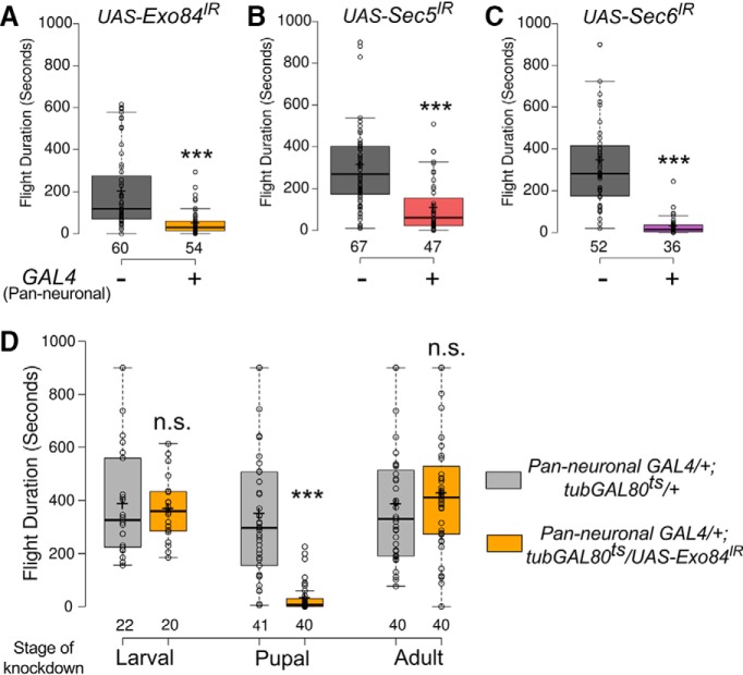
Inhibition or knockdown of the small GTPase Ral in pupal neurons leads to significant flight deficits in adult flies (Richhariya et al., 2017). To test if Ral function in pupal neurons is exocyst dependent, three components of the exocyst complex, Sec5, Sec6, and Exo84 (Finger and Novick, 1998; Guo et al., 1999) were independently knocked down by pan-neuronal expression of RNA interference constructs with the GAL4 driver ElavC155 (Lin and Goodman, 1994). Knockdown of each of the three exocyst components resulted in significantly shortened flight durations compared to the corresponding RNAi controls (Fig. 1A–C). This indicates a role for the exocyst complex in flight, as none of the RNAis used have any predicted off-targets (Dietzl et al., 2007). Furthermore, significant flight defects were also obtained with independent RNAi lines for Exo84 and Sec6 (Fig 1-1A,B).
Figure 1.
Exocyst components are required in pupal neurons for maintaining the duration of adult flight bouts (refer to Figs. 1-1 and 1-2 and Table 1-1). A–C, Box plots represent durations of flight bouts in flies from the indicated genotypes measured by the single flight assay. D, Box plots represent durations of flight bouts in flies with knockdown of Exo84 in neurons during the indicated stages of development. In the box plots, horizontal lines represent medians, crosses indicate means, box limits indicate 25th and 75th percentiles, whiskers extend 1.5 times the interquartile range from the 25th and 75th percentiles, individual data points are represented as open circles, and the numbers below represent the n number for each box. ***, p < 0.001, n.s., not significant at p < 0.05 by two-tailed Student’s t test. For exact p-values, refer to Table 1-1.
The role of exocyst components in flight. A, B, Box plots represent flight durations of flies of the indicated genotypes. Knockdown of Exo84 and Sec6 with alternate RNAi lines resulted in similar phenotypes as shown in Fig. 1. These flies were grown at 25°C throughout. C, D, Box plots represent flight durations of control flies with RNAi-mediated knockdown of Exo84 (Exo84IR) in neurons raised at the indicated temperatures throughout development. At the permissive temperature of 18°C, there is no knockdown. GAL4-mediated knockdown of Exo84 occurs at the restrictive temperature of 29°C. Box plot symbols are as described in Fig. 1. *, p < 0.05, **, p < 0.01, ***, p < 0.001, n.s., not significant at p < 0.05 by two-tailed Student’s t test (for A and B) or one-way ANOVA followed by post hoc Tukey’s test (for C and D). All comparisons for significance were with the control values except where marked by a horizontal line. For exact p-values, refer to Table 1-1. Download Figure 1-1, TIF file (654.7KB, tif) .
Expression of exocyst components is not regulated by SOCE. A–D, Bar graphs represent the level of expression of the indicated genes using values for fragments per kilobase gene per million reads (FPKM) in control and dStim knockdown pupal brains. These data were obtained from the transcriptomic screen performed in Richhariya et al. (2017). q-values are as obtained by CuffDiff. Download Figure 1-2, TIF file (321.5KB, tif) .
The temporal requirement of the exocyst complex for flight was investigated by knockdown of Exo84, as a proxy for the complex, at different developmental stages using the TARGET system (McGuire et al., 2004). Knockdown in the pupal stage alone resulted in flight deficits equivalent to those observed by knockdown throughout development, suggesting that Exo84 expression is exclusively required during pupal development for maturation of the flight circuit. Indeed, Exo84 knockdown exclusively in either the larval or adult stage did not alter flight durations significantly from the respective controls (Figs. 1D and 1-1C,D). This temporal requirement of Exo84 in the context of flight coincides with that of Ral and SOCE observed earlier (Richhariya et al., 2017). Therefore, regulation of exocyst components by SOCE was tested next. There was no change in mRNA levels of the three exocyst components, Exo84, Sec5, and Sec6, in pupal neurons with knockdown of a core SOCE component, the ER-Ca2+ sensor, dStim, when Ral levels were found to be reduced (Richhariya et al., 2017; Fig. 1-2A–D). These data demonstrate a requirement for SOCE, Ral, and Exo84 in pupal neurons for adult flight maintenance and suggest that SOCE modulates exocyst-dependent vesicular release in maturing pupal neurons, most likely by regulating Ral expression as identified earlier (Richhariya et al., 2017).
Ral functions downstream of dStim to mediate vesicular release in pupal neurons
To measure vesicular release in pupal neurons, we used the rat atrial natriuretic peptide tagged with emerald GFP (ANF::GFP). ANF::GFP is recognized by the Drosophila protein-sorting machinery, and its expression is detected in all neurons after embryonic stage 17 when driven by a pan-neuronal GAL4 (Rao et al., 2001). Vesicular release by ANF::GFP expression has been measured from peptidergic (Kula et al., 2006) and nonpeptidergic (Levitan et al., 2007; Liu et al., 2012) neurons in vivo. The vesicular release assay in pupal neurons was standardized by expression of a temperature-sensitive dynamin mutant, Shibirets (Shits1), that reversibly blocks vesicular release in Drosophila neurons at 29°C but not at 22°C (Kitamoto, 2001) and can also block peptide release at the restrictive temperature (Wong et al., 2015). In neurons expressing both ANF::GFP and Shits1, depolarization by KCl at 22°C resulted in a decrease in ANF::GFP intensity at the soma of cultured neurons over time (Fig. 2-1A–C). To rule out effects of elevated temperature on neuronal activity–mediated release (Tomchik, 2013), or on GFP fluorescence, we performed the same experiment without Shits1 at 29°C and observed release comparable to that at the permissive temperature of 22°C with Shits1 (Fig. 2-1B,C). The small rise observed just before stimulation could be either basal release or possibly an artifact of photobleaching, as it was similar under all conditions (Fig. 2-1B,D). Furthermore, the drop in intensity of ANF::GFP was specific to a depolarization stimulus and not due to osmotic shock, because replacement of KCl with NaCl (at the same concentration as KCl) did not result in reduction of ANF::GFP intensity above baseline levels (Fig. 2-1C,D). Thus, we interpret reduction in GFP intensity after KCl addition as vesicular release from the soma due to depolarization. Blocking vesicular recycling by subjecting neurons expressing Shits1 to the restrictive temperature (29°C) while imaging led to a significant reduction in release of ANF::GFP compared to the permissive temperature (Fig. 2-1A–C), validating this assay for assessing vesicular release in primary cultures of pupal neurons.
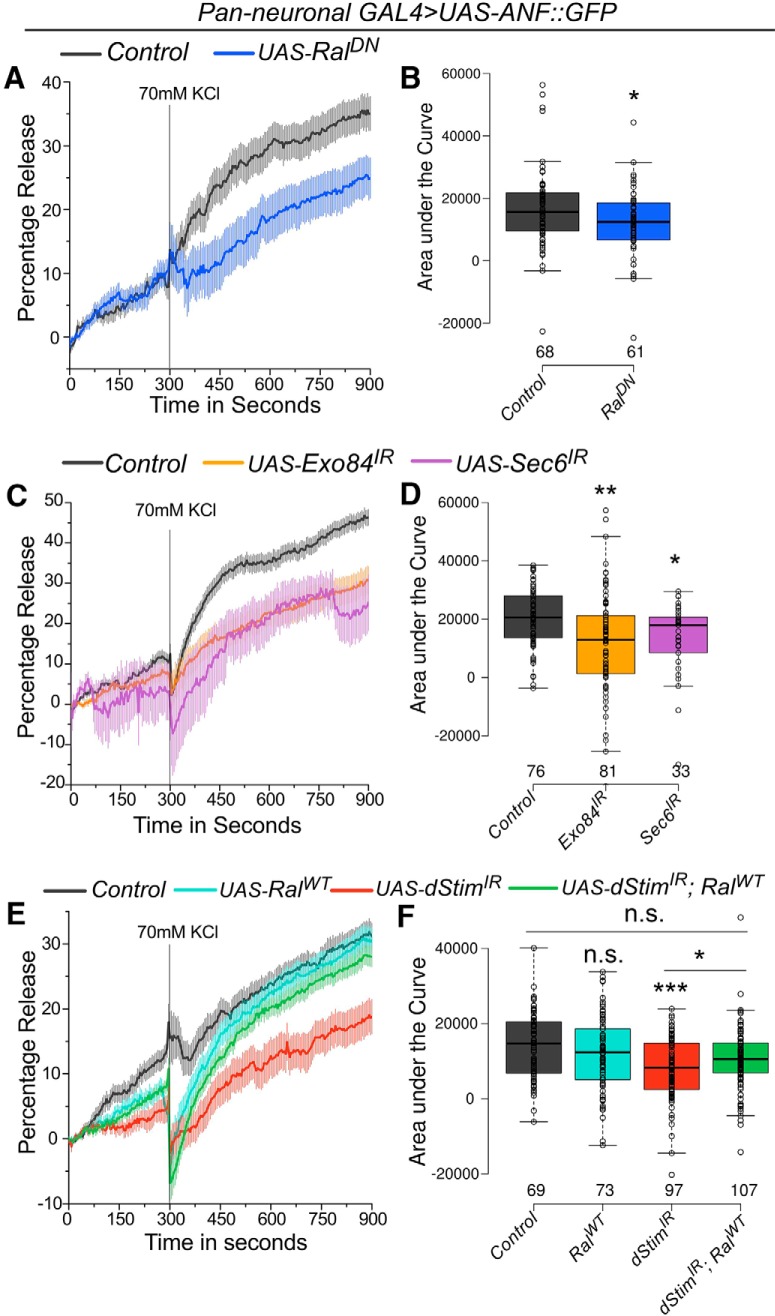
Inhibiting Ral function in cultured pupal neurons by expression of a dominant-negative form of Ral, RalDN, led to a significant reduction in release of ANF::GFP (Fig. 2A,B). A similar reduction in release was observed on knockdown of either Exo84 or Sec6 (Fig. 2C,D), which correlates with the strong flight defects observed on their knockdown (Fig. 1A,C). Knockdown of an essential neuronal SOCE component, dStim, also led to reduction in ANF::GFP release, as predicted from previous data demonstrating that Ral expression in pupal neurons requires SOCE (Richhariya et al., 2017, Fig. 2E,F). Furthermore, reduced release of ANF::GFP by dStim knockdown could be rescued by overexpressing RalWT (Fig. 2E,F), indicating that Ral function is downstream of SOCE in mediating vesicular release in pupal neurons.
Figure 2.
dStim regulates vesicular release in pupal neurons through Ral (refer to Fig. 2-1 and Table 1-1). A, C, E, Traces represent release of ANF::GFP, estimated by reduction in GFP fluorescence, on depolarization with KCl. Lines represent means and the error bars, standard error of means, from cells as mentioned in the box plots on the right. Point of KCl addition is indicated with a line. B, D, F, Amount of release quantified as area under the curve from 300 to 900 s is represented as box plots for the indicated genotypes. Box plots symbols are as described in Fig. 1. Numbers below each box represent number of cells analyzed from at least three different culture dishes. *, p < 0.05, **, p < 0.01, ***, p < 0.001, n.s., not significant at p < 0.05 by two-tailed Student’s t test (for A) or one-way ANOVA followed by post hoc Tukey’s test (for D and F). All comparisons for significance were with the control values except where marked by a horizontal line. For exact p-values, refer to Table 1-1.
Assay to measure vesicular release from cultured pupal neurons. A, Representative images of single somata of cultured pupal neurons expressing ANF::GFP and Shits. The images were obtained over time upon depolarization with KCl at permissive (22°C) and restrictive (29°C) temperatures. Scale bar represents 10 µm. B, D, Traces represent release of ANF::GFP, estimated by the reduction in GFP fluorescence, upon depolarization with KCl, or addition of NaCl over time. The control trace in D is the same as that in Fig. 2A. C, E, Amount of release quantified as area under the curve from 300 to 900 s is represented as box plots for the indicated genotypes. Box plot symbols are as described in Fig. 1. ***, p < 0.001, n.s., not significant at p < 0.05 by one-way ANOVA followed by post hoc Tukey’s test (for C) or two-tailed Student’s t test (for E). All comparisons for significance were with the control values except where marked by a horizontal line. For exact p-values, refer to Table 1-1. Download Figure 2-1, TIF file (2.5MB, tif) .
Ral function in pupal dopaminergic neurons is required for adult flight maintenance
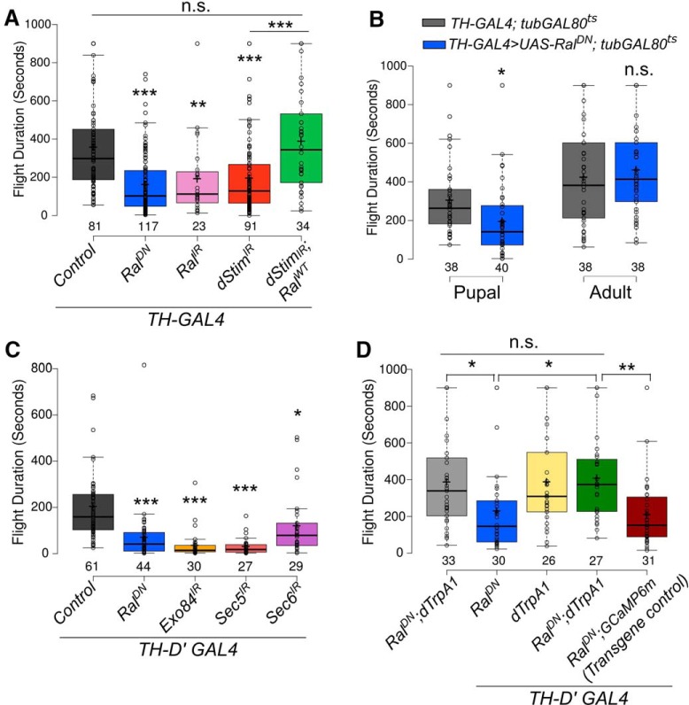
Next, we tested the spatio-temporal requirement for Ral in previously identified neuronal subsets where loss of SOCE is known to affect the maintenance of flight bouts. These include dopaminergic (Pathak et al., 2015) and glutamatergic (Venkiteswaran and Hasan, 2009) neurons. Blocking Ral function either in all glutamatergic neurons by the OK371-GAL4 (Mahr and Aberle, 2006) or in the motor neurons using the OK6-GAL4 (Sanyal, 2009) resulted in significantly reduced flight durations (Fig. 3-1A,B). Similarly, reducing either Ral levels or function in dopaminergic cells, with TH-GAL4 (Friggi-Grelin et al., 2003a), resulted in significantly reduced flight durations that were comparable to flight deficits observed by knockdown of dStim (Fig. 3A). Importantly, the flight deficits observed on dStim knockdown were rescued to control levels by restoring Ral function, when RalWT was overexpressed in this background (Fig. 3A). Ral knockdown exclusively during the pupal stage, but not in the adult stage, resulted in flight durations significantly lower than those of control flies (Figs. 3B and 3-1C,D).
Figure 3.
Extended flight bout durations in adults requires Ral and exocyst function in pupal dopaminergic neurons (refer to Fig. 3-1 and Table 1-1). A, C, Box plots represent flight bout durations of flies from the indicated genotypes measured by the single flight assay. B, Box plots represent flight bout durations of flies with expression of a dominant negative mutant of Ral (RalDN) in dopaminergic neurons during the indicated stages of development. D, Box plots represent flight bout durations of flies from the indicated genotypes that were grown at 22°C and transferred to 29°C from 0 to 72 h after Puparium formation for activation of dTrpA1. Box plot symbols are as described in Fig. 1. *, p < 0.05, **, p < 0.01, ***, p < 0.001, n.s., not significant at p < 0.05 by two-tailed Student’s t test (for B) or one-way ANOVA followed by post hoc Tukey’s test (for A, C, and D). All comparisons for significance were with the control values except where marked by a horizontal line. For exact p-values, refer to Table 1-1.
Neuronal subsets where Ral function is required for maintenance of flight bouts. A–G, Box plots represent flight durations of flies from the indicated genotypes measured by the single flight assay. Box plot symbols are as described in Fig. 1. Statistically distinguishable groups are indicated by different alphabets over the box plots (two-tailed Student’s t test, p < 0.05). *, p < 0.05, **, p < 0.01, ***, p < 0.001, n.s., not significant by two-tailed Student’s t test. For exact p-values, refer to Table 1-1. Download Figure 3-1, TIF file (1.3MB, tif) .
Because TH-GAL4 expresses in both neuronal and non-neuronal cells (Friggi-Grelin et al., 2003b), we further refined the spatial requirement of Ral for flight using GAL4 drivers that mark dopaminergic cell subsets. Significantly shortened flight durations were observed on reducing Ral function with the neuronal-specific TH-D′ (Fig. 3C) and TH-D1 (Fig. 3-1F) GAL4s (Liu et al., 2012; Pathak et al., 2015). However, attenuating Ral function in hypodermal cells with TH-A GAL4 (Liu et al., 2012) did not significantly alter the duration of flight (Fig 3-1G). Blocking Ral function in serotonergic neurons with the TRH-GAL4 also did not affect flight duration (Fig. 3-1E). Thus, Ral is required in pupal dopaminergic neurons for a functional adult flight circuit. These data are in agreement with previous findings demonstrating that loss of SOCE by inhibition of dOrai in similar subsets of pupal dopaminergic neurons impairs flight in adult Drosophila (Pathak et al., 2015).
Knockdown of the exocyst components, Exo84, Sec5, and Sec6, in TH-D′GAL4-marked dopaminergic neurons also resulted in flight deficits (Fig. 3C). Importantly, the flight deficits observed on reducing Ral function in dopaminergic neurons could be rescued by expression of the heat-activated cation channel dTrpA1 in TH-D′ marked neurons in pupae (Fig. 3D). Expression of a neutral transgene, GCaMP6m, had no effect on the fight deficit shown by animals expressing RalDN in TH-D′-marked neurons (Fig 3D). dTrpA1 enhances neuronal activity (Hamada et al., 2008) and thus, possibly, the extent of vesicular release in pupal dopaminergic neurons. This was tested next.
dStim regulates synaptic vesicle release from pupal dopaminergic neurons through Ral
Vesicular release from neurons can occur either through small synaptic vesicles (SSVs) or large dense core vesicles (LDCVs). SSVs are small, clear vesicles that primarily carry neurotransmitters whereas LDCVs are large, granular vesicles that transport neuropeptides, hormones and aminergic neurotransmitters (Park and Kim, 2009). Mammalian Ral colocalizes with both SSVs (Volknandt et al., 1993) and LDCVs (De Leeuw et al., 1999) and hence, regulation by Ral of both classes of vesicles is possible in Drosophila. The assay with ANF::GFP in cultured neurons (Figs. 2 and 2-1) most likely assayed for release from LDCVs at the soma, and although such vesicles can corelease a monoamine neurotransmitter like dopamine (Nusbaum et al., 2017), it would not test the ability of Ral to affect neurotransmitter release from SSVs. To measure release from SSVs at synaptic release sites in the maturing pupal brain, we used spH, a pH-sensitive GFP, that localizes to synaptic vesicles and whose fluorescence is quenched in acidic conditions but fluoresces on fusion of the vesicle with the pre-synaptic membrane (Miesenböck et al., 1998). SSV release was important to assess, because SOCE-regulated expression of TH, a dopamine-synthesizing enzyme, in pupal stages is required for adult flight (Pathak et al., 2015), suggesting that formation of appropriate functional connectivity between central dopaminergic neurons and their postsynaptic targets requires dopamine release. We hypothesized that SOCE and Ral-dependent exocyst function could be a mechanism underlying such dopamine release in developing synapses of the pupal brain.
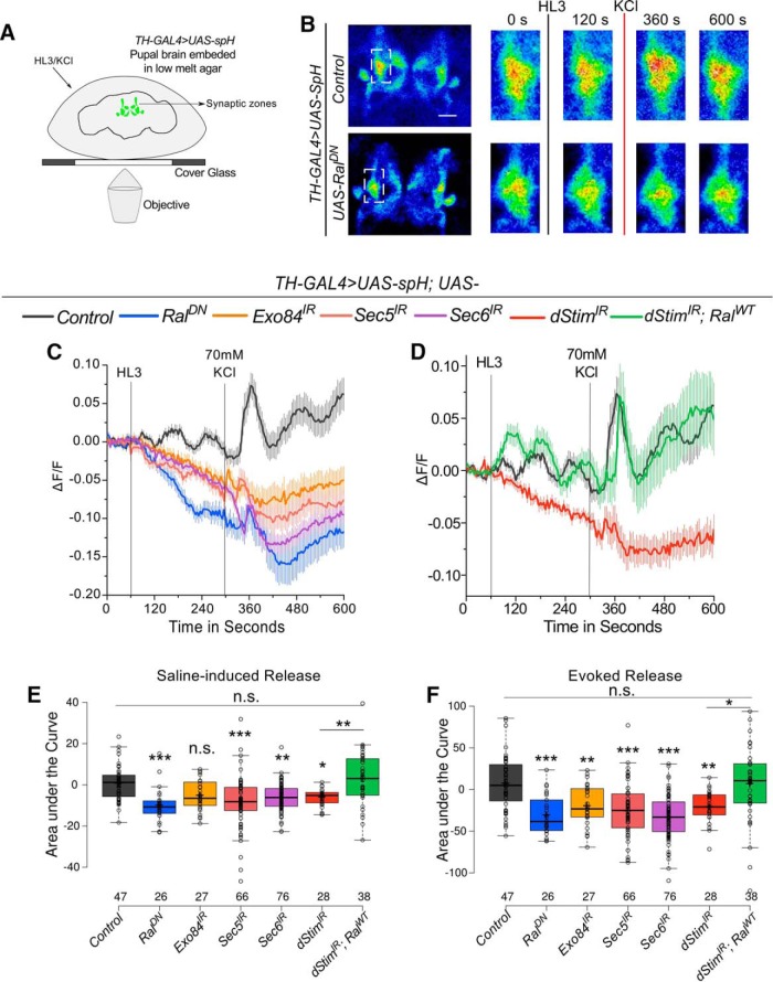
Synaptic release from dopaminergic neurons in pupae was visualized in ex vivo preparations of 48–72-h old pupal brains expressing UAS-spH (Ng et al., 2002) using TH-GAL4 (Fig. 4A). The TH-GAL4 marks synaptic zones in parts of the mushroom body (MB) viz. the α and α′ vertical lobes, upper stalk, lower stalk and junction, and the distal heel and peduncle deriving from the PPL1 neurons among others (Mao and Davis, 2009). Thus, synaptic release was measured at maturing synaptic zones of the MB (Fig. 4A). Depolarization by KCl evoked vesicle release, as evidenced by a visible and quantifiable change in spH fluorescence (Video 1 and Fig. 4B–D,F). The amplitude and kinetics of the change in fluorescence observed were similar to what have been observed earlier using spH in mammalian (Burrone et al., 2007) and Drosophila neurons (Slawson et al., 2014). Interestingly, addition of hemolymph-like saline (HL3) caused a measurable level of synaptic release at the MB synaptic zones even without KCl-induced depolarization (Video 1 and Fig. 4C,E). This saline-induced release could result from basal activity of the endogenous circuit activity, because the recordings were performed from an intact live brain without any inhibitors. Both saline and depolarization evoked release from pupal dopaminergic neurons were severely affected on either, reducing Ral function or knockdown of exocyst components as well as knockdown of dStim (Figs. 4B–F and 4-1D–F). Importantly, overexpression of wild-type Ral in the background of dStim knockdown rescued both saline and KCl evoked release (Fig. 4D–F). Expression of a neutral transgene, UAS-H2BmRFP in dStim knockdown background, did not rescue either kind of release (Fig. 4-1A–C), demonstrating that rescue was indeed because of restoring Ral levels and not insufficient knockdown of dStim caused by expression of multiple transgenes.
Figure 4.
dStim regulates evoked synaptic release from pupal dopaminergic neurons through Ral (Refer to Fig. 4-1 and Table 1-1). A, Schematic representation of the experimental setup used to image vesicular release from pupal brains. B, Representative images of central regions of pupal brains expressing spH in dopaminergic neurons. Boxed images on the right were obtained by zooming into the inset (white box in the images on the left) at the indicated time points of the time series. C, D, Traces represent average (±SEM) change in fluorescence of spH over time from brains of the indicated genotypes. Points of addition of HL3 and KCl are denoted by vertical lines. E, F, Amount of release quantified as area under the curve from 60 to 300 s (E-Saline–induced) and 300 to 600 s (F-KCl–evoked) from traces in C and D is represented as box plots for the indicated genotypes. Box plot symbols are as described in Fig. 1. Numbers below the boxes represent number of ROIs in which fluorescence changes were measured. They were obtained from a minimum of five brains per genotype. *, p < 0.05, **, p < 0.01, ***, p < 0.001, n.s., not significant at p < 0.05 by one-way ANOVA followed by post hoc Tukey’s test. All comparisons for significance were with the control values except where marked by a horizontal line. For exact p-values, refer to Table 1-1.
dStim and Ral regulate synaptic release from pupal dopaminergic neurons. A, D, Traces represent change in fluorescence of spH over time from brains of the indicated genotypes. Lines represent means and the error bars, standard error of means. Points of addition of HL3 and KCl are denoted by vertical lines. B, E, Saline-induced release as quantified by the area under the curve from 60 to 300 s of the respective traces. C, F, Evoked release as quantified by the area under the curve from 300 to 600 s of the respective traces. Box plot symbols are as described in Fig. 1. Numbers below the boxes represent the number of ROIs measured for change in fluorescence from a minimum of five brains per genotype. *, p < 0.05, **, p < 0.01, ***, p < 0.001, n.s., not significant at p < 0.05 by two-tailed Student’s t test (for B and C) or one-way ANOVA followed by post hoc Tukey’s test (for E and F). For exact p-values, refer to Table 1-1. Download Figure 4-1, TIF file (1.7MB, tif) .
Visualizing synaptic release from dopaminergic neurons. Changes in fluorescence of spH driven by the TH-GAL4 indicative of synaptic release from dopaminergic neurons in the mushroom body. Gray flash indicates addition of HL3 (mock); red flash indicates addition of 70 mm KCl.
Discussion
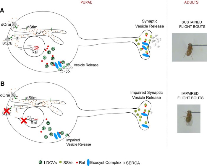
Neuronal Ral was recently identified as a regulator of Drosophila flight (Richhariya et al., 2017). In this study, we have identified dopaminergic neurons as a cellular focus for Ral function during flight circuit maturation in pupae. In dopaminergic neurons Ral functions through Exo84, and presumably the exocyst, to regulate synaptic release during pupal development. Moreover, we identify a novel role for the SOCE gene dStim in regulating synaptic vesicular release through positive control of Ral gene expression, identified previously in a screen for SOCE-regulated genes (Richhariya et al., 2017). Importantly, both flight deficits and reduced synaptic vesicular release observed as a consequence of dSTIM knockdown were rescued by Ral over-expression in dopaminergic neurons. These findings support an essential requirement for dopamine release during synapse formation and maturation of the Drosophila flight circuit (Pathak et al., 2015; Fig. 5).
Figure 5.
Vesicle release regulated by SOCE, Ral, and the exocyst in pupal neurons regulates flight in adults. A, Cartoon representation of a pupal neuron where SOCE regulates Ral expression. Ral is required for tagging secretory vesicles toward the exocyst complex, thus marking them for both somatic and synaptic release. Such regulated vesicular release of dopamine from pupal neurons appears essential for maturation of a neural circuit required in adults for maintaining the duration of flight bouts. B, In conditions of dStim knockdown, when SOCE is attenuated, Ral levels are reduced, and this in turn has a negative impact on vesicular release in dopaminergic neurons. Consequently, flight bout durations in adult flies are also reduced.
We observed a loss of somatic and synaptic vesicular release on knockdown of dStim, Ral, and the exocyst components tested (Figs. 2 and 4). Because Ral and the exocyst have established roles in regulating pre-synaptic vesicle dynamics and exocytosis (Moskalenko et al., 2002; Wang et al., 2004) and Ral expression is regulated by dStim (Richhariya et al., 2017), we propose that dStim regulates vesicular release via Ral. However, the reduction in vesicular release on dStim knockdown might also be mediated by other factors such as expression levels and densities of ion channels (Voglis and Tavernarakis, 2006) and altered signaling in response to neurotransmitters (Huang and Thathiah, 2015), which have not been tested here. Because Ral almost completely rescues the release defects of dStim knockdown (Figs. 2E,F and 4E,F), we believe that a significant component of dStim mediated vesicular release in pupal dopaminergic neurons is by regulating expression of Ral.
Activity-dependent calcium entry is essential for synaptic release (Südhof, 2012). However, this is the first report where calcium entry through SOCE regulates synaptic release. Our work demonstrates an important distinction between how the two modes of calcium entry regulate synaptic vesicle release, viz., that SOCE-regulated synaptic release is indirect by regulation of Ral expression. Thus, neuromodulators that stimulate SOCE very likely change synaptic function over time scales that are much greater than activity-based synaptic release. We favor the dSTIM/dOrai mode of SOCE in this context primarily because very similar flight deficits are observed by knockdown of dOrai and these can also be rescued by overexpression of Ral (Richhariya et al., 2017).
Data in this paper suggest that dStim, by regulation of Ral expression (Richhariya et al., 2017), impacts both spontaneous and evoked synaptic vesicle release in pupal dopaminergic neurons. Neurotransmitter release from SSVs and neuropeptide as well as neurotransmitter release from LDCVs, regulates multiple steps during the development of neural circuits, including synapse formation and maturation (Lochner et al., 2008; Park and Kim, 2009; Tau and Peterson, 2010). Neurotransmitter release during circuit development can affect both synapse formation and strength (Bleckert and Wong, 2011). Moreover, spontaneous release of neurotransmitters, similar to an earlier observation in larval neurons (Choi et al., 2014), could be important for synaptic maturation. The absence of flight deficits by adult-specific knockdown or abrogation of Ral/Exo84 (Richhariya et al., 2017; Figs. 1D and 3B) function agrees with an earlier finding where exocyst function was dispensable for synaptic release from mature neurons (Murthy et al., 2003).
In pupal dopaminergic neurons, Ral-modulated vesicle release is a likely mechanism by which SOCE affects synaptic maturation and thus function of adult flight circuit neurons. Regulation of circadian activity by Ral has been demonstrated in Drosophila (Klose et al., 2016). Our results suggest that such regulation may be initiated by receptor-stimulated SOCE. Moreover, SOCE-regulated Ral expression and vesicle release at presynaptic sites may act as co-incident detectors of neuromodulatory signals that enhance synaptic efficacy and network drive for optimal function of cognate neural circuits in other organisms as well, including vertebrates.
Acknowledgments
Acknowledgments: We thank Gero Miesenböck, Mark Wu, and Boris Egger for fly strains. We thank the Central Imaging and Flow Facility at NCBS for the use of its microscopes. Flies from the Vienna Drosophila RNAi Center (VDRC) and the Bloomington Drosophila Stock Center (National Institutes of Health P40OD018537) were used in this study.
Synthesis
Reviewing Editor: Margaret McCarthy, Univ of Maryland School of Medicine
Decisions are customarily a result of the Reviewing Editor and the peer reviewers coming together and discussing their recommendations until a consensus is reached. When revisions are invited, a fact-based synthesis statement explaining their decision and outlining what is needed to prepare a revision will be listed below. The following reviewer(s) agreed to reveal their identity: Hiromu Tanimoto.
The manuscript improved after the revision by addressing reviewers' criticisms.
One last thing to clarify before publication is the measurement of basal fluorescence (comment 12 in the decision letter). The authors' claim on spontaneous release relies on the basal fluorescence, but as the absolute fluorescent level before applying any stimuli is not shown anywhere in the manuscript. As different levels of basal fluorescence may affect the speed of photobleaching. the measurement of “basal release” is not possible, and the claim must be moderated accordingly.
Author Response
May 06, 2018
Dear Dr. McCarthy,
We are now ready to upload a revised version of our manuscript "dSTIM and Ral/exocyst mediated synaptic release from pupal dopaminergic neurons sustains Drosophila flight".
Both text and figures have been revised appropriately to address the concerns of reviewers. Details of the revisions made are described in the Response to Reviewers. For ease of the reviewers we have also appended a track change version of the manuscript. We hope that with these changes, the manuscript will be acceptable for publication.
Yours Sincerely,
Gaiti Hasan
References
- Agrawal N, Venkiteswaran G, Sadaf S, Padmanabhan N, Banerjee S, Hasan G (2010) Inositol 1,4,5-trisphosphate receptor and dSTIM function in Drosophila insulin-producing neurons regulates systemic intracellular calcium homeostasis and flight. J Neurosci 30:1301–1313. 10.1523/JNEUROSCI.3668-09.2010 [DOI] [PMC free article] [PubMed] [Google Scholar]
- Blankenship AG, Feller MB (2010) Mechanisms underlying spontaneous patterned activity in developing neural circuits. Nat Rev Neurosci 11:18–29. [DOI] [PMC free article] [PubMed] [Google Scholar]
- Bleckert A, Wong ROL (2011) Identifying roles for neurotransmission in circuit assembly: insights gained from multiple model systems and experimental approaches. Bioessays 33:61–72. 10.1002/bies.201000095 [DOI] [PMC free article] [PubMed] [Google Scholar]
- Burrone J, Li Z, Murthy VN (2007) Studying vesicle cycling in presynaptic terminals using the genetically encoded probe synaptopHluorin. Nat Protoc 1:2970–2978. 10.1038/nprot.2006.449 [DOI] [PubMed] [Google Scholar]
- Choi BJ, Imlach WL, Jiao W, Wolfram V, Wu Y, Grbic M, Cela C, Baines RA, Nitabach MN, McCabe BD (2014) Miniature neurotransmission regulates drosophila synaptic structural maturation. Neuron 82:618–634. 10.1016/j.neuron.2014.03.012 [DOI] [PMC free article] [PubMed] [Google Scholar]
- Consoulas C, Restifo LL, Levine RB (2002) Dendritic remodeling and growth of motoneurons during metamorphosis of Drosophila melanogaster. J Neurosci 22:4906–4917. [DOI] [PMC free article] [PubMed] [Google Scholar]
- De Leeuw HPJC, Wijers-Koster PM, Van Mourik JA, Voorberg J (1999) Small GTP-binding protein RalA associates with Weibel-Palade bodies in endothelial cells. Thromb Haemost 82:1177–1181. [PubMed] [Google Scholar]
- Dietzl G, Chen D, Schnorrer F, Su K-C, Barinova Y, Fellner M, Gasser B, Kinsey K, Oppel S, Scheiblauer S, Couto A, Marra V, Keleman K, Dickson BJ (2007) A genome-wide transgenic RNAi library for conditional gene inactivation in Drosophila. Nature 448:151–156. 10.1038/nature05954 [DOI] [PubMed] [Google Scholar]
- Feske S (2007) Calcium signalling in lymphocyte activation and disease. Nat Rev Immunol 7:690–702. 10.1038/nri2152 [DOI] [PubMed] [Google Scholar]
- Finger FP, Novick P (1998) Spatial regulation of exocytosis: lessons from yeast. J Cell Biol [DOI] [PMC free article] [PubMed] [Google Scholar]
- Friggi-Grelin F, Coulom H, Meller M, Gomez D, Hirsh J, Birman S (2003a) Targeted gene expression in Drosophila dopaminergic cells using regulatory sequences from tyrosine hydroxylase. J Neurobiol 54:618–627. 10.1002/neu.10185 [DOI] [PubMed] [Google Scholar]
- Friggi-Grelin F, Iché M, Birman S (2003b) Tissue-specific developmental requirements of Drosophila tyrosine hydroxylase isoforms. Genesis 35:175–184. 10.1002/gene.10178 [DOI] [PubMed] [Google Scholar]
- Gentry LR, Martin TD, Reiner DJ, Der CJ (2014) Ral small GTPase signaling and oncogenesis: more than just 15 minutes of fame. Biochim Biophys Acta 1843:2976–2988. [DOI] [PMC free article] [PubMed] [Google Scholar]
- Guo W, Grant A, Novick P (1999) Exo84p is an exocyst protein essential for secretion. J Biol Chem 274:23558–23564. [DOI] [PubMed] [Google Scholar]
- Hamada FN, Rosenzweig M, Kang K, Pulver SR, Ghezzi A, Jegla TJ, Garrity PA (2008) An internal thermal sensor controlling temperature preference in Drosophila. Nature 454:217–220. 10.1038/nature07001 [DOI] [PMC free article] [PubMed] [Google Scholar]
- Huang Y, Thathiah A (2015) Regulation of neuronal communication by G protein-coupled receptors. FEBS Lett 589:1607–1619. [DOI] [PubMed] [Google Scholar]
- Kitamoto T (2001) Conditional modification of behavior in Drosophila by targeted expression of a temperature-sensitive shibire allele in defined neurons. J Neurobiol 47:81–92. 10.1002/neu.1018 [DOI] [PubMed] [Google Scholar]
- Klose M, Duvall LB, Li W, Liang X, Ren C, Steinbach JH, Taghert PH (2016) Functional PDF signaling in the Drosophila circadian neural circuit is gated by Ral A-dependent modulation. Neuron 90:1–14. 10.1016/j.neuron.2016.04.002 [DOI] [PMC free article] [PubMed] [Google Scholar]
- Kula E, Levitan ES, Pyza E, Rosbash M (2006) PDF cycling in the dorsal protocerebrum of the Drosophila brain is not necessary for circadian clock function. J Biol Rhythms 21:104–117. 10.1177/0748730405285715 [DOI] [PubMed] [Google Scholar]
- Lalli G, Hall A (2005) Ral GTPases regulate neurite branching through GAP-43 and the exocyst complex. J Cell Biol 171:857–869. 10.1083/jcb.200507061 [DOI] [PMC free article] [PubMed] [Google Scholar]
- Langevin J, Le Borgne R, Rosenfeld F, Gho M, Schweisguth F, Bellaïche Y (2005) Lethal giant larvae controls the localization of Notch-signaling regulators Numb, neuralized, and Sanpodo in Drosophila sensory-organ precursor cells. Curr Biol 15:955–962. 10.1016/j.cub.2005.04.054 [DOI] [PubMed] [Google Scholar]
- Levitan ES, Lanni F, Shakiryanova D (2007) In vivo imaging of vesicle motion and release at the Drosophila neuromuscular junction. Nat Protoc 2:1117–1125. 10.1038/nprot.2007.142 [DOI] [PubMed] [Google Scholar]
- Li G, Han L, Chou T-C, Fujita Y, Arunachalam L, Xu A, Wong A, Chiew S-K, Wan Q, Wang L, Sugita S (2007) RalA and RalB function as the critical GTP sensors for GTP-dependent exocytosis. J Neurosci 27:190–202. 10.1523/JNEUROSCI.2537-06.2007 [DOI] [PMC free article] [PubMed] [Google Scholar]
- Lin DM, Goodman CS (1994) Ectopic and increased expression of Fasciclin II alters motoneuron growth cone guidance. Neuron 13:507–523. [DOI] [PubMed] [Google Scholar]
- Liu Q, Liu S, Kodama L, Driscoll MR, Wu MN (2012) Two dopaminergic neurons signal to the dorsal fan-shaped body to promote wakefulness in Drosophila. Curr Biol 22:2114–2123. 10.1016/j.cub.2012.09.008 [DOI] [PMC free article] [PubMed] [Google Scholar]
- Lochner JE, Spangler E, Chavarha M, Jacobs C, McAllister K, Schuttner LC, Scalettar BA (2008) Efficient copackaging and cotransport yields postsynaptic colocalization of neuromodulators associated with synaptic plasticity. Dev Neurobiol 68:1243–1256. 10.1002/dneu.20650 [DOI] [PMC free article] [PubMed] [Google Scholar]
- Lohmann C (2009) Calcium signaling and the development of specific neuronal connections. Prog Brain Res 175:443–452. [DOI] [PubMed] [Google Scholar]
- Mahr A, Aberle H (2006) The expression pattern of the Drosophila vesicular glutamate transporter: a marker protein for motoneurons and glutamatergic centers in the brain. Gene Expr Patterns 6:299–309. 10.1016/j.modgep.2005.07.006 [DOI] [PubMed] [Google Scholar]
- Majewski L, Kuznicki J (2015) SOCE in neurons: signaling or just refilling? Biochim Biophys Acta - 1853:1940–1952. [DOI] [PubMed] [Google Scholar]
- Makos MA, Han K-A, Heien ML, Ewing AG (2010) Using in vivo electrochemistry to study the physiological effects of cocaine and other stimulants on the Drosophila melanogaster dopamine transporter. ACS Chem Neurosci 1:74–83. 10.1021/cn900017w [DOI] [PMC free article] [PubMed] [Google Scholar]
- Mao Z, Davis R (2009) Eight different types of dopaminergic neurons innervate the Drosophila mushroom body neuropil: anatomical and physiological heterogeneity. Front Neural Circuits 3:5. 10.3389/neuro.04.005.2009 [DOI] [PMC free article] [PubMed] [Google Scholar]
- McGuire SE, Le PT, Osborn AJ, Matsumoto K, Davis RL (2003) Spatiotemporal rescue of memory dysfunction in Drosophila. Science 302:1765–1768. 10.1126/science.1089035 [DOI] [PubMed] [Google Scholar]
- McGuire SE, Mao Z, Davis RL (2004) Spatiotemporal gene expression targeting with the TARGET and gene-switch systems in Drosophila. Sci STKE 2004:pl6. 10.1126/stke.2202004pl6 [DOI] [PubMed] [Google Scholar]
- Miesenböck G, De Angelis DA, Rothman JE (1998) Visualizing secretion and synaptic transmission with pH-sensitive green fluorescent proteins. Nature 394:192–195. 10.1038/28190 [DOI] [PubMed] [Google Scholar]
- Moskalenko S, Henry DO, Rosse C, Mirey G, Camonis JH, White MA (2002) The exocyst is a Ral effector complex. Nat Cell Biol 4:66–72. 10.1038/ncb728 [DOI] [PubMed] [Google Scholar]
- Murthy M, Garza D, Scheller RH, Schwarz TL (2003) Mutations in the exocyst component Sec5 disrupt neuronal membrane traffic, but neurotransmitter release persists. Neuron 37:433–447. [DOI] [PubMed] [Google Scholar]
- Nedelman JR, Gibiansky E (1996) The variance of a better AUC estimator for sparse, destructive sampling toxicokinetics. J Pharm Sci 85:884–886. 10.1021/js960083a [DOI] [PubMed] [Google Scholar]
- Ng M, Roorda RD, Lima SQ, Zemelman BV, Morcillo P, Miesenböck G (2002) Transmission of olfactory information between three populations of neurons in the antennal lobe of the fly. Neuron 36:463–474. [DOI] [PubMed] [Google Scholar]
- Nusbaum MP, Blitz DM, Marder E (2017) Functional consequences of neuropeptide and small-molecule co-transmission. Nat Rev Neurosci 18:389–403. [DOI] [PMC free article] [PubMed] [Google Scholar]
- Park Y, Kim K-T (2009) Short-term plasticity of small synaptic vesicle (SSV) and large dense-core vesicle (LDCV) exocytosis. Cell Signal 21:1465–1470. 10.1016/j.cellsig.2009.02.015 [DOI] [PubMed] [Google Scholar]
- Pathak T, Agrawal T, Richhariya S, Sadaf S, Hasan G (2015) Store-operated calcium entry through orai is required for transcriptional maturation of the flight circuit in Drosophila. J Neurosci 35:13784–13799. 10.1523/JNEUROSCI.1680-15.2015 [DOI] [PMC free article] [PubMed] [Google Scholar]
- Polzin A, Shipitsin M, Goi T, Feig LA, Turner TJ (2002) Ral-GTPase influences the regulation of the readily releasable pool of synaptic vesicles. Mol Cell Biol 22:1714–1722. [DOI] [PMC free article] [PubMed] [Google Scholar]
- Prakriya M, Lewis RS (2015) Store-operated calcium channels. Physiol Rev 95:1383–1436. 10.1152/physrev.00020.2014 [DOI] [PMC free article] [PubMed] [Google Scholar]
- Qin L, Liu Y, Wang J, Li S, Sato Y (2009) Neural and behavioral discrimination of sound duration by cats. J Neurosci 29:15650–15659. 10.1523/JNEUROSCI.2442-09.2009 [DOI] [PMC free article] [PubMed] [Google Scholar]
- Rao S, Lang C, Levitan ES, Deitcher DL (2001) Fast track: visualization of neuropeptide expression, transport, and exocytosis in Drosophila melanogaster. J Neurobiol 49:159–172. 10.1002/neu.1072 [DOI] [PubMed] [Google Scholar]
- Richhariya S, Jayakumar S, Abruzzi K, Rosbash M, Hasan G (2017) A pupal transcriptomic screen identifies Ral as a target of store-operated calcium entry in Drosophila neurons. Sci Rep 7:42586. 10.1038/srep42586 [DOI] [PMC free article] [PubMed] [Google Scholar]
- Rosenberg SS, Spitzer NC (2011) Calcium signaling in neuronal development. Cold Spring Harb Perspect Biol 3:a004259. 10.1101/cshperspect.a004259 [DOI] [PMC free article] [PubMed] [Google Scholar]
- Sadaf S, Birman S, Hasan G (2012) Synaptic activity in serotonergic neurons is required for air-puff stimulated flight in Drosophila melanogaster. PLoS One 7:e46405. 10.1371/journal.pone.0046405 [DOI] [PMC free article] [PubMed] [Google Scholar]
- Sanyal S (2009) Genomic mapping and expression patterns of C380, OK6 and D42 enhancer trap lines in the larval nervous system of Drosophila. Gene Expr Patterns 9:371–380. 10.1016/j.gep.2009.01.002 [DOI] [PubMed] [Google Scholar]
- Schindelin J, et al. (2012) Fiji: an open source platform for biological image analysis. Nat Methods 9:676–682. 10.1038/nmeth.2019 [DOI] [PMC free article] [PubMed] [Google Scholar]
- Slawson JB, Kuklin EA, Mukherjee K, Pírez N, Donelson NC, Griffith LC (2014) Regulation of dopamine release by CASK-β modulates locomotor initiation in Drosophila melanogaster. Front Behav Neurosci 8:394. 10.3389/fnbeh.2014.00394 [DOI] [PMC free article] [PubMed] [Google Scholar]
- Somasundaram A, Shum AK, McBride HJ, Kessler JA, Feske S, Miller RJ, Prakriya M (2014) Store-operated CRAC channels regulate gene expression and proliferation in neural progenitor cells. J Neurosci 34:9107–9123. 10.1523/JNEUROSCI.0263-14.2014 [DOI] [PMC free article] [PubMed] [Google Scholar]
- Spitzer M, Wildenhain J, Rappsilber J, Tyers M (2014) BoxPlotR: a web tool for generation of box plots. Nat Methods 11:121–122. 10.1038/nmeth.2811 [DOI] [PMC free article] [PubMed] [Google Scholar]
- Südhof TC (2012) Calcium control of neurotransmitter release. Cold Spring Harb Perspect Biol 4:a011353. 10.1101/cshperspect.a011353 [DOI] [PMC free article] [PubMed] [Google Scholar]
- Tau GZ, Peterson BS (2010) Normal development of brain circuits. Neuropsychopharmacol Rev 35:147–168. 10.1038/npp.2009.115 [DOI] [PMC free article] [PubMed] [Google Scholar]
- Teodoro RO, Pekkurnaz G, Nasser A, Higashi-Kovtun ME, Balakireva M, McLachlan IG, Camonis J, Schwarz TL (2013) Ral mediates activity-dependent growth of postsynaptic membranes via recruitment of the exocyst. EMBO J 32:2039–2055. 10.1038/emboj.2013.147 [DOI] [PMC free article] [PubMed] [Google Scholar]
- Tissot M, Stocker RF (2000) Metamorphosis in Drosophila and other insects: the fate of neurons throughout the stages. Prog Neurobiol 62:89–111. [DOI] [PubMed] [Google Scholar]
- Tomchik SM (2013) Dopaminergic neurons encode a distributed, asymmetric representation of temperature in Drosophila. J Neurosci 33:2166–2176. 10.1523/JNEUROSCI.3933-12.2013 [DOI] [PMC free article] [PubMed] [Google Scholar]
- Truman JW (1990) Metamorphosis of the central nervous system of Drosophila. J Neurobiol 21:1072–1084. 10.1002/neu.480210711 [DOI] [PubMed] [Google Scholar]
- Truman JW, Bate M (1988) Spatial and temporal patterns of neurogenesis in the central nervous system of Drosophila melanogaster. Dev Biol 125:145–157. [DOI] [PubMed] [Google Scholar]
- Venkiteswaran G, Hasan G (2009) Intracellular Ca2+ signaling and store-operated Ca2+ entry are required in Drosophila neurons for flight. Proc Natl Acad Sci U S A 106:10326–10331. 10.1073/pnas.0902982106 [DOI] [PMC free article] [PubMed] [Google Scholar]
- Voglis G, Tavernarakis N (2006) The role of synaptic ion channels in synaptic plasticity. EMBO Rep 7:1104–1110. 10.1038/sj.embor.7400830 [DOI] [PMC free article] [PubMed] [Google Scholar]
- Volknandt W, Pevsne J, Elferink LA, Scheller RH (1993) Association of three small GTP-binding proteins with cholinergic synaptic vesicles. FEBS Lett 317:53–56. [DOI] [PubMed] [Google Scholar]
- Wang L, Li G, Sugita S (2004) RalA-exocyst interaction mediates GTP-dependent exocytosis. J Biol Chem 279:19875–19881. 10.1074/jbc.M400522200 [DOI] [PubMed] [Google Scholar]
- Wong MY, Cavolo SL, Levitan ES (2015) Synaptic neuropeptide release by dynamin-dependent partial release from circulating vesicles. Mol Biol Cell 26:2466–2474. 10.1091/mbc.E15-01-0002 [DOI] [PMC free article] [PubMed] [Google Scholar]
Associated Data
This section collects any data citations, data availability statements, or supplementary materials included in this article.
Supplementary Materials
Statistical tests and p-values for all figures. Download Table 1-1, XLSX file (29.7KB, xlsx) .
The role of exocyst components in flight. A, B, Box plots represent flight durations of flies of the indicated genotypes. Knockdown of Exo84 and Sec6 with alternate RNAi lines resulted in similar phenotypes as shown in Fig. 1. These flies were grown at 25°C throughout. C, D, Box plots represent flight durations of control flies with RNAi-mediated knockdown of Exo84 (Exo84IR) in neurons raised at the indicated temperatures throughout development. At the permissive temperature of 18°C, there is no knockdown. GAL4-mediated knockdown of Exo84 occurs at the restrictive temperature of 29°C. Box plot symbols are as described in Fig. 1. *, p < 0.05, **, p < 0.01, ***, p < 0.001, n.s., not significant at p < 0.05 by two-tailed Student’s t test (for A and B) or one-way ANOVA followed by post hoc Tukey’s test (for C and D). All comparisons for significance were with the control values except where marked by a horizontal line. For exact p-values, refer to Table 1-1. Download Figure 1-1, TIF file (654.7KB, tif) .
Expression of exocyst components is not regulated by SOCE. A–D, Bar graphs represent the level of expression of the indicated genes using values for fragments per kilobase gene per million reads (FPKM) in control and dStim knockdown pupal brains. These data were obtained from the transcriptomic screen performed in Richhariya et al. (2017). q-values are as obtained by CuffDiff. Download Figure 1-2, TIF file (321.5KB, tif) .
Assay to measure vesicular release from cultured pupal neurons. A, Representative images of single somata of cultured pupal neurons expressing ANF::GFP and Shits. The images were obtained over time upon depolarization with KCl at permissive (22°C) and restrictive (29°C) temperatures. Scale bar represents 10 µm. B, D, Traces represent release of ANF::GFP, estimated by the reduction in GFP fluorescence, upon depolarization with KCl, or addition of NaCl over time. The control trace in D is the same as that in Fig. 2A. C, E, Amount of release quantified as area under the curve from 300 to 900 s is represented as box plots for the indicated genotypes. Box plot symbols are as described in Fig. 1. ***, p < 0.001, n.s., not significant at p < 0.05 by one-way ANOVA followed by post hoc Tukey’s test (for C) or two-tailed Student’s t test (for E). All comparisons for significance were with the control values except where marked by a horizontal line. For exact p-values, refer to Table 1-1. Download Figure 2-1, TIF file (2.5MB, tif) .
Neuronal subsets where Ral function is required for maintenance of flight bouts. A–G, Box plots represent flight durations of flies from the indicated genotypes measured by the single flight assay. Box plot symbols are as described in Fig. 1. Statistically distinguishable groups are indicated by different alphabets over the box plots (two-tailed Student’s t test, p < 0.05). *, p < 0.05, **, p < 0.01, ***, p < 0.001, n.s., not significant by two-tailed Student’s t test. For exact p-values, refer to Table 1-1. Download Figure 3-1, TIF file (1.3MB, tif) .
dStim and Ral regulate synaptic release from pupal dopaminergic neurons. A, D, Traces represent change in fluorescence of spH over time from brains of the indicated genotypes. Lines represent means and the error bars, standard error of means. Points of addition of HL3 and KCl are denoted by vertical lines. B, E, Saline-induced release as quantified by the area under the curve from 60 to 300 s of the respective traces. C, F, Evoked release as quantified by the area under the curve from 300 to 600 s of the respective traces. Box plot symbols are as described in Fig. 1. Numbers below the boxes represent the number of ROIs measured for change in fluorescence from a minimum of five brains per genotype. *, p < 0.05, **, p < 0.01, ***, p < 0.001, n.s., not significant at p < 0.05 by two-tailed Student’s t test (for B and C) or one-way ANOVA followed by post hoc Tukey’s test (for E and F). For exact p-values, refer to Table 1-1. Download Figure 4-1, TIF file (1.7MB, tif) .
Visualizing synaptic release from dopaminergic neurons. Changes in fluorescence of spH driven by the TH-GAL4 indicative of synaptic release from dopaminergic neurons in the mushroom body. Gray flash indicates addition of HL3 (mock); red flash indicates addition of 70 mm KCl.


