Description
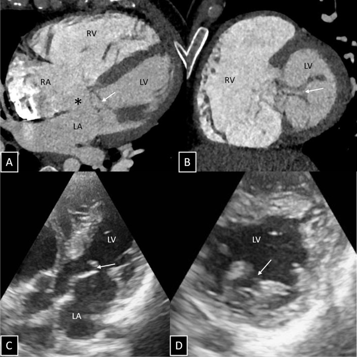
A 30-year-old man was presented to the cardiology outpatient department with complains of easy fatigability and recurrent chest infections since childhood. There was no history of cyanosis, palpitations or effort intolerance. Chest radiograph was unremarkable. Transthoracic echocardiography (ECHO) showed ostium primum atrial septal defect (ASD), trifoliate mitral valve (MV) with mitral regurgitation and presence of two papillary muscles. Cardiac CT was done using a third-generation dual-source 192-slice CT system (SOMATOM Force, Siemens Healthcare, Forchheim, Germany) to look for extracardiac vascular anatomy as part of preoperative workup. It clearly demonstrated the partial atrioventricular septal defect (ostium primum ASD without any shunt at the level of ventricles) with anterior MV cleft (figure 1A,B). CT findings corroborated with the ECHO findings (figure 1C, D). No other associated malformations were seen. The patient is now planned for direct suturing of mitral valve cleft with concomitant mitral annuloplasty and ASD repair.
Figure 1.
CT angiogram with multiplanar reconstructed images in the four-chamber view (panel A) and short-axis view (panel B) reveals ostium primum atrial septal defect (indicated by * in panel A) with the presence of cleft in the anterior mitral valve leaflet (indicated by arrows in A and B). Transthoracic echocardiography (four-chamber view, panel C and short-axis view, panel D) depicts the cleft in the anterior mitral valve leaflet (indicated by arrows in C and D). LA, left atrium; LV, left ventricle; RA, right atrium; RV, right ventricle.
Ostium primum ASD results from lack of fusion between the septum primum and the endocardial cushions.1 It can be seen as an abnormal communication between the right and left atria immediately posterior to the MV annulus on CT. Various forms of atrioventricular valve deformations (valves with up to seven leaflets) including MV cleft and papillary muscle abnormalities may be seen associated with it. The presence of MV cleft has important implications when ASD surgical correction is attempted due to associated mitral regurgitation, thus affecting left ventricular size and function.2 3 Echocardiography remains the diagnostic modality of choice to diagnose MV cleft along with other associated cardiac shunts. Cross-sectional imaging like, current generation dual-source CT or MRI may serve as additional diagnostic modalities to assess extracardiac vascular anatomy and ventricular function, thus mapping ideal treatment option.
Learning points.
Meticulous search for an associated anterior mitral leaflet cleft should be performed in cases of primum atrial septal defect (ASD).
Presence of a mitral valve cleft has important implications in surgical correction of ASD.
Footnotes
MS, AS, NNP and SK contributed equally.
Contributors: All authors have participated sufficiently in the conception of the idea, development of the intellectual content, design, writing and final approval of the manuscript.
Funding: The authors have not declared a specific grant for this research from any funding agency in the public, commercial or not-for-profit sectors.
Competing interests: None declared.
Patient consent: Obtained.
Provenance and peer review: Not commissioned; externally peer reviewed.
References
- 1.Sulafa AK, Tamimi O, Najm HK, et al. . Echocardiographic differentiation of atrioventricular septal defects from inlet ventricular septal defects and mitral valve clefts. Am J Cardiol 2005;95:607–10. 10.1016/j.amjcard.2004.11.007 [DOI] [PubMed] [Google Scholar]
- 2.Johri AM, Rojas CA, El-Sherief A, et al. . Imaging of atrial septal defects: echocardiography and CT correlation. Heart 2011;97:1441–53. 10.1136/hrt.2010.205732 [DOI] [PubMed] [Google Scholar]
- 3.Chui J, Anderson RH, Lang RM, et al. . The Trileaflet Mitral Valve. Am J Cardiol 2018;121:513–9. 10.1016/j.amjcard.2017.11.018 [DOI] [PubMed] [Google Scholar]


