Abstract
The gamut of gallium labeled radiopharmaceuticals contributes to augmented variety in molecular imaging approach for in vivo identification of tumor characteristics. The spectrum ranges from somatostatin receptor based target-specific imaging agents, to those used for tumor imaging based on specific receptor types extending into ones used for therapeutic monitoring. The versatility of gallium chemistry provides the needed advantage for imaging which is further exploited in clinical practice influences the specificity of tumor imaging. Ga-68 has revealed applicability in labeling compounds from nanoparticles to micro as well as macromolecules. We in this image, present variety of frequently and infrequently used gallium labeled radiopharmaceuticals, for imaging diverse malignancies other than conventional established tracers.
Keywords: Ga-68, positron emission tomography/computed tomography, CPCR4, exendin, prostate-specific membrane antigen, RGD, αvβ3 integrin
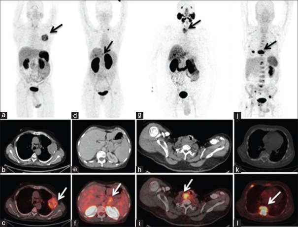
The use of Ga-68 in clinical practice has continually evolved owing to its notable flexibility in labeling properties with antibodies, peptides, and drug conjugates.[1,2] We hereby depict a composite image of four cases showing the usefulness of 68-Ga radiopharmaceuticals for imaging diverse malignancies [Figure 1]. Integrin expression in lung carcinoma can be imaged by Ga-68-RGD which targets the αvβ3 integrin[3] as represented by maximum intensity projection (MIP) positron emission tomography image [Figure 1a] of a patient diagnosed to have squamous cell lung carcinoma, showing intense uptake of Ga-68-DOTA-RGD2 (SUVmax 7.8) in the left hemithorax corresponding to a heterogeneously enhancing pleural based soft-tissue mass on the transaxial images [Figure 1b and c] in left lung with involvement of adjacent rib. Note is also made of mild tracer uptake in the left lower paratracheal and sub-cranial lymph nodes. Ga-68 RGD uptake correlates with the degree of angiogenesis, and therefore it may be a useful marker for identifying metastatic mediastinal lymph nodes in comparison to F-18 FDG. The identification of mediastinal lymph nodes noninvasively would guide mediastinoscopy in countries with an epidemic of infectious disease like tuberculosis. The Ga-68-NOTA-RGD was initially used in breast cancer[4] depicting the variability in integrin (αvβ3) expression with respect to estrogen dependent and independent tumors which can be further extended to other cancers like lung carcinoma.
Figure 1.
(a-c) Ga-68-DOTA-RGD2 positron emission tomography images, maximum intensity projection (a) acquired of a 62-year-old male patient with squamous cell lung carcinoma showing intense uptake of radiotracer in the left hemi-thorax corresponding to a heterogeneously enhancing pleural based soft-tissue mass lesion (SUVmax 7.8) on the transaxial computed tomography (b) and fused positron emission tomography-computed tomography (c) images in left lung with involvement of adjacent rib. (d-f) Ga-68-DOTA-Exendin-4 positron emission tomography images, maximum intensity projection (d) and transaxial computed tomography (e) and fused positron emission tomography-computed tomography (f) of a 57-year-old female patient with G1 neuroendocrine tumor of the pancreas showing uptake of radiotracer (SUVmax 5.8) in pancreatic tumor mass (arrow) with metastasis to liver and left supraclavicular lymph node. (g-i): Ga-68-HBED-CC-PSMA positron emission tomography images, maximum intensity projection (g), transaxial computed tomography (h) and fused positron emission tomography-computed tomography (i) of 58-year-old male patient with differentiated thyroid carcinoma posttotal thyroidectomy, showing intense tracer uptake in the right trachea-esophageal groove (SUVmax 11.2), representing local recurrence in the neck in a case of thyroglobulin elevated negative iodine scan. (j-l) Ga-68-CPCR4 trifluoroacetate positron emission tomography images, maximum intensity projection (j), transaxial computed tomography (k) and fused positron emission tomography-computed tomography (l) of a 63-year-old male patient diagnosed with multiple myeloma, showing abnormal foci of tracer uptake in multiple ribs, cervico dorso lumbar vertebra (D9 vertebra SUVmax 22.42), sternum and multiple sites in pelvis
Glucagon-like peptide-1 receptor (GLP-1R) imaging using Ga-68-DOTA-Exendin-4 is a novel receptor-targeted imaging technique for benign insulinomas, but GLP-1R is also overexpressed in pancreatic neuroendocrine tumors[5] The MIP and transaxial images [Figure 1d-f] show uptake of Ga-68-DOTA-Exendin-4 (SUVmax 5.8) in a G1 neuroendocrine tumour (Ki index <2%) of the pancreas with metastasis to liver and left supraclavicular lymph node.
Prostate-specific membrane antigen (PSMA) expression is noted in solid malignancies other than prostate, namely, melanoma, NET, breast, and colon cancer.[6] PSMA expression in the cells of neovasculature is well documented and have been depicted on thyroid adenomas.[7] Figure 1g-i] shows intense tracer uptake of 68Ga labeled HBED-CC-PSMA in the right tracheoesophageal groove (SUVmax 11.2) in a post total thyroidectomy patient of thyroglobulin elevated negative iodine scan differentiated thyroid carcinoma representing local recurrence in the neck, potentially opening the role for Lu-177 based PSMA therapy.
The chemokine receptors CXCR4 are expressed in either nucleus or cytoplasm and play a noticeable role in metastasis serves to predict poor survival.[8] 68Ga-CPCR4 trifluoroacetate, a cyclic pentapeptide targeting the CXCR4 receptor, shows abnormal foci of tracer uptake in multiple ribs, cervico dorso lumbar vertebra (D9; SUVmax 22.42), sternum and multiple sites in the pelvis [Figure 1j-l]. The targeted receptor may potentially serve as sites of the therapeutic target which may be further useful for assessing response to therapy. Thus, diverse Ga-68 labeled radiopharmaceuticals can be used to target-specific receptor sites leading to better identification and characterization of tumors.
Declaration of patient consent
The authors certify that they have obtained all appropriate patient consent forms. In the form the patient(s) has/have given his/her/their consent for his/her/their images and other clinical information to be reported in the journal. The patients understand that their names and initials will not be published and due efforts will be made to conceal their identity, but anonymity cannot be guaranteed.
Financial support and sponsorship
Nil.
Conflicts of interest
There are no conflicts of interest.
References
- 1.Velikyan I. Prospective of Ga-68-radiopharmaceutical development. Theranostics. 2014;4:47–80. doi: 10.7150/thno.7447. [DOI] [PMC free article] [PubMed] [Google Scholar]
- 2.Jalilian AR. An overview on Ga-68 radiopharmaceuticals for positron emission tomography applications. Iran J Nucl Med. 2016;24:1–10. [Google Scholar]
- 3.Zheng K, Liang N, Zhang J, Lang L, Zhang W, Li S, et al. 68Ga-NOTA-PRGD2 PET/CT for integrin imaging in patients with lung cancer. J Nucl Med. 2015;56:1823–7. doi: 10.2967/jnumed.115.160648. [DOI] [PMC free article] [PubMed] [Google Scholar]
- 4.Yoon HJ, Kang KW, Chun IK, Cho N, Im SA, Jeong S, et al. Correlation of breast cancer subtypes, based on estrogen receptor, progesterone receptor, and HER2, with functional imaging parameters from 68Ga-RGD PET/CT and 18F-FDG PET/CT. Eur J Nucl Med Mol Imaging. 2014;41:1534–43. doi: 10.1007/s00259-014-2744-4. [DOI] [PubMed] [Google Scholar]
- 5.Wild D, Wicki A, Mansi R, Béhé M, Keil B, Bernhardt P, et al. Exendin-4-based radiopharmaceuticals for glucagonlike peptide-1 receptor PET/CT and SPECT/CT. J Nucl Med. 2010;51:1059–67. doi: 10.2967/jnumed.110.074914. [DOI] [PubMed] [Google Scholar]
- 6.Verburg FA, Krohn T, Heinzel A, Mottaghy FM, Behrendt FF. First evidence of PSMA expression in differentiated thyroid cancer using [68Ga] PSMA-HBED-CC PET/CT. Eur J Nucl Med Mol Imaging. 2015;42:1622–3. doi: 10.1007/s00259-015-3065-y. [DOI] [PubMed] [Google Scholar]
- 7.Moore M, Panjwani S, Mathew R, Crowley M, Liu YF, Aronova A, et al. Well-differentiated thyroid cancer neovasculature expresses prostate-specific membrane antigen-a possible novel therapeutic target. Endocr Pathol. 2017;28:339–44. doi: 10.1007/s12022-017-9500-9. [DOI] [PubMed] [Google Scholar]
- 8.Herrmann K, Lapa C, Wester HJ, Schottelius M, Schiepers C, Eberlein U, et al. Biodistribution and radiation dosimetry for the chemokine receptor CXCR4-targeting probe 68Ga-pentixafor. J Nucl Med. 2015;56:410–6. doi: 10.2967/jnumed.114.151647. [DOI] [PubMed] [Google Scholar]


