

Abstract
A life-long turnover of sensory and interneuronal populations has been documented in the olfactory pathways of both vertebrates and invertebrates, creating a situation where the axons of new afferent and interneuronal populations must insert into a highly specialized glomerular neuropil. A dense serotonergic innervation of the primary olfactory processing areas where these neurons synapse also is a consistent feature across species. Prior studies in lobsters have shown that serotonin promotes the branching of olfactory projection neurons. This paper presents evidence that serotonin also regulates the proliferation and survival of projection neurons in lobsters, and that the serotonergic effects are associated with a transient uptake of serotonin into newborn neurons.
The olfactory pathways of vertebrates and invertebrates show a remarkable degree of life-long structural plasticity. The basis of this plasticity is the turnover of olfactory receptor neurons (1, 2) as well as of interneuronal populations (3–5). The new afferents and interneurons insert their axons into existing synaptic regions, which are organized into a highly ordered array of glomeruli. These glomeruli are thought to be arranged odotopically, with each glomerulus having a specific role in the coding of odor quality (6–8). Throughout life, as the processes of new sensory neurons and interneurons are added to the glomeruli, their structural integrity is stable: there is no evidence for the formation of new glomeruli or the loss of existing glomeruli (9–11). The intercalation of the processes of new neurons into the glomeruli, in which pre- and postsynaptic populations of neurons need to be matched, presents major challenges for the nervous system in preserving odotopic order.
A feature that is common to olfactory systems across phylogenetic lines is a dense serotonergic innervation of the regions where the olfactory receptor neurons synapse onto second-order cells. In vertebrates, the olfactory bulb is one of the major forebrain targets of serotonergic neurons ascending from the brainstem (12). In molluscs, serotonergic interneurons innervate the protocerebral olfactory neuropil where serotonin is thought to be involved in the storage of odor memory (13, 14). Among the arthropods, giant serotonergic neurons innervate the antennal lobes of insects (15) and the olfactory lobes (OLs) of crustaceans (16). The present study examines the role this serotonergic innervation may play in the assembly and maintenance of olfactory areas in the lobster, Homarus americanus, by testing whether serotonin depletion influences the proliferation and survival of projection neurons and by documenting a transient uptake of serotonin by the newborn cells.
The crustacean olfactory pathway consists of primary sensory neurons that synapse with local and projection interneurons within the glomeruli of the OLs. These two categories of interneurons are functionally analogous to the local (periglomerular and granule) and projection (mitral and tufted) neurons of the vertebrate olfactory bulb (17). The projection neurons of lobsters, which are the primary focus of this paper, innervate the OL, accessory lobe (AL) and olfactory globular tract neuropil (OGTN), and possess axons that project to neuropil regions in the lateral protocerebrum (arrows in Fig. 1A). The AL is involved with the higher order integration of olfactory, visual, and mechanosensory information (16). Beyond a presumed role in the processing of olfactory information (18), the functional roles of the OGTN are unclear. However, the OGTN is interesting in the context of the present study because it is the site of a major synaptic input to the paired dorsal giant neurons (DGNs), which provide the primary serotonergic input to the olfactory neuropils of lobsters and crayfish (ref. 19; Fig. 1 B and C). The DGNs have been the focus of extensive anatomical, physiological, and ultrastructural investigations because of their massive projections to the OLs and ALs where they innervate each and every glomerulus (16, 19–22). The complex circuitry, sheer size, and geometry of the DGNs and their ubiquitous inputs throughout the OLs and ALs pose unusual challenges in understanding the function of the DGNs within the context of the olfactory system. Thus far, no satisfactory explanation of their role has been offered.
Figure 1.

The olfactory pathway in the brain of Homarus americanus. (A) Confocal image of the OLs and ALs in a hemi-brain of an embryonic lobster stained with an antibody against Drosophila synapsin (E. Buchner, Würzburg, Germany). Tissues were processed by standard methods (22). The diverging arrows mark the path of the primary neurites of the olfactory projection neurons (somata located in cluster 10) that innervate the OL and AL. These processes form the OGT (converging arrows) that passes through the OGTN (*) en route to the lateral protocerebrum. (B) Schematic diagram illustrating the position of the neuropils that are altered by serotonin depletion during embryonic development: OL, AL, and OGTN. (Left Inset) The area of synapsin labeling shown in A. (Right Inset) The area of serotonin labeling shown in C. (C) Serotonin immunostaining (for method, see ref. 21) of the DGN, which innervates the ipsilateral OL, AL, and OGTN (not shown). The intense labeling of the OL and AL is due to the massive DGN projection into these areas (16, 22). (Bars: A = 20 μm; C = 200 μm.) [A is reprinted with permission from B. S. Beltz and colleagues (23) (Copyright 2000, Society for Neuroscience).]
Serotonin has been implicated as a developmental architect in the olfactory pathway of lobster embryos. Pharmacological depletion of serotonin during embryogenesis with 5,7-dihydroxytryptamine (5,7-DHT) dramatically reduces the size of the target neuropils of the DGN: the OL, AL, and OGTN (21, 22). However, the morphology and size of the serotonergic DGNs are not affected by 5,7-DHT treatment (22). These results suggest that the proliferation, growth, or morphogenesis of the local and projection neurons may be affected by serotonin depletion. Dye-injection studies have shown that the differentiation of a subset of the olfactory projection neurons is indeed affected by serotonin depletion: ∼14% of the projection neurons sampled in serotonin-depleted embryos send axons toward the lateral protocerebrum via the olfactory globular tract (OGT), as expected, but do not innervate the OLs or ALs, as in normal embryos (23). Hence, it would seem that serotonin depletion in vivo inhibits the branching of olfactory projection neurons in lobsters, suggesting that one of the functions of serotonin during normal development is to promote, directly or indirectly, the growth of these neurons into the olfactory lobe glomeruli and associated brain neuropils.
Neurogenesis within cluster 10 (the cell body cluster of the projection neurons) ceases during late embryonic development in lobsters, but resumes after hatching and thereafter continues throughout the animal's life (5). Even in decades-old lobsters, a proliferation zone exists adjacent to the AL at the posteromedial margin of cell cluster 10 (5). Studies in adult (sexually mature) lobsters using the substitute nucleotide bromodeoxyuridine (BrdUrd) have shown that newly born projection neurons move anterolaterally away from the proliferation zone and disperse throughout the cell cluster (Fig. 2; ref. 5).
Figure 2.

Persistent cell proliferation in cluster 10, which contains the somata of the olfactory projection neurons, in juvenile and adult brains. (A) Newly born projection neurons are labeled in 0.5-year-old juveniles by injecting BrdUrd 3 weeks before killing and with a second pulse on the day of killing. The dividing cells labeled by the 24-h pulse are located in the proliferation zone (double arrowheads), whereas the cells labeled by the injection 3 weeks before killing have migrated anterolaterally (single arrowhead; ref. 5). (B) BrdUrd injection of adult lobsters followed by killing 6 weeks later demonstrates a band of labeled cells in cluster 10 (arrowhead) that has moved anterolaterally from their origin at the posteromedial margin of the cluster (5). (C) When BrdUrd was injected into adult lobsters at 6 months and again at 24 h before killing, nuclei are labeled posteromedially in the proliferation zone (double arrowheads), whereas individual labeled cells have dispersed among the cluster 10 neurons. (D) Higher magnification of labeled cells outlined in C. (Bars: A–C = 50 μm; D = 20 μm.) [A and B are reprinted with permission from B. S. Beltz and colleagues (5) (Copyright 1999, Society for Neuroscience).]
In the present study, we used BrdUrd-labeling methods to examine the effects of serotonin depletion on neurogenesis in cell cluster 10 of the lobster. These studies indicate that serotonin may regulate both proliferation and survival of newly born projection neurons. Further experiments showed that, although mature projection neurons are not serotonin-immunoreactive in lobsters (24, 25), newborn olfactory projection neurons are transiently able to take up serotonin. This phenomenon may be related to the role serotonin plays in promoting the ingrowth of new projection neurons into the olfactory neuropils of lobsters (23) and/or to the effects of serotonin on the regulation of neurogenesis that are reported here.
Our results suggest specific roles for the massive serotonergic innervation of olfactory centers in lobsters in regulating the development and maintenance of interneuronal populations. Because of the strong parallels between the function and organization of olfactory systems in a broad range of species, these findings also may be relevant to the serotonergic innervation of olfactory areas in other types of organisms where interneuronal populations turn over throughout life.
Materials and Methods
Embryonic and juvenile lobsters (Homarus americanus) were obtained from the New England Aquarium (Boston, MA) and maintained at 14°C in aquaria with circulating artificial seawater and a 12-h light/12-h dark cycle. Embryos were staged by using the system of Helluy and Beltz (1991), in which egg extrusion is defined as 0% and hatching as 100% (26). Methods related to the use of adult lobsters to demonstrate life-long neurogenesis in cluster 10 are published in Harzsch et al. (5).
Effects of Serotonin Depletion on Neuronal Proliferation and Survival.
Embryonic lobsters.
Embryos at 58% of embryonic development (E58%) were assigned to either the experimental or control group. Experimental embryos were injected once a week for 2 weeks with 0.13–0.26 μl of 10−2 M 5,7-DHT (Sigma) in a vehicle solution (saline, 1% ascorbate, 0.05% visual marker) by using the procedure of Benton et al. (21). Control embryos were injected only with vehicle solution. Three days after the final injection, proliferating cells were labeled by immersing the embryos for 2 h in BrdUrd (Amersham Pharmacia) diluted in seawater (14°C) to a concentration of 0.1 mg/ml. Experimental and control embryos were killed either 24 h or 2 weeks after exposure to BrdUrd to examine the effects of serotonin depletion on neuronal proliferation and longer-term survival, respectively. Brains were dissected from the embryos, fixed in 4% (wt/vol) paraformaldehyde, and processed by using avidin-biotin-peroxidase immunocytochemistry according to the procedure of Harzsch et al. (5). Preparations were examined with a Nikon compound microscope, and the numbers of BrdUrd-labeled profiles were counted blindly by two independent observers using a camera lucida.
Juvenile lobsters.
Five-month-old juvenile lobsters (≈10 g) were injected (into the ventral hemolymph sinus of the abdomen) twice a week for 4 weeks with either 5,7-DHT diluted in vehicle solution (experimental animals) or with the vehicle solution alone (control animals). Circulating levels of 5,7-DHT immediately after injection were calculated to be ≈10−3 M; this concentration would be rapidly lowered as 5,7-DHT is taken up by cells, metabolized, and oxidized (21). Three days after the final injection, proliferating cells were labeled by injecting the lobsters with solutions of BrdUrd (0.3 mg of BrdUrd per 10 g of body weight) diluted in physiological saline. To examine the effects of serotonin depletion on neuronal proliferation and survival, experimental and control animals were killed 24 h (proliferation) or 3 weeks (survival) after the BrdUrd injection. Brains were dissected, fixed in 4% (wt/vol) paraformaldehyde, then processed immunocytochemically as whole mounts. The mouse anti-BrdUrd antibody (1:100; Amersham Pharmacia) was visualized by using a Texas red conjugated-rabbit anti-mouse antibody (1:50; Molecular Probes), and preparations were examined with a Leica scanning confocal microscope. The numbers of labeled cells in cluster 10 of control and experimental animals were counted from the series of confocal images.
Serotonin Uptake by Newborn Olfactory Projection Neurons.
Juvenile lobsters (8–26 g, 3 months–2 years old) were injected with BrdUrd (0.3 mg of BrdUrd per 10 g of body weight) diluted in saline. Animals were killed 6–24 h after injection, and their brains were dissected free from the head. As an assay for neurons possessing the serotonin transporter, the brains then were incubated for 2–4 h at 10–12°C in the dark in 10−5 M serotonin (Sigma) in L-15 culture medium (Sigma), with salts adjusted to the concentrations in physiological saline. After this treatment, tissues were rinsed in saline, fixed in 4% (wt/vol) paraformaldehyde, and processed immunocytochemically for serotonin and BrdUrd by using standard methods (5, 21). The mouse anti-BrdUrd antibody (1:100; Amersham Pharmacia) was visualized by using an Alexa green-conjugated goat anti-mouse antibody (1:50; Molecular Probes); the rabbit anti-serotonin antibody (1:1,000; Diasorin) was visualized with a Texas red-conjugated goat anti-rabbit antibody (1:50; Molecular Probes). To test whether the serotonin transporter also is present in embryonic neurons, brains were dissected from embryonic lobsters and incubated for 4 h in 10−5 M serotonin in L-15 culture medium with salts adjusted to be iso-osmotic with lobster saline. Brains then were processed immunocytochemically for serotonin (22). Preparations were examined by using a Leica TCS SP confocal microscope equipped with argon and krypton lasers and spectrophotometer-based emission filters. Fluorescence is represented in the images with contrasting pseudocolors.
Results and Discussion
Effects of Serotonin Depletion on Neuronal Proliferation and Survival.
Serotonin depletion in both embryonic and juvenile lobsters was found to have profound effects on neuronal proliferation and the longer-term survival of newly born cells (Fig. 3). Studies examining the effects of serotonin depletion on neurogenesis in embryos showed that the number of BrdUrd-labeled cells in cluster 10 present 1 day after a 2-hr exposure to BrdUrd was significantly reduced in serotonin-depleted brains (by 35%, compared with controls; Fig. 3A). As previous studies have shown that neuroblasts in the lobster brain persist and continue to proliferate after 5,7-DHT treatment (22), the current results suggest that serotonin depletion may slow the mitotic cycles of the neuroblasts and/or the intermediate precursor cells of the projection neurons. The number of BrdUrd-labeled cells present in the brains of serotonin-depleted embryos 2 weeks after a 2-hr exposure to BrdUrd was reduced by 52% compared with the number labeled in control embryos (Fig. 3B). This further reduction in the numbers of labeled cells in the 5,7-DHT-treated embryos suggests that serotonin depletion also may influence the survival of intermediate precursor cells and/or newborn olfactory projection neurons. However, because the clearing time for BrdUrd is 2–3 days in lobster embryos, and the sequence of proliferation of projection neurons involves three distinct cell types (ref. 22, and J.L.B. and B.S.B., unpublished results), it is likely that the labeled profiles seen after 2 weeks (surviving cells) are largely a different population of cells than those labeled after just 24 h (proliferating cells). The effects of serotonin depletion in embryos are likely to involve regulatory mechanisms that persist throughout the animal's life, as shown by experiments in the brains of juvenile lobsters where proliferation was dramatically reduced by serotonin depletion (Fig. 3C), and where no BrdUrd-labeled neurons were present at the end of a 3-week assessment period (Fig. 3D).
Figure 3.

A comparison of the levels of proliferation (A and C) and survival (B and D) of BrdUrd-labeled cells in cluster 10 of control and 5,7-DHT-treated (experimental) embryonic and juvenile lobsters. The numbers of BrdUrd-labeled profiles were counted and compared with two-tailed t tests. (A, B, and D) *, statistically significant differences (P < 0.0001) between experimental and control groups. Statistical tests were not valid for C because of the low control–sample size. The numbers of labeled cells are represented as means ± SD. (A) To examine the effects of serotonin depletion on neuronal proliferation during embryogenesis, control (n = 10 cell clusters) and 5,7-DHT-treated (n = 10 cell clusters) embryos were exposed to BrdUrd for 2 h and killed after 24 h. The number of BrdUrd-labeled cells in cluster 10 in 5,7-DHT-treated embryos was found to be on average 35% smaller than that observed in control embryos. (B) To study the longer term effects of serotonin depletion, control (n = 10 cell clusters) and 5,7-DHT-treated (n = 10 cell clusters) embryos were killed 2 weeks after a 2-h period of exposure to BrdUrd. By using this protocol, the number of BrdUrd-labeled cells observed in 5,7-DHT-treated embryos was found to be 52% smaller than the number of labeled cells in control embryos. (C and D) Serotonin depletion in juvenile lobsters was found to reduce the numbers of labeled profiles when assessed 24 h (C) (control, n = 2 cell clusters; 5,7-DHT-treated, n = 4 cell clusters) and 3 weeks (D) (control, n = 4 cell clusters; 5,7-DHT-treated, n = 4 cell clusters) after BrdUrd injection. (C) Whereas an average of 78 neurons was labeled in each cluster 10 of control lobsters 24 h after BrdUrd injection, an average of only 4 neurons was labeled in serotonin-depleted lobsters. (D) No labeled neurons were observed in the brains of 5,7-DHT-treated lobsters after a 3-week survival time after BrdUrd injection.
Serotonin Uptake by Newborn Olfactory Projection Neurons.
We were interested in the mechanisms by which serotonin regulates the proliferation, survival, and branching (23) of new projection neurons. Olfactory projection neurons in mature crustaceans are not immunoreactive for serotonin (24, 25), indicating that they do not synthesize or take up significant levels of this amine. However, studies in a variety of species suggest that some neurons lacking the enzymes involved in the biosynthesis of serotonin are capable of taking up serotonin for use as an intracellular signal (27, 28). Therefore, we hypothesized that serotonin could exert its effects on olfactory projection neurons by means of such an uptake mechanism. To test this hypothesis, we performed double labeling experiments in which juvenile lobsters initially were injected with BrdUrd to label newborn projection neurons, after which brains were dissected free and incubated in serotonin as an assay for neurons possessing the serotonin transporter. In these preparations, we observed BrdUrd-labeled cells in cluster 10 toward the anterolateral margin of the proliferation zone (white arrows, Fig. 4A), as reported (ref. 5; see also Fig. 2), as well as an occasional serotonin–BrdUrd double-labeled cell (yellow arrowhead, Fig. 4A). Serotonin antibodies also selectively labeled a single row of cells in the proliferation zone (white arrowheads, Fig. 4A) adjacent to the BrdUrd-labeled profiles that have migrated anterolaterally. Older projection neurons that lie even more anterolaterally are unlabeled by these treatments. The position of the serotonin-immunoreactive neurons immediately posteromedial to the BrdUrd-labeled cells indicates that these neurons were born after the BrdUrd-labeled cells; therefore, the uptake of serotonin must occur within the first few hours after the neurons are born. Because the majority of serotonin-labeled cells are not double-labeled with BrdUrd, the serotonin-labeled cells must have been born after the animal was killed and when BrdUrd was no longer available, i.e., during the in vitro incubation time. The fact that such small numbers of cells are labeled by serotonin treatment relative to the numbers of proliferating neurons suggests that the serotonin transporter is active for only a short period after birth, and/or that only a subset of newborn projection neurons takes up serotonin. A group of fine-caliber fibers entering the projection neuron cluster and ending blindly at the proliferation zone also label for serotonin (Fig. 4B), and these are a likely source of the serotonin taken up by the newly proliferated neurons in vivo. We have traced some of these fibers to their origin at the primary axonal trunks of the DGN.
Figure 4.

Confocal images of the brains of juvenile lobsters labeled for BrdUrd (red) and serotonin (green) immunoreactivities. (A) Lobsters were killed 24 h after BrdUrd injection, and dissected brains were incubated in serotonin, rinsed, fixed, and processed by standard methods. BrdUrd-labeled nuclei are found at the anterior margin of the proliferation zone in cluster 10. Serotonin labeling is seen in the AL and in a row of six labeled cells (arrowheads) that lie between the BrdUrd profiles and the AL; one of these cells is double-labeled (yellow arrowhead). The positions of the serotonin-labeled cells relative to the BrdUrd-labeled nuclei suggest that these cells were born after the BrdUrd-labeled neurons that have migrated anterolaterally during the 24-h survival time (n = 4 cell clusters). (B) After a 6-h survival time after BrdUrd injection, labeled nuclei are found in the proliferation zone immediately adjacent to the AL. Serotonin antibodies label a group of fine fibers (arrow) that terminate blindly at the proliferation zone. These fibers have been traced back to the DGN in adult, juvenile, and larval brains. In embryos, the narrow region to which these fibers project is obscured by the intense labeling for serotonin in newborn cells, and, therefore, it has not been possible to trace the fibers to the DGN. (Bars: A = 100 μm; B = 50 μm.)
Uptake of serotonin into newly proliferated projection neurons also is observed in embryonic brains (Fig. 5). The neurons that take up serotonin are found again at the posteromedial margin of cluster 10, near the site where the AL will form later in development. Fibers can be readily traced from the labeled neurons directly to the OL (Fig. 5B), demonstrating that the transiently labeled cells are indeed olfactory projection neurons. The finding of transient serotonin uptake by newborn olfactory projection neurons in embryonic as well as juvenile brains is evidence that this mechanism is likely to be important throughout the lobster's life.
Figure 5.
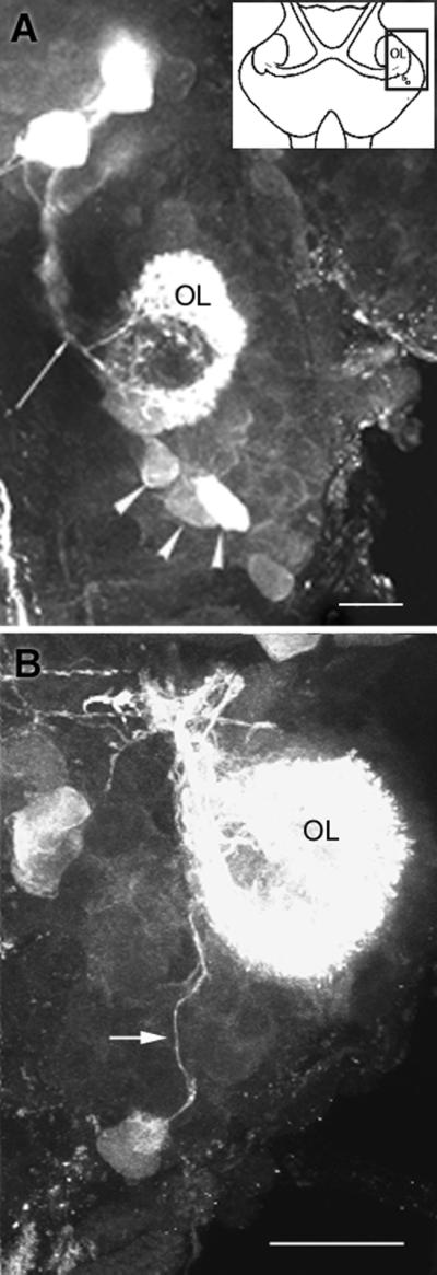
Confocal images of serotonin labeling in embryonic brains. (A) In lobsters at 35% of embryonic development, projection neurons with somata located at the posterior margin of cluster 10 (arrowheads) take up serotonin transiently (n = 12 cell clusters). Unlabeled projection neuron somata can be seen just anterior and lateral to the stained cells. The AL has not emerged at this time in development. Fibers from local interneurons (arrow), including the DGN, also label for serotonin. (B) The axons of serotonin-labeled cluster 10 neurons can be traced to the embryonic OL. (Bars = 20 μm.)
Conclusions
On the basis of these data, we propose that the uptake of serotonin by newborn olfactory projection neurons may underlie, directly or indirectly, the effects of serotonin that we have demonstrated on the proliferation, survival (Fig. 3), or branching (23) of these neurons in lobsters. A wealth of supporting evidence in the literature suggests that serotonin can regulate the proliferation, growth, and connectivity of serotonin-sensitive neurons (22, 29–34). In a striking example, serotonin has been shown to play a role in the development of the primary somatosensory map (28). In the somatosensory cortex, a transient, dense serotonergic innervation appears shortly after birth in mice and rats and disappears abruptly in a matter of days. During this brief period, however, the cortical barrels are delineated. Serotonin is intimately involved in the cortical ingrowth and arborization of thalamic axons, apparently by means of a high-affinity uptake of serotonin into the thalamic neurons, which do not contain the enzymes necessary to synthesize serotonin. It has been proposed that the internalized serotonin might be used for extracellular signaling or, alternatively, could exert intraneuronal control as a growth factor or transcriptional regulator (28).
The present results in the lobster olfactory system are compelling because several important elements come together in vivo: serotonin uptake by newborn projection neurons; effects of serotonin reduction on proliferation, survival and branching of newborn neurons; and the persistence of neurogenesis in the projection neuron cluster throughout life. Thus, serotonin may be important not only in shaping the developing olfactory pathway, but also in the long-term maintenance of the olfactory system. We propose that two life-long roles of the serotonergic DGNs in the lobster may be (i) to match the sizes of the pre- and postsynaptic populations of neurons by means of the regulation of interneuronal proliferation, and (ii) to direct the insertion of branches of new projection neurons into the highly specialized olfactory lobe neuropil.
The example provided here of serotonin uptake by a select group of newly born olfactory projection neurons suggests critical intracellular roles for this amine among neurons in the olfactory pathway. Serotonin has been implicated, at least indirectly, as an intracellular signaling molecule or transcriptional regulator in a variety of other systems (28, 35–37). It remains to be determined whether the neurons that take up serotonin in lobsters are a subset of newly proliferated cells, or whether all new olfactory projection neurons transport serotonin. Our data indicate that the former case is true, suggesting a degree of developmental specificity that may relate to the functional heterogeneity of the projection neurons (18).
Life-long neurogenesis and the intercalation of new interneurons into highly specialized synaptic areas are processes that are shared by olfactory systems in an evolutionarily diverse group of animals. The serotonergic regulation of these events in lobsters may be one example of a more general mechanism for directing neuronal turnover and influencing axonal outgrowth in systems where neurons are constantly turning over. Therefore, these phenomena may be associated with the presence of a dense and persistent serotonergic innervation of primary olfactory areas in a variety of species.
Acknowledgments
We thank S. Harzsch and S. Kohler for technical assistance, E. Buchner for antibody, and D. Sandeman for critical readings of this manuscript. This work was supported by National Science Foundation Grants IBN 9709514 and 0091092.
Abbreviations
- OL
olfactory lobe
- AL
accessory lobe
- OGT
olfactory globular tract
- OGTN
olfactory globular tract neuropil
- DGN
dorsal giant neuron
- 5,7-DHT
5,7-dihydroxytryptamine
- BrdUrd
bromodeoxyuridine
References
- 1.Graziadei P P C, Monti Graziadei G A. Neuroscience. 1986;19:1025–1035. doi: 10.1016/0306-4522(86)90119-3. [DOI] [PubMed] [Google Scholar]
- 2.Harrison P J, Cate H S, Swanson E S, Derby C D. J Neurobiol. 2001;47:51–66. doi: 10.1002/neu.1015. [DOI] [PubMed] [Google Scholar]
- 3.Lois C, Alvarez-Buylla A. Science. 1994;264:1145–1148. doi: 10.1126/science.8178174. [DOI] [PubMed] [Google Scholar]
- 4.Sandeman R, Clarke D, Sandeman D, Manly M. J Neurosci. 1998;18:6195–6206. doi: 10.1523/JNEUROSCI.18-16-06195.1998. [DOI] [PMC free article] [PubMed] [Google Scholar]
- 5.Harzsch S, Miller J, Benton J, Beltz B S. J Neurosci. 1999;19:3472–3485. doi: 10.1523/JNEUROSCI.19-09-03472.1999. [DOI] [PMC free article] [PubMed] [Google Scholar]
- 6.Xu F, Greer C A, Shepherd G M. J Comp Neurol. 2000;422:489–495. doi: 10.1002/1096-9861(20000710)422:4<489::aid-cne1>3.0.co;2-#. [DOI] [PubMed] [Google Scholar]
- 7.Laurent G. Curr Opin Neurobiol. 1997;7:547–553. doi: 10.1016/s0959-4388(97)80035-9. [DOI] [PubMed] [Google Scholar]
- 8.Cinelli A R, Hamilton K A, Kauer J S. J Neurophysiol. 1995;73:2053–2071. doi: 10.1152/jn.1995.73.5.2053. [DOI] [PubMed] [Google Scholar]
- 9.LaMantia A S, Pomeroy S L, Purves D. J Neurosci. 1992;12:976–988. doi: 10.1523/JNEUROSCI.12-03-00976.1992. [DOI] [PMC free article] [PubMed] [Google Scholar]
- 10.Baier H, Korsching S. J Neurosci. 1994;14:219–230. doi: 10.1523/JNEUROSCI.14-01-00219.1994. [DOI] [PMC free article] [PubMed] [Google Scholar]
- 11.Helluy S M, Benton J L, Langworthy K A, Ruchhoeft M L, Beltz B S. J Neurobiol. 1996;29:459–472. doi: 10.1002/(SICI)1097-4695(199604)29:4<459::AID-NEU4>3.0.CO;2-7. [DOI] [PubMed] [Google Scholar]
- 12.Halasz N, Shepherd G M. Neuroscience. 1983;3:579–619. doi: 10.1016/0306-4522(83)90206-3. [DOI] [PubMed] [Google Scholar]
- 13.Sudlow L C, Jing J, Moroz L L, Gillette R. J Comp Neurol. 1998;395:466–480. [PubMed] [Google Scholar]
- 14.Gelperin A. J Exp Biol. 1999;202:1855–1864. doi: 10.1242/jeb.202.14.1855. [DOI] [PubMed] [Google Scholar]
- 15.Sun X J, Tolbert L P, Hildebrand J G. J Comp Neurol. 1993;338:5–16. doi: 10.1002/cne.903380103. [DOI] [PubMed] [Google Scholar]
- 16.Sandeman D, Beltz B S, Sandeman R. J Comp Neurol. 1995;352:263–279. doi: 10.1002/cne.903520209. [DOI] [PubMed] [Google Scholar]
- 17.Strausfeld N J, Hildebrand J G. Curr Opin Neurobiol. 1999;5:634–639. doi: 10.1016/S0959-4388(99)00019-7. [DOI] [PubMed] [Google Scholar]
- 18.Beltz B S. Microsc Res Tech. 1999;44:105–120. doi: 10.1002/(SICI)1097-0029(19990115/01)44:2/3<105::AID-JEMT5>3.0.CO;2-K. [DOI] [PubMed] [Google Scholar]
- 19.Sandeman D C, Sandeman R E. J Comp Neurol. 1994;341:130–144. doi: 10.1002/cne.903410111. [DOI] [PubMed] [Google Scholar]
- 20.Sandeman R E, Sandeman D C. Brain Res. 1987;403:371–374. doi: 10.1016/0006-8993(87)90078-3. [DOI] [PubMed] [Google Scholar]
- 21.Benton J, Helluy S, Huber R, Beltz B S. J Neurobiol. 1997;33:357–373. doi: 10.1002/(sici)1097-4695(199710)33:4<357::aid-neu2>3.0.co;2-9. [DOI] [PubMed] [Google Scholar]
- 22.Benton J L, Beltz B S. J Neurobiol. 2001;46:193–205. doi: 10.1002/1097-4695(20010215)46:3<193::aid-neu1002>3.0.co;2-8. [DOI] [PubMed] [Google Scholar]
- 23.Sullivan J M, Benton J L, Beltz B S. J Neurosci. 2000;20:7716–7721. doi: 10.1523/JNEUROSCI.20-20-07716.2000. [DOI] [PMC free article] [PubMed] [Google Scholar]
- 24.Langworthy K S, Helluy S, Beltz B S. Cell Tissue Res. 1997;288:191–206. doi: 10.1007/s004410050806. [DOI] [PubMed] [Google Scholar]
- 25.Schmidt M, Ache B W. Cell Tissue Res. 1997;287:541–563. doi: 10.1007/s004410050778. [DOI] [PubMed] [Google Scholar]
- 26.Helluy S, Beltz B S. Biol Bull (Woods Hole, Mass.) 1991;180:355–371. doi: 10.2307/1542337. [DOI] [PubMed] [Google Scholar]
- 27.Cases O, Lebrand C, Giros B, Vitalis T, DeMaeyer E, Caron M G, Price D J, Gaspar P, Seif I. J Neurosci. 1998;18:6914–6927. doi: 10.1523/JNEUROSCI.18-17-06914.1998. [DOI] [PMC free article] [PubMed] [Google Scholar]
- 28.Lebrand C, Cases O, Adelbrecht C, Doye A, Alvarez C, Mestikawy S W, Seif I, Gaspar P. Neuron. 1996;17:823–835. doi: 10.1016/s0896-6273(00)80215-9. [DOI] [PubMed] [Google Scholar]
- 29.Whitaker-Azmitia P M, Druse M, Walker P, Lauder J M. Behav Brain Res. 1996;73:19–29. doi: 10.1016/0166-4328(96)00071-x. [DOI] [PubMed] [Google Scholar]
- 30.Lipton S A, Kater S B. Trends Neurosci. 1989;12:265–270. doi: 10.1016/0166-2236(89)90026-x. [DOI] [PubMed] [Google Scholar]
- 31.Lessmann V, Dietzel I D. J Neurosci. 1991;11:800–809. doi: 10.1523/JNEUROSCI.11-03-00800.1991. [DOI] [PMC free article] [PubMed] [Google Scholar]
- 32.Haydon P G, McCobb D P, Kater S B. Science. 1984;226:561–564. doi: 10.1126/science.6093252. [DOI] [PubMed] [Google Scholar]
- 33.Gould E. Neuropsychopharmacology. 1999;21:46S–51S. doi: 10.1016/S0893-133X(99)00045-7. [DOI] [PubMed] [Google Scholar]
- 34.Mercer A R, Kirchhof B S, Hildebrand J G. J Neurobiol. 1996;29:49–64. doi: 10.1002/(SICI)1097-4695(199601)29:1<49::AID-NEU4>3.0.CO;2-7. [DOI] [PubMed] [Google Scholar]
- 35.Liu Q R, Hattar S, Endo S, MacPhee K, Zhang H, Cleary L J, Byrne J H, Eskin A. J Neurosci. 1997;17:755–764. doi: 10.1523/JNEUROSCI.17-02-00755.1997. [DOI] [PMC free article] [PubMed] [Google Scholar]
- 36.Torres G, Horowitz J M, Laflamme N, Rivest S. Neuroscience. 1998;87:463–477. doi: 10.1016/s0306-4522(98)00147-x. [DOI] [PubMed] [Google Scholar]
- 37.Laflamme N, Feuvrier E, Richard D, Rivest S. Neuroscience. 1999;88:223–240. doi: 10.1016/s0306-4522(98)00369-8. [DOI] [PubMed] [Google Scholar]