. 2018 Jun 22;7:e34700. doi: 10.7554/eLife.34700

Figure 3. Turnover of spines on basal dendrites of CA1 pyramidal neurons in vivo.

(A) Repetitive imaging of basal dendrites in CA1 area using 2P-STED microscopy. The upper panel shows low-magnification overviews containing the dendrite of interest highlighted with a white box. The lower panel shows the corresponding 2P-STED images of the dendrite over time. The images represent single z-planes. Dendritic spines with blue arrowheads were stable between imaging sessions. Red arrowheads mark lost spines, and green arrowheads mark new ones. The axonal bouton (AB) is marked by a white arrowhead. The numbering of spines is continuous. (B) Quantification of spine density over 4 days (n = 14 dendrites, 3 mice). (C) Quantification of the 4-day survival fraction of dendritic spines. (D) Fraction of lost spines and (E) fraction of new spines. Thin grey lines represent the measurements of single dendrites.

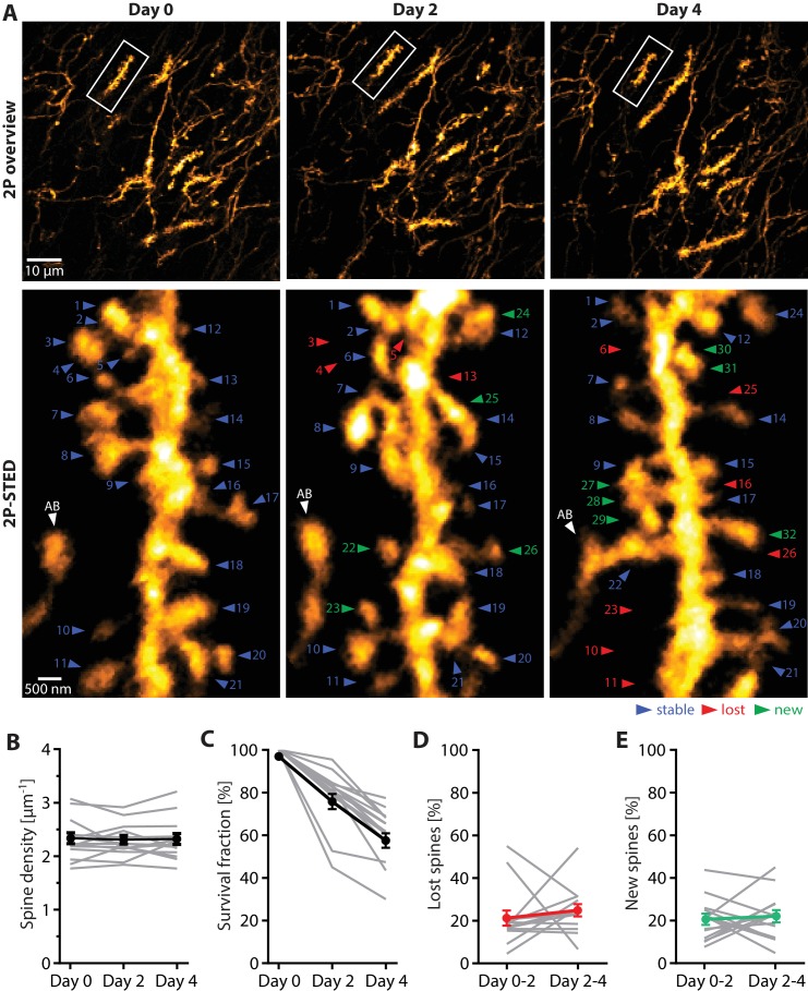
Figure 3—source data 1. Source data of the parameters underlying Figure 3 extracted from the turnover data set.
DOI: 10.7554/eLife.34700.017
Figure 3—source data 2.
DOI: 10.7554/eLife.34700.018
Figure 3—source data 3. Data for Figure 3—figure supplement 1.
DOI: 10.7554/eLife.34700.019

Figure 3.

Figure 3—figure supplement 1. 2P vs 2P-STED measurement of spine turnover in vivo.

Figure 3—figure supplement 1.

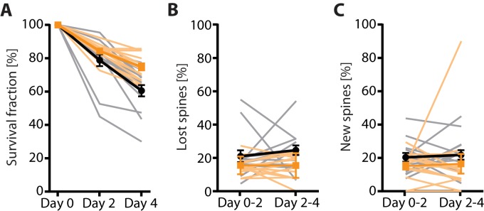
(A) Quantification of the 4-day survival fraction in 2P and 2P-STED (n = 14 dendrites, 3 mice). (B) Fraction of lost spines and (C) fraction of new spines in 2P and 2P-STED (n = 14 dendrites, 3 mice). Thin grey lines represent the measurement of single dendrites. In all panels, orange and black lines represent 2P and 2P-STED, respectively.
Figure 3—figure supplement 2. 2P-STED time-lapse imaging of hippocampal dendrites in vivo.

Figure 3—figure supplement 2.

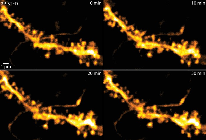
2P-STED could be used for time-lapse imaging of hippocampal dendrites as bleaching of the volume-filling fluorophore was negligible and no apparent phototoxicity was induced. MIP of three z-sections of a YFP-labeled dendrite imaged every 10 min.