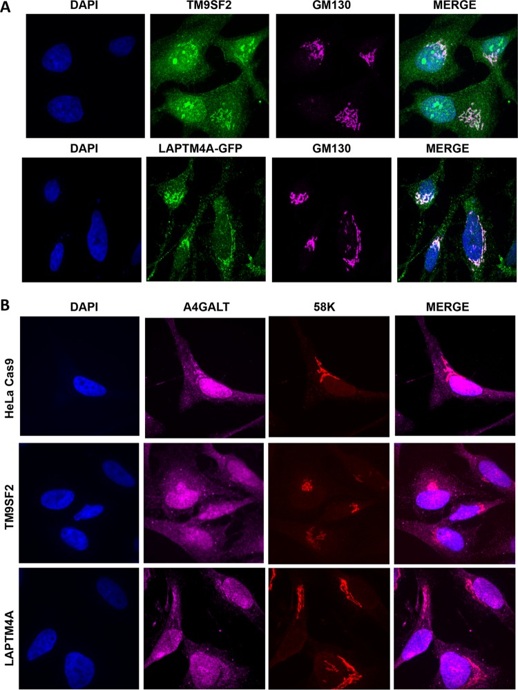
FIG 5 .
Subcellular localization of TM9SF2, LAPTM4A, and A4GALT in wt and mutant HeLa cells. (A) Confocal immunofluorescence microscopy of HeLa cells stained with anti-TM9SF2 (green), anti-GM130 to label the Golgi complex (pink) and DAPI. For LAPTM4A localization, HeLa cells were transfected with GFP-tagged LAPTM4A, which was imaged directly after counterstaining as described above. (B) Confocal immunofluorescence microscopy of control and mutant HeLa Cas9 cells labeled with anti-A4GALT antibody (pink), anti-58K to label the Golgi complex (pink) and DAPI. GM130 and 58K stain similar populations of Golgi complex membranes and were used interchangeably to accommodate the primary antibodies of interest.

