Abstract
Evidence exists to suggest that melatonin (MT) is important to abiotic stress tolerance in plants. Here, we investigated whether exogenous MT reduces heat damage on biological parameters and gene expression in kiwifruit (Actinidia deliciosa) seedlings. Pretreatment with MT alleviates heat-induced oxidative harm through reducing H2O2 content and increasing proline content. Moreover, MT application raised ascorbic acid (AsA) levels and the activity of antioxidant enzymes, including superoxide dismutase (SOD), catalase (CAT), and peroxidase (POD). We also observed elevation in the activity of enzymes related to the AsA-GSH cycle, such as ascorbate peroxidase (APX), monodehydroascorbate reductase (MDHAR), dehydroascorbate reductase (DHAR), and glutathione reductase (GR). Furthermore, MT application increased the expression of 28/31 glutathione S-transferase (GST) genes, reducing oxidative stress. These results clearly indicate that in kiwifruit, MT exerts a protective effect against heat-related damage through regulating antioxidant pathways.
Keywords: antioxidant enzymes, glutathione S-transferase, kiwifruit, melatonin, high temperature stress
1. Introduction
Temperatures 5 °C above optimal growing conditions induces heat shock or stress in plants, causing growth inhibition and crop failure [1,2]. These negative effects occur because cellular homeostasis is disrupted through mass formation of reactive oxygen species (ROS) in plant cells. These compounds include singlet oxygen (1O2), superoxide radical (O2•−), hydrogen peroxide (H2O2), and hydroxyl radical (OH•) are responsible for oxidative stress [3]. As a result, lipid peroxidation increases to cause oxidative stress, damaging membrane protein polymerization and cross-linking, as well as lowering membrane mobility, permeability, and thermal stability [4,5]. Like other aerobic organisms [6], plants have evolved defense systems that are well equipped with different antioxidant components to scavenge over-produced ROS, thus protecting plants from oxidative injury. An important aspect of these systems are antioxidant enzymes such as superoxide dismutase (SOD), catalase (CAT), peroxidase (POD), ascorbate peroxidase (APX), glutathione reductase (GR), monodehydroascorbate reductase (MDHAR), and dehydroascorbate (DHAR), as well as non-enzyme antioxidants such as ascorbic acid (AsA) and glutathione (GSH) [7,8]. In particular, glutathione S-transferases (EC 2.5.1.18) are a diverse, multifunctional group of stress-response enzymes, catalyzing GSH-dependent peroxidase reactions that scavenge toxic organic hydroperoxides. According to the genetic structure and protein homology, plant GSTs can be divided into 6 categories: Phi, Tau, Zeta, Lambda, Theta and Dehydroascorbate reductases (DHAR) [9,10].
Melatonin (MT) has received much recent attention in plant research because of its role as a growth regulator and a biostimulator for stress resistance [11]. The molecule enhances photosystem (PS) II activity [12]; alleviates growth inhibition and leaf senescence [13]; improves germination percentage [14], raise antioxidative enzymatic activity, antioxidant content [15,16]; and nitrogen metabolic enzyme activity [17]; as well as improve overall growth and rooting [18].
Kiwifruit (Actinidia deliciosa) is a perennial vine that is commercially cultivated in China, New Zealand, Chile, Japan, and Italy. Its heat-sensitivity is a major obstacle to crop productivity, however [19]. Long-term high temperatures cause flower and fruit dropping, quality deterioration, and storage decline [20]. Although a few studies on kiwifruit heat resistance are available [21,22], few researchers have examined how exogenous MT applications may improve antioxidation systems in kiwifruit seedlings under heat stress. Thus, the present study investigated the effectiveness of exogenous MT as an antioxidant-pathway regulator and an enhancer of heat-stress tolerance in kiwifruit.
2. Results
2.1. Seedling Morphology, H2O2 and Proline Content in Heat-Stressed Kiwifruit
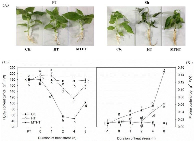
Before the experiment, seedlings were nearly identical across all three treatments (control [CK, 25 °C], high-temperature [HT, 45 °C], melatonin-pretreated high-temperature [MTHT, 45 °C) (Figure 1A). After 8 h treatment, HT seedlings exhibited dried leaves and water loss, whereas the MTHT group showed significantly fewer heat-stress symptoms (Figure 1A).
Figure 1.
(A) Seedling morphology at PT (25 °C starting temperature) and after 8 h at 45 °C; (B) H2O2 content in leaves under heat stress; (C) Proline content in leaves under heat stress. CK, control; HT, high temperature treatment (45 °C); melatonin-pretreated high-temperature (MTHT), high temperature with melatonin pre-treatment. Data are means of three biological replicates (n = 3). Lowercase letters indicate significant differences (p < 0.05).
Under the first 2 h of HT, H2O2 content in kiwifruit seedling increased, but then rapidly decreased until 4 h of HT (Figure 1B). Notably, MTHT seedlings had significantly lower H2O2 levels than HT seedlings.
Proline prevents plant cell dehydration and protects cytoplasmic membrane integrity. In HT and MTHT kiwifruits, proline content gradually increased over time (Figure 1C), but by 8 h, MTHT seedlings contained 1.36 and 2.1 times more proline than CK and HT seedlings, respectively.
2.2. POD, CAT, and SOD Activities under Heat Stress
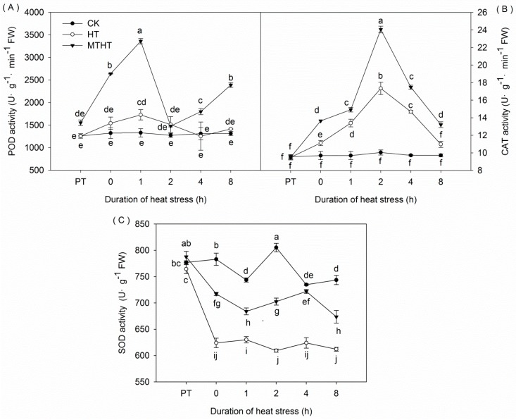
We observed a significant increase in POD activity that peaked at 1727.4 U·g−1·min−1 FW after 1 h of HT, before falling to CK levels after 4 h. Additionally, MT pretreatment dramatically increased POD activity, peaking after 1 h at 3360 U·g−1·min−1 FW, over two times greater than activity in HT leaves. After a drop at 2 h, POD activity in MTHT seedlings continued to increase (Figure 2A).
Figure 2.
Antioxidant enzyme activity under heat stress. (A) Peroxidase (POD); (B) catalase (CAT); and (C) superoxide dismutase (SOD). Lowercase letters indicate significant differences (p < 0.05).
We recorded a persistent increase in CAT activity under heat stress, peaking at 2 h of treatment (HT: 17.35 U·g−1·min−1 FW, MTHT: 24.06 U·g−1·min−1 FW) (Figure 2B). Subsequently, CAT activity decreased in both MTHT and HT seedlings. Throughout the experiment, CAT activity was significantly higher in MTHT than in HT.
Heat stress immediately (at 0 h) caused a marked and rapid decrease of 20.31% in SOD activity among HT leaves compared with CK leaves (Figure 2C). This decline was halved in MTHT leaves.
2.3. Ascorbic Acid Content and AsA-GSH-Cycle Enzymatic Activity under Heat Stress
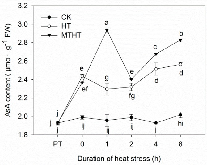
Compared with the steady levels in CK, AsA content exhibited two peaks in both HT and MTHT (Figure 3). In MTHT, AsA peaked at 1 h, reaching a value that was 19.19% greater than corresponding values in HT. Overall, except at 0 h, MTHT seedlings had higher AsA content than HT seedlings.
Figure 3.
Ascorbic acid (AsA) content in kiwi leaves under heat stress. Lowercase letters indicate significant differences (p < 0.05).
Under heat stress, APX activity in HT increased steadily until 4 h, when activity began to fluctuate slightly. In contrast, APX activity in MTHT persistently increased over time, peaking at 8 h with 3.32 U·g−1·min−1 FW, twice as high as corresponding HT values (Figure 4A). Heat stress also increased MDHAR activity, most noticeably in MTHT. In these leaves, enzyme levels exhibited a wavelike pattern that peaked at 5.76 U·g−1·min−1 FW after 8 h, a 193.31% increase from levels at 0 h (Figure 4B). In both HT and MTHT, DHAR activity first rose steadily beyond CK levels, before decreasing after 2 h. At the 2 h peak level, DHAR activity in MTHT was 20.85% higher than in HT, and both values were higher than CK. (Figure 4C). Finally, GR activity in HT significantly increased, peaking at 2 h (2.18 U·g−1·min−1 FW) before decreasing. In contrast, GR activity rose continuously in MTHT, increasing by 143.89% at 8 h (Figure 4D).
Figure 4.
Activity of key enzymes from the AsA-GSH cycle in heat-stressed kiwi leaves. (A) Ascorbate peroxidase (APX); (B) monodehydroascorbate reductase (MDHAR); (C) dehydroascorbate (DHAR); and (D) glutathione reductase (GR). Lowercase letters indicate significant differences (p < 0.05).
2.4. Expression Profile of GST under Heat Stress
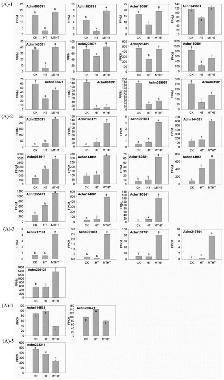
Using RNA-seq data, we discovered GST gene expression patterns differed significantly between MTHT and HT (adjusted p < 0.05; Figure 5A). There were 25 Tau GSTs, 2 Lambda GSTs, 2 Theta GSTs, 1 Phi GST and 1 unknown GST. The 31 differentially expressed GST genes were classified in five groups based on their deviation from CK expression level. (1) GST was down-regulated in HT and up-regulated in MTHT (12 transcripts); (2) GST was up-regulated in both HT and MTHT (11 transcripts); (3) GST remained unchanged in HT and up-regulated in MTHT (5 transcripts); (4) GST was up-regulated in HT and down-regulated in MTHT (2 transcripts); (5) Finally, GST was down-regulated in HT and MTHT (1 transcript). Overall, MT significantly up-regulated 28 GST genes, and only down-regulated 3.
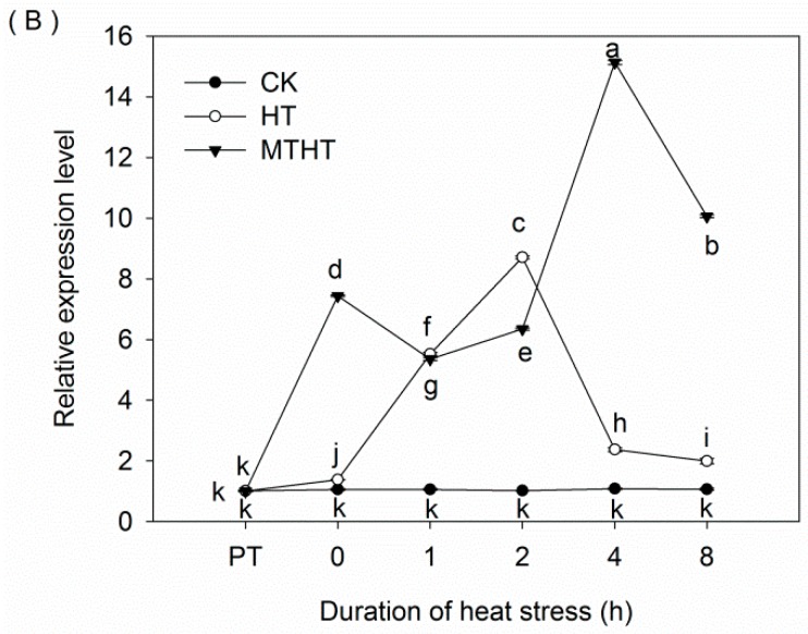
Figure 5.
(A) Results of RNA-seq showing significant differences in GST gene expression between MTHT and HT; (B) Data from qRT-PCR showing GST25 (ACHN160841 expression profile under heat stress. Lowercase letters indicate significant differences (p < 0.05).
Next, we selected GST25 (ACHN160841) in Group 2 for quantitative real-time PCR (qRT-PCR) analysis, to understand how GST expression changed over time (Figure 5B). Compared with CK, GST expression in HT first increased and then decreased. Additionally, GST gene expression in MTHT increased by 439.36%, 539.09%, and 495.04% at 0, 4, and 8 h, respectively, compared with HT (Figure 5).
3. Discussion
Melatonin is a well-documented antioxidant in plants that is critical to alleviating environmental stress [23,24,25,26]. Here, we observed that one key way exogenous MT increased kiwifruit heat resistance was through decreasing H2O2 content, in accordance with other work on Malus, Cynodon dactylon, and cucumber [27,28,29]. The mechanism underlying H2O2 reduction is likely the fact that MT acts as an electron donor [24]. Additionally, SOD catalyzes the removal of O2•− by dismutating it into O2 and H2O2 [30]; CAT and POD are involved in scavenging H2O2 to H2O and O2 [31]. In our study, we found H2O2 content in HT is lower than in CK, which may be because of the SOD reduction activity was not enough to counteract the occurring oxidative load. We, observed that under heat stress, MT enhanced the activity of major antioxidant enzymes (SOD, CAT, POD), possibly through upregulation of relevant genes. These results are similar to findings in cold-stressed cucumber and pepper seeds, showing that MT increased SOD activity through various physiological and molecular mechanisms in response to decreased H2O2 [15,16]. Likewise, our data correspond to results of MT treatment on stressed tea [32] and wheat [33]. Furthermore, as observed in wheat seedling [34,35], we found that heat stress increased proline content in kiwi leaves, perhaps because stress abolished feedback inhibition in the proline biosynthetic pathway [36]. Furthermore, MT pretreatment magnified this increase, as reported in cherry [37] and tomatoes [38]. These patterns may be attributable to the maintenance of low cell osmotic potential and reduced water loss through MT-induced proline accumulation, allowing improved adaption to a hot environment [39,40].
In our study, exogenous MT increased AsA content through elevating MDHAR and DHAR activity. Moreover, MT treatment increased GR activity more than it increased DHAR activity. These compounds are all part of the AsA-GSH cycle, an important antioxidant pathway that generates the small-molecule, non-enzymatic antioxidants AsA and GSH [41]. Fluctuation in AsA content is dependent on APX, MDHAR, and DHAR activities, with the latter two responsible for recycling AsA. Additionally, DHAR oxidizes GSH to GSSG during ROS scavenging, while GR recycles GSH. Our data thus suggest that exogenous MT is important to AsA and GSH biosynthesis/regeneration. Overall, we demonstrated that enhancing the AsA-GSH cycle is another way exogenous MT can protect plant tissues from oxidative damage [42,43].
Finally, we discovered 31 differentially expressed GST genes between MTHT and HT kiwifruit. They had five expression patterns which may be due to different functions of the gene family [9,44]. Glutathione S-transferases are critical to plant development and stress response through their scavenging of peroxides and other electrophiles [45,46,47,48,49,50]. Indeed, GST over-expression improves abiotic stress tolerance in tobacco and Arabidopsis [51,52]. The fact that we observed more up-regulated than down-regulated genes suggest that MT may dramatically decrease free-radical production and improve plant heat tolerance through elevating GST transcript abundance [53,54]. In general, our results proved that MT can improve the heat tolerance of heat-sensitive plants, moreover provided a way to make heat-sensitive plants grow better under heat stress.
4. Materials and Methods
4.1. Plant Materials and Treatment
Kiwifruit seeds were first disinfected for 5 min using 5% sodium hypochlorite and rinsed with distilled water. Cleaned seeds were grown at 4 °C and 60–70% relative humidity for 60 days. After a week-long poikilothermic treatment at 4 °C for 10 h and 25 °C for 14 h, germinated seeds were planted in plastic pots (diameter: 18 cm; height: 23 cm) filled with sand. They were then moved to a phytotron at Sichuan Agricultural University, Chengdu, China (30°42′ N, 103°51′ E), under conditions of 25/20 °C (day/night) and a 12/12 h (day/night) photoperiod. At the two-true-leaf stage, seedlings were watered in 2 days intervals with 1/2 Hoagland’s nutrient solution (pH adjusted to 6.5 ± 0.1 with diluted HCl or NaOH).
Treatments began at the 10-true-leaf stage. First, CK plants were maintained at 25 °C throughout the entire experiment. Second, HT seedlings were transferred to an incubator that increased from 25 °C to 45 °C across 2 h, and then maintained at the latter temperature for 8 h. Third, MTHT seedlings were pretreated 5 times with 200 µM MT solution, every two days, and then subjected to the same conditions as HT plants. Each treatment was performed in triplicate. The moment when the incubator temperature at 25 °C, was designed as PT; the moment when the incubator temperature just reached at 45 °C, was designed as 0 h. Five to eight middle leaves per plant were sampled at PT, 0, 1, 2, 4, and 8 h. All collected tissues were immediately frozen in liquid nitrogen and stored at −80 °C.
4.2. Assays of H2O2 Content and Antioxidant Enzyme Activity
Determination of H2O2 and proline levels followed previously described methods [55,56].
The photochemical reduction of NBT [57] was used to assay SOD activity. The guaiacol colorimetric method [58] was employed for measuring POD activity. Finally, CAT activity was calculated as the decline in A240 [59].
4.3. Extraction and Assay of AsA Content and AsA-GSH Cycle Enzymes
Ascorbic acid content was measured following existing methods [60]. Briefly, leaves (0.3 g) were ground in a prechilled mortar, then homogenized in 5 mL of ice-cold 6% (v/v) trichloroacetic acid (TCA) and 1 mM EDTA- Na2 solution. Crude extract was centrifuged at 2 °C and 12,000 g for 10 min; the supernatant was collected for analysis. We neutralized 50 μL extract with 250 μL 10% (w/v) TCA, 200 μL 42% H3PO4, and 200 μL 2% (w/v) 2,2-Dipyridyl (C10H8N2). The assay was performed using a spectrophotometer at 525 nm in 200 mm sodium phosphate buffer (pH 7.4), both before and after a 60 min incubation at 42 °C with 100 μL of 3% (w/v) FeCl3.
To determine the activity of enzymes involved in the AsA-GSH cycle, leaves (0.5 g) were ground in a chilled mortar with 4% (w/v) polyvinylpolypyrrolidone, then homogenized with 8 mL of 50 mM potassium phosphate buffer (pH 7.5) containing 1 mM EDTA-Na2 and 0.3% Triton X-100. The activity of APX was determined via monitoring absorbance decreases at 290 nm as reduced H2O2 was oxidized [61]. Similarly, MDHAR activity was assayed through monitoring absorbance decreases at 340 nm as NADH was oxidized [62]. Next, DHAR activity was determined through absorbance increases at 265 nm due to dehydroascorbate (DHA) formation [61]. Finally, GR activity was assayed through absorbance decreases at 340 nm from NADPH oxidation [63].
4.4. Quantitative Real-Time PCR for Profiling GST Expression
Total RNA was extracted from frozen fresh leaves using a modified CTAB method and treated with RNase-free DNase I (Takara, Dalian, China) to remove genomic DNA contamination. The NanoPhotometer® spectrophotometer (IMPLEN, Westlake Village, CA, USA) was used to check RNA purity. Quantitative real-time PCR was used to determine one selected GST gene. These reactions were performed on the CFX96 Real-Time System C1000 Thermal Cycler (Bio-RAD, Hercules, CA, USA), following manufacturer protocol in a SYBR Premix Ex Taq kit (TaKaRa, Dalian, China), and analyzed with 2−ΔΔCT. Relative gene expression was normalized with kiwifruit Actin1 and Actin2 [64]. Table 1 contains the primer sequences used for PCR. Three replicates were performed for three separate RNA extracts from three samples.
Table 1.
qRT-PCR primer sequences.
| Gene Locus | Forward Primer | Reverse Primer |
|---|---|---|
| ACHN160841 | GGTGTTGATACATAACGGAAAG | TGGACAATGATGAGGGACT |
| Actin1 | GCAGGAATCCATGAGACTACC | GTCTGCGATACCAGGGAACAT |
4.5. Expression Analysis of GST Genes Based on Transcriptome Data
Six cDNA libraries were constructed for three 8 h treatments (CK, HT, and MTHT), each with two biological replicates. Sequencing libraries were generated using the NEBNext Ultra RNA Library Prep Kit for Illumina (NEB, Ipswich, MA, USA) and sequenced on an Illumina Hiseq 2000 platform. Paired-end reads (150 bp) were generated by Novogene (Beijing, China).
Reads numbers mapped to each gene were counted in HTSeq version 0.6.1. The FPKM per gene was calculated based on gene length and read count. Differential expression analysis of two biological replicates was performed in R with the DESeq package (version 1.18.0, European Molecular Biology Laboratory, Heidelberg, Germany). The resultant P-values were adjusted using the Benjamini-Hochberg procedure for controlling false discovery rate [65]. Adjusted p < 0.05 was considered significant differential expression.
Acknowledgments
This work was financially supported by the Sichuan Science and Technology Project Program (2016NZ0105, 2017JY0054).
Author Contributions
Dong Liang, Fan Gao and Hui Xia conceived the idea and designed the research; Dong Liang, Fan Gao, Zhiyou Ni, Lijin Lin, Qunxian Deng, Yi Tang, Xun Wang, Xian Luo performed the experiments and analyzed the data; Hui Xia supervised the study; Dong Liang and Fan Gao wrote the manuscript with contributions from other coauthors.
Conflicts of Interest
The authors declare no conflict of interest.
Footnotes
Sample Availability: Samples of the compounds are not available from the authors.
References
- 1.Rodriguez V.M., Soengas P., Alonso-Villaverde V., Sotelo T., Cartea M.E., Velasco M.P. Effect of temperature stress on the early vegetative development of Brassica oleracea L. BMC Plant Biol. 2015;15:145. doi: 10.1186/s12870-015-0535-0. [DOI] [PMC free article] [PubMed] [Google Scholar]
- 2.Shah F., Huang J., Cui K., Nie L., Shah T., Chen C., Wang K. Impact of high temperature stress on rice plant and its traits related to tolerance. J. Agric. Sci. 2011;149:545–556. doi: 10.1017/S0021859611000360. [DOI] [Google Scholar]
- 3.Vasseur F., Pantin F., Vile D. Changes in light intensity reveal a major role for carbon balance in Arabidopsis responses to high temperature. Plant Cell Environ. 2011;34:1563–1576. doi: 10.1111/j.1365-3040.2011.02353.x. [DOI] [PubMed] [Google Scholar]
- 4.Mishkind M., Vermeer J.E., Darwish E., Munnik T. Heat stress activates phospholipase D and triggers PIP accumulation at the plasma membrane and nucleus. Plant J. 2009;60:10–21. doi: 10.1111/j.1365-313X.2009.03933.x. [DOI] [PubMed] [Google Scholar]
- 5.Narayanan S., Tamura P.J., Roth M.R., Prasad P.V., Welti R. Wheat leaf lipids during heat stress, high day and night temperatures result in major lipid alterations. Plant Cell Environ. 2016;39:787–803. doi: 10.1111/pce.12649. [DOI] [PMC free article] [PubMed] [Google Scholar]
- 6.Mittler R., Vanderauwera S., Suzuki N., Miller G., Tognetti V.B., Vandepoele K., Gollery M., Shulaev V., Van Breusegem F. ROS signaling, the new wave? Trends Plant Sci. 2011;16:300–309. doi: 10.1016/j.tplants.2011.03.007. [DOI] [PubMed] [Google Scholar]
- 7.Allakhverdiev S.I., Kreslavski V.D., Klimov V.V., Los D.A., Carpentier R., Mohanty P. Heat stress, an overview of molecular responses in photosynthesis. Photosynth. Res. 2008;98:541–550. doi: 10.1007/s11120-008-9331-0. [DOI] [PubMed] [Google Scholar]
- 8.Hasanuzzaman M., Nahar K., Alam M.M., Roychowdhury R., Fujita M. Physiological, biochemical, and molecular mechanisms of heat stress tolerance in plants. Int. J. Mol. Sci. 2013;14:9643–9684. doi: 10.3390/ijms14059643. [DOI] [PMC free article] [PubMed] [Google Scholar]
- 9.Dixon D.P., Davis B.G., Edwards R. Functional divergence in the glutathione transferase superfamily in plants. Identification of two classes with putative functions in redox homeostasis in Arabidopsis thaliana. J. Biol. Chem. 2002;277:30859–30869. doi: 10.1074/jbc.M202919200. [DOI] [PubMed] [Google Scholar]
- 10.Moons A. Regulatory and Functional Interactions of Plant Growth Regulators and Plant Glutathione S-Transferases (GSTs) Vitam. Horm. 2005;72:155–202. doi: 10.1016/S0083-6729(05)72005-7. [DOI] [PubMed] [Google Scholar]
- 11.Arnao M.B., Hernández-Ruiz J. Functions of melatonin in plants, a review. J. Pineal Res. 2015;59:133–150. doi: 10.1111/jpi.12253. [DOI] [PubMed] [Google Scholar]
- 12.Fan J., Hu Z., Xie Y., Chan Z., Chen Z., Amombo E., Chen L., Fu J. Alleviation of cold damage to photosystem II and metabolisms by melatonin in Bermudagrass. Front. Plant Sci. 2015;6:925. doi: 10.3389/fpls.2015.00925. [DOI] [PMC free article] [PubMed] [Google Scholar]
- 13.Zhang J., Shi Y., Zhang X.Z., Du H.M., Bin X., Bingru H. Melatonin suppression of heat-induced leaf senescence involves changes in abscisic acid and cytokinin biosynthesis and signaling pathways in perennial ryegrass (Lolium perenne, L.) Environ. Exp. Bot. 2017;138:36–54. doi: 10.1016/j.envexpbot.2017.02.012. [DOI] [Google Scholar]
- 14.Tiryaki I., Keles H. Reversal of the inhibitory effect of light and high temperature on germination of Phacelia tanacetifolia seeds by melatonin. J. Pineal Res. 2012;52:332–339. doi: 10.1111/j.1600-079X.2011.00947.x. [DOI] [PubMed] [Google Scholar]
- 15.Marta B., Szafrańska K., Posmyk M.M. Exogenous melatonin improves antioxidant defense in cucumber seeds (Cucumis sativus L.) germinated under chilling stress. Front. Plant Sci. 2016;7:575. doi: 10.3389/fpls.2016.00575. [DOI] [PMC free article] [PubMed] [Google Scholar]
- 16.Korkmaz A., Karaca A., Kocaçinar F., Cuci Y. The effects of seed treatment with melatonin on germination and emergence performance of pepper seeds under chilling stress. Tarim Bilimleri Dergisi J. Agric. Sci. 2017;23:167–176. [Google Scholar]
- 17.Zhang R., Sun Y., Liu Z., Jin W., Sun Y. Effects of melatonin on seedling growth, mineral nutrition, and nitrogen metabolism in cucumber under nitrate stress. J. Pineal Res. 2017;62 doi: 10.1111/jpi.12403. [DOI] [PubMed] [Google Scholar]
- 18.Arnao M.B., Hernández-Ruiz J. Melatonin promotes adventitious- and lateral root regeneration in etiolated hypocotyls of Lupinus albus L. J. Pineal Res. 2007;42:147–152. doi: 10.1111/j.1600-079X.2006.00396.x. [DOI] [PubMed] [Google Scholar]
- 19.Laing W.A. Temperature and light response curves for photosynthesis in kiwifruit (Actinidia chinensis) cv. Hayward. N. Z. J. Agric. Res. 1985;28:117–124. doi: 10.1080/00288233.1985.10427004. [DOI] [Google Scholar]
- 20.Richardson A.C., Marsh K.B., Boldingh H.L., Pickering A.H., Bulley S.M., Frearson N.J., Ferguson A.R., Thornber S.E., Bolitho K.M., Macrae E.A. High growing temperatures reduce fruit carbohydrate and vitamin C in kiwifruit. Plant Cell Environ. 2004;27:423–435. doi: 10.1111/j.1365-3040.2003.01161.x. [DOI] [Google Scholar]
- 21.Greer D.H., Laing W.A., Kipnis T. Photoinhibition of photosynthesis in intact kiwifruit (Actinidia deliciosa) leaves, Effect of temperature. Planta. 1988;174:152–158. doi: 10.1007/BF00394766. [DOI] [PubMed] [Google Scholar]
- 22.Luo H.T., Zhang J.Y., Wang G. Functional characterization of waterlogging and heat stresses tolerance gene Pyruvate decarboxylase 2 from Actinidia deliciosa. Int. J. Mol. Sci. 2017;18:2377. doi: 10.3390/ijms18112377. [DOI] [PMC free article] [PubMed] [Google Scholar]
- 23.Li H., He J., Yang X., Li X., Luo D., Wei C., Ma J., Zhang Y., Yang J., Zhang X. Glutathione-dependent induction of local and systemic defense against oxidative stress by exogenous melatonin in cucumber (Cucumis sativus L.) J. Pineal Res. 2016;60:206–216. doi: 10.1111/jpi.12304. [DOI] [PubMed] [Google Scholar]
- 24.Pieri C., Marra M., Moroni F., Recchioni R., Marcheselli F. Melatonin, A peroxyl radical scavenger more effective than vitamin E. Life Sci. 1994;55:271–276. doi: 10.1016/0024-3205(94)00666-0. [DOI] [PubMed] [Google Scholar]
- 25.Tan D.X., Chen L.D., Poeggeler B., Manchester L.D., Reiter R.J. Melatonin, a potent, endogenous hydroxyl radical scavenger. Endocr. J. 1993;1:57–60. [Google Scholar]
- 26.Tan D.X., Poeggeler B., Reiter R.J. The pineal hormone melatonin inhibits DNA adduct formation induced by the chemical carcinogen safrole in vivo. Cancer Lett. 1993;70:65–71. doi: 10.1016/0304-3835(93)90076-L. [DOI] [PubMed] [Google Scholar]
- 27.Shi H., Wang X., Tan D.X., Reiter R.J., Chan Z. Comparative physiological and proteomic analyses reveal the actions of melatonin in the reduction of oxidative stress in Bermuda grass (Cynodon dactylon (L). Pers.) J. Pineal Res. 2015;59:120–131. doi: 10.1111/jpi.12246. [DOI] [PubMed] [Google Scholar]
- 28.Li C., Tan D.X., Liang D., Chang C., Jia D.F., Ma F.W. Melatonin mediates the regulation of ABA metabolism, free-radical scavenging, and stomatal behaviour in two malus species under drought stress. J. Exp. Bot. 2015;66:669–680. doi: 10.1093/jxb/eru476. [DOI] [PubMed] [Google Scholar]
- 29.Zhao H., Ye L., Wang Y., Zhou X.T., Yang J.W., Wang J.W., Cao K., Zou Z. Melatonin Increases the Chilling Tolerance of Chloroplast in Cucumber Seedlings by regulating photosynthetic electron flux and the ascorbate-glutathione cycle. Front. Plant Sci. 2016;7:1814. doi: 10.3389/fpls.2016.01814. [DOI] [PMC free article] [PubMed] [Google Scholar]
- 30.Srinivasa-Rao N.K., Shivashankara K.S., Laxman R.H. Abiotic Stress Physiology of Horticultural Crops. Springer; Berlin/Heidelberg, Germany: 2016. [Google Scholar]
- 31.Mittler R. Oxidative stress, antioxidants and stress tolerance. Trends Plant Sci. 2002;7:405–410. doi: 10.1016/S1360-1385(02)02312-9. [DOI] [PubMed] [Google Scholar]
- 32.Li X., Wei J.P., Scott E.R., Liu J.W., Guo S., Li Y., Zhang L., Han W.Y. Exogenous Melatonin Alleviates Cold Stress by Promoting Antioxidant Defense and Redox Homeostasis in Camellia sinensis L. Molecules. 2018;23:165. doi: 10.3390/molecules23010165. [DOI] [PMC free article] [PubMed] [Google Scholar]
- 33.Li X.N., Brestic M., Tan D.X., Zivcak M., Zhu X.C., Liu S.Q., Song F.B., Reiter R.J., Liu F.L. Melatonin alleviates low PS I-limited carbon assimilation under elevated CO2 and enhances the cold tolerance of offspring in chlorophyll b-deficient mutant wheat. J. Pineal Res. 2018;64:e12453. doi: 10.1111/jpi.12453. [DOI] [PubMed] [Google Scholar]
- 34.Wang C., Wen D., Sun A., Han X.Y., Zhang J.B., Wang Z.J., Yin Y.P. Differential activity and expression of antioxidant enzymes and alteration in osmolyte accumulation under high temperature stress in wheat seedlings. J. Cereal Sci. 2014;60:653–659. doi: 10.1016/j.jcs.2014.05.004. [DOI] [Google Scholar]
- 35.Ahmed J.U., Hassan M.A. Evaluation of seedling proline content of wheat genotypes in relation to heat tolerance. Bangladesh J. Bot. 2011;40:17–22. doi: 10.3329/bjb.v40i1.7991. [DOI] [Google Scholar]
- 36.Boggess S.F., Stewart C.R. The relationship between water stress induced proline accumulation and inhibition of protein synthesis in tobacco leaves. Plant Sci. Lett. 1980;17:245–252. doi: 10.1016/0304-4211(80)90154-6. [DOI] [Google Scholar]
- 37.Sarropoulou V., Dimassi-Theriou K., Therios L., Koukourekou-Petridou M. Melatonin enhances root regeneration, photosynthetic pigments, biomass, total carbohydrates and proline content in the cherry rootstock PHL-C (Prunus avium × Prunus cerasus) Plant Physiol. Biochem. 2012;61:162–168. doi: 10.1016/j.plaphy.2012.10.001. [DOI] [PubMed] [Google Scholar]
- 38.Ding F., Liu B., Zhang S. Exogenous melatonin ameliorates cold-induced damage in tomato plants. Sci. Hortic. 2017;219:264–271. doi: 10.1016/j.scienta.2017.03.029. [DOI] [Google Scholar]
- 39.Turk H., Erdal S., Genisel M., Atici O., Demir Y., Yanmis D. The regulatory effect of melatonin on physiological, biochemical and molecular parameters in cold-stressed wheat seedlings. Plant Growth Regul. 2014;74:139–152. doi: 10.1007/s10725-014-9905-0. [DOI] [Google Scholar]
- 40.Meng J.F., Xu T.F., Wang Z.Z., Fang Y.L., Xi Z.M., Zhang Z.W. The ameliorative effects of exogenous melatonin on grape cuttings under water-deficient stress: Antioxidant metabolites, leaf anatomy, and chloroplast morphology. J. Pineal Res. 2014;57:200–212. doi: 10.1111/jpi.12159. [DOI] [PubMed] [Google Scholar]
- 41.Noctor G., Foyer C.H. Ascorbate and glutathione, keeping active oxygen under control. Annu. Rev. Plant Physiol. Plant Mol. Biol. 1998;49:249–279. doi: 10.1146/annurev.arplant.49.1.249. [DOI] [PubMed] [Google Scholar]
- 42.Bonnefont-Rousselot D., Collin F., Jore D., Gardèsalbert M. Reaction mechanism of melatonin oxidation by reactive oxygen species in vitro. J. Pineal Res. 2011;50:328–335. doi: 10.1111/j.1600-079X.2010.00847.x. [DOI] [PubMed] [Google Scholar]
- 43.Karam E.A., Maresca V., Sorbo S., Keramat B., Basile A. Effects of triacontanol on ascorbate-glutathione cycle in Brassica napus L. exposed to cadmium-induced oxidative stress. Ecotoxicol. Environ. Saf. 2017;144:268–274. doi: 10.1016/j.ecoenv.2017.06.035. [DOI] [PubMed] [Google Scholar]
- 44.Dixon D.P., Adrian L., Robert E. Plant glutathione transferases. Genome Biol. 2005;401:169–186. doi: 10.1016/S0076-6879(05)01011-6. [DOI] [PubMed] [Google Scholar]
- 45.Fujita M., Hossain M.Z. Modulation of pumpkin glutathione S-transferases by aldehydes and related compounds. Plant Cell Physiol. 2003;44:481–490. doi: 10.1093/pcp/pcg060. [DOI] [PubMed] [Google Scholar]
- 46.Dean J.D., Goodwin P.H., Hsiang T. Induction of glutathione S-transferase genes of nicotiana benthamiana following infection by colletotrichum destructivum and C. orbiculare and involvement of one in resistance. J. Exp. Bot. 2005;56:1525–1533. doi: 10.1093/jxb/eri145. [DOI] [PubMed] [Google Scholar]
- 47.Gong H., Jiao Y., Hu W.W., Pua E.C. Expression of glutathione-S-transferase and its role in plant growth and development in vivo and shoot morphogenesis in vitro. Plant Mol. Biol. 2005;57:53–66. doi: 10.1007/s11103-004-4516-1. [DOI] [PubMed] [Google Scholar]
- 48.Kunieda T., Fujiwara T., Amano T., Shioi Y. Molecular Cloning and characterization of a senescence-induced Tau-class glutathione S-transferase from barley leaves. Plant Cell Physiol. 2005;46:1540–1548. doi: 10.1093/pcp/pci167. [DOI] [PubMed] [Google Scholar]
- 49.Urbanek H., Majorowicz H., Zalewski M., Saniewski M. Induction of glutathione S-transferase and glutathione by toxic compounds and elicitors in reed canary grass. Biotechnol. Lett. 2005;27:911–914. doi: 10.1007/s10529-005-7181-9. [DOI] [PubMed] [Google Scholar]
- 50.Wagner U., Edwards R., Dixon D.P., Mauch F. Probing the diversity of the Arabidopsis glutathione S-transferase gene family. Plant Mol. Biol. 2002;49:515–532. doi: 10.1023/A:1015557300450. [DOI] [PubMed] [Google Scholar]
- 51.Le M.B., Poage M., Shiel K., Nugent G.D., Dix P.J. Tobacco chloroplast transformants expressing genes encoding dehydroascorbate reductase, glutathione reductase, and glutathione-S-transferase, exhibit altered anti-oxidant metabolism and improved abiotic stress tolerance. Plant Biotechnol. J. 2011;9:661–673. doi: 10.1111/j.1467-7652.2011.00611.x. [DOI] [PubMed] [Google Scholar]
- 52.Xu J., Tian Y.S., Xing X.J., Peng R.H., Zhu B., Gao J.J., Yao Q.H. Over-expression of AtGSTU19 provides tolerance to salt, drought and methyl viologen stresses in Arabidopsis. Physiol. Plant. 2015 doi: 10.1111/ppl.12347. [DOI] [PubMed] [Google Scholar]
- 53.Anwar M.M., Meki A.R. Oxidative stress in streptozotocin-induced diabetic rats: Effects of garlic oil and melatonin. Comp. Biochem. Physiol. Part A Mol. Integr. Physiol. 2003;135:539–547. doi: 10.1016/S1095-6433(03)00114-4. [DOI] [PubMed] [Google Scholar]
- 54.Reiter R.J., Tan D.X. Melatonin: An antioxidant in edible plants. Ann. N. Y. Acad. Sci. USA. 2002;957:341–344. doi: 10.1111/j.1749-6632.2002.tb02938.x. [DOI] [PubMed] [Google Scholar]
- 55.Sergiev L., Alexieva V., Karanova E. Effect of spermine, atrazine and combination between them on some endogenous protective systems and stress markers in plants. C. R. Acad. Bulg. Sci. 1997;51:121–124. [Google Scholar]
- 56.Bates L.S., Waldeen R.P., Teare I.D. Rapid determination of free proline for water stress studies. Plant Soil. 1973;39:205–207. doi: 10.1007/BF00018060. [DOI] [Google Scholar]
- 57.Giannopolitis C.N., Ries S.K. Superoxide Dismutases, II. purification and quantitative relationship with water-soluble protein in seedlings. Plant Physiol. 1977;59:315–318. doi: 10.1104/pp.59.2.315. [DOI] [PMC free article] [PubMed] [Google Scholar]
- 58.Scebba F., Sebastiani L., Vitagliano C. Activities of antioxidant enzymes during senescence of Prunus Armeniaca leaves. Biol. Plant. 2001;44:41–46. doi: 10.1023/A:1017962102950. [DOI] [Google Scholar]
- 59.Patra H.K., Kar M., Mishra D. Catalase activity in leaves and cotyledons during plant development and senescence. Biochem. Physiol. Pflanz. 1978;172:385–390. doi: 10.1016/S0015-3796(17)30412-2. [DOI] [Google Scholar]
- 60.Kampfenkel K., Montagu M.C., Inzè D. Extraction and determination of ascorbate and dehydroascorbate from plant tissue. Anal. Biochem. 1995;225:165–167. doi: 10.1006/abio.1995.1127. [DOI] [PubMed] [Google Scholar]
- 61.Nakano Y., Asada K. Hydrogen peroxide is scavenged by ascorbate-specific peroxidase in spinach chloroplasts. Plant Cell Physiol. 1981;22:867–880. doi: 10.1093/oxfordjournals.pcp.a076232. [DOI] [Google Scholar]
- 62.Arrigoni O., Dipierro S., Borraccino G. Ascorbate free radical reductase, a key enzyme of the ascorbic acid system. FEBS Lett. 1981;125:242–244. doi: 10.1016/0014-5793(81)80729-6. [DOI] [Google Scholar]
- 63.Edwards E.A., Rawsthorne S., Mullineux P.M. Subcellular distribution of multiple forms of glutathione reductase in leaves of pea (Pisum sativum L.) Planta. 1990;180:278–284. doi: 10.1007/BF00194008. [DOI] [PubMed] [Google Scholar]
- 64.Ferradás Y., Rey L., Troncoso O.M., Rey M., González M.V. Identification and validation of reference genes for accurate normalization of real-time quantitative PCR data in kiwifruit. Plant Physiol. Biochem. 2016;102:27–36. doi: 10.1016/j.plaphy.2016.02.011. [DOI] [PubMed] [Google Scholar]
- 65.Benjamini Y., Hochberg Y. Controlling the false discovery rate: A practical and 580 powerful approach to multiple testing. J. R. Stat. Soc. 1995;57:289–300. doi: 10.2307/2346101. [DOI] [Google Scholar]


