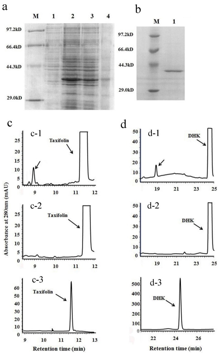
Figure 3.
Recombinant protein expression and enzymatic analysis of VbDFR. (a) a SDS-PAGE image shows recombinant VbDFR protein induced in E. coli BL21 (DE3) plysS strain. Lane 1: 20 μg crude protein extracts from BL21 (DE3) plysS/pET28a (+) vector control; lane 2: 20 μg crude protein extracts from BL21 (DE3) plysS/pET28a (+)-VbDFR. Lane 3: insoluble crude protein extracts from BL21 (DE3) plysS/pET28a (+)-VbDFR. Lane 4: soluble crude protein extracts from BL21 (DE3) plysS/pET28a (+)-VbDFR. M: protein molecular weight marker; (b) an image shows recombinant VbDFR purified; (c) HPLC profiles show one product formed from the incubation of taxifolin and recombinant VbDFR (c-1) but not denatured VbDFR (c-2); c-3, taxifolin standard; (d) HPLC profiles show one product formed from the incubation of dihydrokaempferol (DHK) and recombinant VbDFR (d-1) but not denatured recombinant VbDFR (d-2); d-3 DHK standard.

