Abstract
Hyperaccumulator plants have the unique ability to concentrate specific elements in their shoot in concentrations that can be thousands of times greater than in normal plants. Whereas all known zinc hyperaccumulator plants are facultative hyperaccumulators with only populations on metalliferous soils hyperaccumulating zinc (except for Arabidopsis halleri and Noccaea species that hyperaccumulate zinc irrespective of the substrate), the present study discovered that Dichapetalum gelonioides is the only (zinc) hyperaccumulator known to occur exclusively on ‘normal’ soils, while hyperaccumulating zinc. We recorded remarkable foliar zinc concentrations (10 730 µg g−1, dry weight) in Dichapetalum gelonioides subsp. sumatranum growing on ‘normal’ soils with total soil zinc concentrations of only 20 µg g−1. The discovery of zinc hyperaccumulation in this tropical woody plant, especially the extreme zinc concentrations in phloem and phloem-fed tissues (reaching up to 8465 µg g−1), has possible implications for advancing zinc biofortification in Southeast Asia. Furthermore, we report exceptionally high foliar nickel concentrations in D. subsp. tuberculatum (30 260 µg g−1) and >10 wt% nickel in the ash, which can be exploited for agromining. The unusual nickel and zinc accumulation behaviour suggest that Dichapetalum-species may be an attractive model to study hyperaccumulation and hypertolerance of these elements in tropical hyperaccumulator plants.
Introduction
Hyperaccumulators are plants that accumulate exceptional concentrations of certain trace elements in their above-ground biomass when growing in their natural habitats1–3. At present, the hyperaccumulation threshold values are set at: 100 µg g−1 for Cd, Se and Tl; 300 µg g−1 for Co, Cr and Cu; 1000 µg g−1 for As, Ni and Pb; 3000 µg g−1 for Zn; and 10 000 µg g−1 for Mn3. These unique plants have attracted growing attention from science, mainly because of their prospects for agromining (e.g. cultivating high biomass hyperaccumulator plants and recovering valuable products from the harvested biomass)4–8, and similarly for potential use in the phytoremediation of polluted sites9–13. More recently, hyperaccumulators, especially for Zn, have attracted considerable interest for the insights that they may yield for biofortification of food crops for human consumption14,15. Current research in the field of hyperaccumulator plants aims to provide insights into the physiological and biomolecular mechanisms underlying the hyperaccumulation trait, and the ecological significance and function, as well as potential applications16–22.
More than 700 hyperaccumulator plants have been discovered from across the globe, the majority (>70%) of which hyperaccumulate Ni3. Most Ni hyperaccumulator plants are from the Brassicaceae-family originating from Europe and from the Phyllanthaceae-family originating from tropical regions2,23,24. Nickel hyperaccumulator plant species occur on ultramafic outcrops, with some species being ‘obligate’ (i.e. restricted to that substrate and always hyperaccumulating Ni), and other species are able to grow on a range of different soils, with only ultramafic populations hyperaccumulating nickel25,26. In comparison to Ni hyperaccumulator plants, only a small number of Zn hyperaccumulator plant species (15 taxa) are known to date27; about 60% of which are in the Brassicaceae family. Zinc hyperaccumulator species include the herbaceous plants Arabidopsis halleri, Arabis paniculata, Noccaea caerulescens, Noccaea praecox, Noccaea fendleri (Brassicaceae), Picris divaricata (Asteraceae), Potentilla griffithii (Rosaceae) and Sedum plumbizincicola (Crassulaceae)28–34. Among these species, A. halleri and N. caerulescens are key model species for Ni and Zn-Cd hyperaccumulation, and the two most intensively studied hyperaccumulator species globally35–44. Arabidopsis halleri and N. caerulescens can accumulate extremely high concentrations of Cd-Zn (up to 53 450 µg g−1 Zn and 2890 µg g−1 Cd) and/or Ni (16 200 µg g−1) when growing on Zn-Pb-Cd-enriched metalliferous soils or on Ni-Co-Mn-enriched ultramafic soils39–42. However, Zn hyperaccumulation is a species-wide trait in these species29,43, and these two species are also able to hyperaccumulate Zn when growing on ‘normal’ soils with only background concentrations of Zn (i.e. soils with 1.00–300 µg g−1 total Zn)41,44. Reeves et al.41 analysed field collected specimens of N. caerulescens and the corresponding ‘normal’ soils in France and Luxembourg, and reported foliar Zn concentrations of 3230–8890 µg g−1 when occurring on soils with Zn concentrations of only 115–274 µg g−1.
The family Dichapetalaceae has three genera: Dichapetalum, Stephanopodium and Tapura45. The largest genus is Dichapetalum with >150 species46 occurring predominantly in tropical and subtropical regions, with most species distributed in Africa45. Dichapetalum gelonioides occurs throughout Southeast Asia, and has five subspecies: D. subsp. andamanicum, D. subsp. gelonioides, D. subsp. pilosum, D. subsp. sumatranum and D. subsp. tuberculatum46,47. Apart from D. subsp. andamanicum, all the other subspecies occur in Sabah (Malaysian Borneo), in primary lowland mixed Dipterocarp forest. All of the Dichapetalum-species are scandent scrubs, frequently climbing when small, but growing to small trees (<6 m in height with a <20 cm diameter at breast height (DBH) bole) when mature. Whereas D. subsp. tuberculatum and subsp. sumatranum have glabrous leaves, D. subsp. pilosum has pubescent (hairy) leaves. The key taxonomic characteristic is the fruits, which are either three-lobed and smooth (subsp. tuberculatum) or two-lobed and hairy (subsp. sumatranum).
Nearly three decades ago, Baker et al.46 discovered that D. gelonioides subsp. tuberculatum from the Philippines (on Palawan Island) hyperaccumulated Ni when growing on ultramafic soils. This then led to a survey of herbarium specimens of other Dichapetalum gelonioides subspecies from Southeast Asia. The analysis of herbarium specimen fragments confirmed that D. subsp. tuberculatum is a strong Ni hyperaccumulator when growing on ultramafic soils with up to 26 650 µg g−1 foliar Ni, and a strong Zn hyperaccumulator when growing on normal soils with up to 30 000 µg g−1 foliar Zn46. Dichapetalum subsp. sumatranum and D. subsp. pilosum are strong Zn hyperaccumulators when growing on normal soils, reaching up to 15 660 µg g−1 and 26 360 µg g−1 foliar Zn, respectively46. The discovery of Zn hyperaccumulation was unexpected, and unexplained at the time, and even though an important finding, it did not lead to further investigations. Baker et al.46 concluded that exceptional Zn accumulation in the D. gelonioides subsp. tuberculatum specimen from Sumatra (which had 30 000 µg g−1 foliar Zn) must have been due to unknown base metal mineralisation at that site, because of concomitant elevated foliar Cd (9 µg g−1), Pb (88 µg g−1) and Co (200 µg g−1) concentrations in this specimen. The phytochemistry of Ni in D. gelonioides subsp. tuberculatum was studied by Homer et al.48 and revealed that the aqueous plant extracts contain 18% Ni, 24% citric acid and 43% malic acid. Recently, systematic screening of herbarium specimens by our team using handheld X-ray Fluorescence Spectroscopy (XRF) including all specimens (91) in the genus Dichapetalum held at the Forest Research Centre Herbarium in Sepilok, Sabah, Malaysia (see Supplementary Table 1) revealed that:
Dichapetalum gelonioides subsp. tuberculatum is a strong Ni hyperaccumulator with up to 31 700 µg g−1 Ni
Dichapetalum gelonioides subsp. tuberculatum can also attain high Zn concentrations of up to 3990 µg g−1
Dichapetalum gelonioides subsp. pilosum is a strong Zn hyperaccumulator with up to 10 600 µg g−1 Zn
Dichapetalum gelonioides subsp. sumatranum is also a strong Zn hyperaccumulator with up to 10 500 µg g−1 Zn
Dichapetalum grandifolium has elevated Zn concentrations of up to 1300 µg g−1
Herbarium specimens were also measured with handheld XRF at the Queensland Herbarium for several Asia-Pacific species of Dichapetalum, including D. papuanum, D. timoriense, D. tricapsulare and D. sessiliflorum, none of which hyperaccumulated Ni or Zn. However, a specimen of D. vitiense from Fiji (collected in 1947 at Vanua Levu, on the summit ridge of Mt Numbuiloa, east of Lambasa, 500–590 m asl in forest) was confirmed as a Zn hyperaccumulator with 7800 μg g−1 foliar Zn. This finding raises the prospect that Dichapetalum species other than D. gelonioides could be Zn hyperaccumulators.
Dichapetalum gelonioides is a tropical species that occurs throughout Southeast Asia and attains high biomass, thereby overcoming the main drawbacks that limit the use of A. halleri and N. caerulescens for biofortification in tropical areas. Apart from the biofortification potential, D. gelonioides may be useful to advance our knowledge on hyperaccumulation of trace elements, particularly Zn. All Zn hyperaccumulator plant species known to date are facultative hyperaccumulators, and hence the majority occur primarily on normal soils (where they do not hyperaccumulate Zn), and have some populations on metalliferous soils where they do hyperaccumulate Zn41. Therefore, these Zn hyperaccumulators behave essentially as ‘Indicators’ (sensu Baker49) in that the level of accumulation in the shoot is strongly dependant on soil Zn concentrations. The main exceptions are A. halleri and N. caerulescens that have populations on normal soils that are also able to hyperaccumulate Zn41. Dichapetalum gelonioides is the only (Zn) hyperaccumulator known to occur exclusively on normal soils, while hyperaccumulating Zn. As such, Dichapetalum gelonioides provides an attractive model to study Zn tolerance, uptake, translocation, accumulation and detoxification, and may ultimately be useful for applications in the field of biofortification. This study aims to decipher the extent and variability of Ni and Zn hyperaccumulation traits in wild D. gelonioides subspecies occurring in Sabah.
Results
Elemental concentrations in different parts of Dichapetalum gelonioides plants
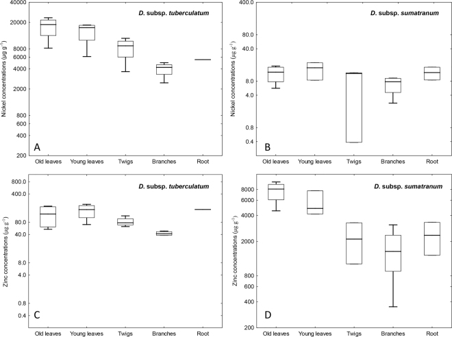
The elemental concentrations in the different plants parts of the various Dichapetalum gelonioides subspecies clearly show that D. gelonioides subsp. tuberculatum hyperaccumulates Ni, whereas D. subsp. pilosum and D. subsp. sumatranum are Zn hyperaccumulators (Table 1). The Ni concentrations in the leaves of D. subsp. tuberculatum are remarkable (the mean concentration is >1wt%), in contrast to that of D. subsp. pilosum and D. subsp. sumatranum, which have low Ni concentrations (mean is 10 and 15 µg g−1 respectively) (Fig. 1). On the contrary, the Zn concentrations are substantially elevated in the leaves of D. subsp. pilosum and D. subsp. sumatranum (>3000 µg g−1), but that of the D. subsp. tuberculatum are below 300 µg g−1 (Fig. 1). The concentrations of the major elements, Ca, K, Mg and S are relatively elevated (mean concentrations >1000 µg g−1), whereas the concentrations of the trace elements Co and Fe are uniformly low (mean < 50 µg g−1) in the leaves of all subspecies. Whereas the concentrations of Mn are elevated in the leaves of D. subsp. pilosum (>500 µg g−1), that of D. subsp. tuberculatum are low (mean <50 µg g−1). The highest Ni and Zn concentrations are recorded in the old leaves of D. subsp. tuberculatum (23 480 µg g−1) and D. subsp. sumatranum (9780 µg g−1) respectively, followed by young leaves with up to 18 230 µg g−1 for D. subsp. tuberculatum and 7720 µg g−1 for D. subsp. sumatranum respectively. The Ni concentrations in the branches and twigs of D. subsp. tuberculatum are substantially enriched (>1000 µg g−1). For D. subsp. pilosum and D. subsp. sumatranum, the Zn concentrations in the branches and twigs are elevated, but the values are below 3000 µg g−1. Unripe and matured fruits of D. subsp. sumatranum are also high in Zn (1955 and 2785 µg g−1 respectively). The bark/phloem of D. subsp. sumatranum has exceptionally high Zn concentrations (8465 µg g−1), whereas that of D. subsp. pilosum is elevated in Zn but below 3000 µg g−1. Whilst the roots of D. subsp. tuberculatum have high Ni concentrations (5510 µg g−1), those of D. subsp. pilosum and D. subsp. sumatranum are enriched in Zn (770 and 1380–3325 µg g−1 respectively). In Dichapetalum subsp. tuberculatum, the mean concentration of Ni in the old leaves (17 190 µg g−1) far exceed that in the roots (5510 µg g−1). Likewise, for D. subsp. pilosum and D. subsp. sumatranum, the Zn concentrations in the old leaves (6600 and 7620 µg g−1 respectively) are higher than in the roots (770 and 2355 µg g−1 respectively).
Table 1.
Bulk elemental concentrations in different plant tissues in Dichapetalum gelonioides subsp. tuberculatum, subsp. sumatranum and subsp. pilosum (values are given in ranges and means in µg g−1).
| Dichapetalum gelonioides subsp. | n | Ni | Zn | Al | Ca | Co | Fe | K | Mg | Mn | P | S |
|---|---|---|---|---|---|---|---|---|---|---|---|---|
| Young leaves | ||||||||||||
| subsp. tuberculatum | 4 | 6135–18230 [14420] | 70–220 [155] | 10–25 [20] | 2350–4510 [3030] | 10–15 [10] | 20–35 [25] | 3070–5920 [4905] | 1460–5370 [3060] | 20–40 [30] | 375–770 [490] | 865–2300 [1690] |
| subsp. sumatranum | 3 | 8.5–20 [15] | 4160–7720 [5570] | 10–15 [15] | 4635–5590 [5110] | 8.0–10 [9.3] | 20–25 [20] | 12650–16770 [14190] | 1610–1820 [1735] | 35–240 [160] | 605–715 [655] | 3740–7150 [5785] |
| subsp. pilosum | 1 | 10 | 4090 | 55 | 5085 | 5.0 | 35 | 13305 | 3320 | 895 | 575 | 4235 |
| Old Leaves | ||||||||||||
| subsp. tuberculatum | 4 | 8210–23480 [17190] | 55–200 [125] | 12–57 [30] | 2660–6420 [4110] | 10–15 [10] | 20–60 [45] | 805–2495 [1470] | 1700–5780 [3300] | 25–70 [40] | 245–460 [380] | 1165–2810 [2180] |
| subsp. sumatranum | 4 | 5.5–15 [10] | 4525–9780 [7620] | 23–81 [40] | 5570–11070 [8235] | 8.0–15 [10] | 25–60 [40] | 6090–11815 [8760] | 1345–1880 [1730] | 60–295 [205] | 430–565 [505] | 5035–7890 [6840] |
| subsp. pilosum | 1 | 5.0 | 6600 | 100 | 5415 | 10 | 45 | 6945 | 1770 | 1085 | 570 | 2765 |
| Branches | ||||||||||||
| subsp. tuberculatum | 4 | 2470–4960 [3965] | 35–50 [45] | 4.5–10 [7.0] | 290–945 [525] | 9.0–10 [9.5] | 8.0–15 [10] | 530–1080 [830] | 365–1525 [685] | 5.5–15 [9.5] | 165–375 [265] | 255–395 [325] |
| subsp. sumatranum | 4 | 2.5–9.5 [6.5] | 350–3115 [1635] | 6.0–25 [15] | 985–2855 [1745] | 9.4–10 [10] | 6.7–25 [15] | 1987–5380 [3825] | 107–445 [275] | 12–50 [25] | 135–555 [310] | 600–1745 [1195] |
| subsp. pilosum | 1 | 5.0 | 495 | 25 | 1090 | 5.0 | 15 | 3095 | 490 | 50 | 285 | 765 |
| Twigs | ||||||||||||
| subsp. tuberculatum | 4 | 3645–11520 [8235] | 65–115 [85] | 9.2–25 [15] | 1015–1785 [1305] | 7.5–10 [9.5] | 15–25 [20] | 1230–3355 [1910] | 1025–3570 [1705] | 8.5–25 [15] | 250–925 [520] | 425–600 [515] |
| subsp. sumatranum | 4 | 0.4–10 [8.1] | 1100–3285 [2170] | 10–60 [30] | 1965–2800 [2515] | 7.6–8.6 [8.2] | 10–40 [25] | 6335–6400 [6355] | 250–860 [475] | 20–75 [40] | 385–510 [435] | 600–1915 [1410] |
| subsp. pilosum | 1 | 15 | 1720 | 85 | 3625 | 10 | 35 | 9875 | 1710 | 175 | 1085 | 2110 |
| Roots | ||||||||||||
| subsp. tuberculatum | 1 | 5515 | 165 | 25 | 220 | 10 | 60 | 1400 | 335 | 10 | 145 | 415 |
| subsp. sumatranum | 2 | 8.5–15 [12] | 1385–3325 [2356] | 20–100 [60] | 1510–4185 [2847] | 9.5–9.9 [9.6] | 30–70 [50] | 1025–3615 [2320] | 140–320 [230] | 5.5–20 [15] | 151–264 [208] | 537–1431 [984] |
| subsp. pilosum | 1 | 10 | 770 | 15 | 1555 | 10 | 15 | 2825 | 135 | 35 | 175 | 535 |
| Stem | ||||||||||||
| subsp. sumatranum | 3 | 7.6–15 [10] | 510–2785 [1470] | 6.8–10 [8.3] | 720–1335 [1095] | 8.4–10 [9.4] | 8.5–10 [9.8] | 2016.5–3615 [3045] | 165–220 [195] | 7.1–20 [15] | 295–520 [415] | 730–1160 [915] |
| subsp. pilosum | 1 | 15 | 455 | 5 | 595 | 10 | 10 | 1210 | 110 | 15 | 155 | 235 |
| Bark/phloem | ||||||||||||
| subsp. sumatranum | 1 | 10 | 8465 | 175 | 11625 | 10 | 250 | 14700 | 580 | 115 | 200 | 1865 |
| subsp. pilosum | 1 | 10 | 2120 | 400 | 9110 | 5.0 | 205 | 6305 | 680 | 380 | 285 | 1405 |
| Wood | ||||||||||||
| subsp. sumatranum | 1 | 5.0 | 1895 | 5.0 | 1335 | 10 | 20 | 1375 | 130 | 5.0 | 140 | 575 |
| subsp. pilosum | 1 | 20 | 505 | 5.0 | 920 | 10 | 5.0 | 1675 | 130 | 40 | 130 | 545 |
| Unripe fruit | ||||||||||||
| subsp. sumatranum | 1 | 45 | 1955 | 10 | 9485 | 35 | 25 | 8075 | 1445 | 180 | 1040 | 4925 |
| Mature fruit | ||||||||||||
| subsp. sumatranum | 1 | 15 | 2785 | 10 | 4525 | 10 | 15 | 13745 | 1625 | 130 | 810 | 2930 |
The digest and extracts were analysed with Inductively Coupled Plasma Atomic Emission Spectroscopy (ICP-AES).
Figure 1.
Differences in the accumulation of Ni and Zn in Dichapetalum-species: (A) Nickel concentrations in D. subsp. tuberculatum, (B) Nickel concentrations in D. subsp. sumatranum, (C) Zinc concentrations in D. subsp. tuberculatum, and (D) Zinc concentrations in D. subsp. sumatranum.
Bio-ore characterisation of Dichapetalum gelonioides plants
The production of ash from the dried biomass of the various subspecies of Dichapetalum gelonioides resulted in mass reduction factors of about 10–12-fold for D. subsp. tuberculatum (leaf fraction), D. subsp. pilosum (twig and leaf fractions) and D. subsp. sumatranum (leaf fraction), and 50-fold for D. subsp. sumatranum (stem fraction), with minimal loss of the contained mineral elements. Table 2 gives the elemental concentrations in the dried biomass and the corresponding ash of D. subsp. tuberculatum (leaf fraction), D. subsp. pilosum (twig and leaf fractions) and D. subsp. sumatranum (separate fractions of leaf and stem). Notably, the Ni concentrations in the dried biomass of D. subsp. tuberculatum (leaf fraction) are exceptionally high, reaching up to 30 265 µg g−1. In addition, an extremely high Zn concentration of 10 730 µg g−1 is recorded in the dried biomass of D. subsp. sumatranum (leaf fraction). Regarding the composition of the ‘bio-ore’, as high as 11.3 wt% Ni is recorded in the ash of D. subsp. tuberculatum, whereas that in the ash of the various plant fractions of D. subsp. pilosum and D. subsp. sumatranum are lower than 1.50 wt% Ni (Table 2). In contrast, very high Zn concentrations (1.00–3.00 wt%) are recorded in the ash of the leaf fractions of D. subsp. pilosum and D. subsp. sumatranum, and even higher concentrations (3.27 wt%) are found in the stem fraction of D. subsp. sumatranum, relative to the low Zn concentrations (mean value of 0.25 wt%) in D. subsp. tuberculatum ash. The concentrations of Ca are significantly high in the ash samples, exceeding 9.50 wt% for all subspecies, and even reaching up to 19.1 wt% in D. subsp. sumatranum ash. Meanwhile, the K and S concentrations in the ash of D. subsp. pilosum and D. subsp. sumatranum (K 8.13–11.8 wt%; S 3.89–7.22 wt%) far exceed that in D. subsp. tuberculatum (K 2.17–2.28 wt%; S 2.57–2.92 wt%). However, the Mg concentrations in D. subsp. tuberculatum ash (2.07–2.48 wt%) exceed that in the D. subsp. pilosum and D. subsp. sumatranum (0.99–1.85 wt%) ash, and finally, relatively moderate P concentrations (0.55–1.44 wt%) are recorded in the ash of all the subspecies. In contrast to the high concentrations of major elements in the ash of all the subspecies, the concentrations of the trace elements Co, Fe and Mn are relatively low (<0.30 wt%) in the ash samples.
Table 2.
Elemental concentrations in the dried biomass and the corresponding ash of Dichapetalum gelonioides subsp. tuberculatum, subsp. sumatranum and subsp. pilosum (values are given in ranges and means).
| Dichapetalum gelonioides subsp. | n | Ni | Zn | Al | Ca | Co | Fe | K | Mg | Mn | P | S |
|---|---|---|---|---|---|---|---|---|---|---|---|---|
| Elemental concentrations in the dried biomass prior to ashing (µg g −1 ) | ||||||||||||
| subsp. tuberculatum (Leaves) | 2 | 29535–30265 [29900] | 255–275 [265] | 40–45 [45] | 13525–15845 [14685] | 9.3–9.5 [9.4] | 175–220 [200] | 1335––1400 [1365] | 1845–2175 [2005] | 40–45 [40] | 3490–3885 [3685] | 3585–3940 [3760] |
| subsp. sumatranum (Leaves) | 1 | 35 | 10730 | 55 | 19285 | 3.4 | 100 | 12190 | 2460 | 190 | 3290 | 11435 |
| subsp. sumatranum (Stems) | 1 | 5.00 | 940 | 45 | 5530 | — | 45 | 2315 | 265 | 50 | 2675 | 1240 |
| subsp. pilosum (Twigs and leaves) | 1 | 15 | 2995 | 45 | 19650 | 0.5 | 115 | 14665 | 1365 | 495 | 3010 | 9395 |
| Elemental concentrations in the ‘bio-ore’ (biomass after ashing) (wt%) | ||||||||||||
| subsp. tuberculatum (Leaves) | 2 | 10.0–11.0 [10.5] | 0.18–0.32 [0.25] | 0.05–0.13 [0.09] | 9.49–10.1 [9.77] | [0.01] | 0.09–0.23 [0.16] | 2.17–2.28 [2.23] | 2.07–2.48 [2.28] | [0.05] | 0.60–0.61 [0.61] | 2.55–2.90 [2.75] |
| subsp. sumatranum (Leaves) | 1 | 1.30 | 2.88 | 0.13 | 13.5 | — | 0.12 | 8.65 | 1.85 | 0.22 | 0.72 | 7.22 |
| subsp. sumatranum (Stems) | 1 | 0.03 | 3.27 | 0.39 | 19.1 | — | 0.14 | 8.13 | 0.99 | 0.17 | 1.14 | 3.89 |
| subsp. pilosum (Twigs and leaves) | 1 | 1.14 | 1.80 | 0.07 | 14.3 | — | 0.07 | 11.8 | 1.14 | 0.29 | 0.55 | 5.80 |
The digest and extracts were analysed with Inductively Coupled Plasma Atomic Emission Spectroscopy (ICP-AES).
Light microscopy of woody stems of Dichapetalum gelonioides
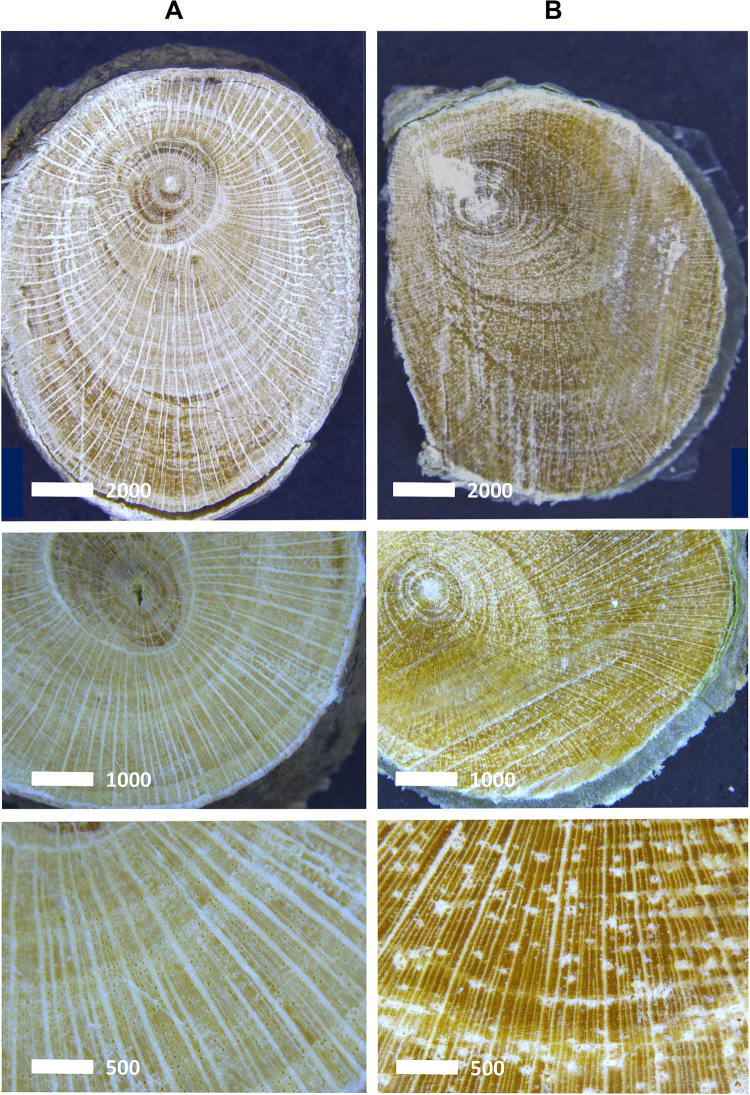
Light microscopy images (Fig. 2) show a whitish phloem in the woody stem of D. subsp. pilosum whilst in D. subsp. tuberculatum the phloem is greenish, mainly from the high concentrations of Zn and Ni ions respectively. The images show that Ca crystals are more abundant in the medullary rays of the stem of D. subsp. pilosum than in D. subsp. tuberculatum (Fig. 2).
Figure 2.
Light electron microscopy of woody stem cross section of (A) D. subsp. pilosum and (B) D. subsp. tuberculatum.
Synchrotron X-ray fluorescence microscopy investigation
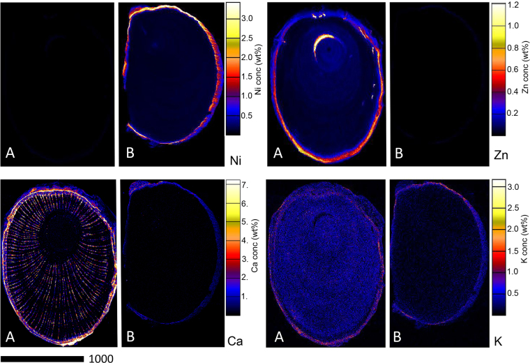
Figure 3 shows the elemental distribution in the stems of D. subsp. tuberculatum and D. subsp. pilosum. In D. subsp. tuberculatum, Ni is strongly enriched in the cortex where it reaches up to 3.00 wt%. Moreover, high concentrations of Ni occur in the phloem and epidermis where it reaches up to 1.00 wt%; there is also minor enrichment in the xylem and pith. However, Ni is notably absent throughout the stem of D. subsp. pilosum. Contrary, Zn is distributed throughout the stem of D. subsp. pilosum, with strong enrichment in the cortex, with up to 1.20 wt%, followed by the phloem and epidermis where it reaches up to 0.40 wt%. The xylem and pith only show minor enrichment in these elements. In the case of D. subsp. tuberculatum, Zn is low throughout the stem except in the cortex where there are slightly higher concentrations. Calcium is highly concentrated in the cortex and the epidermis, as well as in the medullary rays of the stem of D. subsp. pilosum. In D. subsp. tuberculatum, Ca is only enriched in the cortex, but at concentrations much lower than in D. subsp. pilosum. Potassium is distributed throughout the stems of both D. subsp. tuberculatum and D. subsp. pilosum, but more enriched in the latter than the former and more concentrated in the epidermis.
Figure 3.
Individual elemental µXRF maps of Dichapetalum gelonioides wood sections showing Ni, Zn, Ca, and K distribution in (A) D. subsp. pilosum, and (B) D. subsp. tuberculatum.
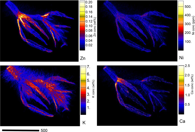
In the growth bud of D. subsp. pilosum (Fig. 4), Zn is strongly enriched in the node and the emerging leaves where it reaches up to 0.20 wt%, whereas there is a minor enrichment in the stem with up to 0.08 wt%. Interestingly, the distribution of Ni mirrors that of Zn, but occurs at much low concentrations (<300 µg g−1). Potassium is strongly enriched in the stem and the young leaves with minor enrichment in the node, whereas Ca is strongly enriched in the node but has relatively low concentration in the stem and in the emerging leaves.
Figure 4.
Individual elemental µXRF maps of freeze-dried D. subsp. pilosum growth bud showing Zn, Ni, K, and Ca distribution.
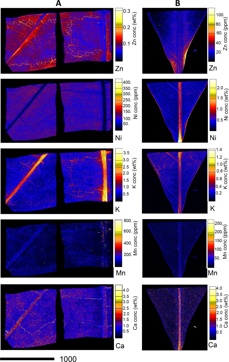
Zinc is distributed throughout the leaf of D. subsp. pilosum with concentrations reaching up to 0.30 wt%, whereas it is distributed at lower concentration in D. subsp. tuberculatum with concentrations below 100 µg g−1 (Fig. 5). Nickel is particularly low in the phloem of the veins of D. subsp. pilosum. In D. subsp. tuberculatum Ni is distributed at low concentrations throughout the leaf, but strongly enriched in the phloem of the mid-vein where it exceeds 2.00 wt%. Potassium is strongly enriched in the phloem of the mid-vein of both D. subsp. pilosum and D. subsp. tuberculatum. Manganese is distributed more or less uniformly in the leaf of both D. subsp. pilosum and D. subsp. tuberculatum at very low concentrations. The distribution of Ca mirrors that of K, albeit with Ca-oxakate deposits lining the veins.
Figure 5.
Individual elemental µXRF maps of freeze-dried Dichapetalum gelonioides leaves showing Zn, Ni, K, Mn, and Ca distribution in (A) D. subsp. pilosum, and (B) D. subsp. tuberculatum.
Rhizosphere soil chemistry of Dichapetalum gelonioides
The rhizosphere soil of D. gelonioides subsp. tuberculatum is near neutral (pH 6.01) as expected for well-buffered ultramafic soils rich in Mg, whereas that of the D. subsp. pilosum and D. subsp. sumatranum is acidic (pH 5.05 and 4.64, respectively), typical for leached tropical soils (Table 3). The total Ni concentration is high in the ultramafic soil on which D. subsp. tuberculatum occurs, unlike that of D. subsp. pilosum and D. subsp. sumatranum growing on sandstone soils, where low total soil Ni concentrations (8.50 and 3.50 µg g−1, respectively) are recorded. Similarly, the phytoavailable Ni concentrations, as extracted by diethylenetriaminepentaacetic acid (DTPA) and Sr(NO3)2 solutions, are relatively high in the rhizosphere soil of D. subsp. tuberculatum (185 µg g−1 and 15 µg g−1, respectively) compared to that of D. subsp. pilosum (1.50 µg g−1 and 0.30 µg g−1, respectively) and D. subsp. sumatranum (0.70 µg g−1 and 0.40 µg g−1, respectively). The soil of D. subsp. tuberculatum also has a higher Mg:Ca ratio and concentrations of Cr, Co, Mn and Fe compared to those of D. subsp. pilosum and D. subsp. sumatranum. However, a low concentration of K is recorded in the soil of D. subsp. tuberculatum, but in the soils of D. subsp. pilosum and D. subsp. sumatranum K concentrations are higher. The phytoavailable P concentrations in all the soils are low (<5.00 µg g−1). In addition, low concentrations of Zn are recorded for all the rhizosphere soils (total Zn concentrations in the rhizosphere soils of D. subsp. tuberculatum, D. subsp. pilosum and D. subsp. sumatranum are 40, 30 and 20 µg g−1, respectively).
Table 3.
Rhizosphere soil chemistry in the natural habitat of Dichapetalum gelonioides subsp. tuberculatum, subsp. sumatranum and subsp. pilosum. Elemental concentrations in µg g−1.
| Dichapetalum gelonioides subsp. | n | pH | Ni | Zn | Mg | P | K | Ca | Cr | Mn | Fe | Co |
|---|---|---|---|---|---|---|---|---|---|---|---|---|
| Total | ||||||||||||
| tuberculatum | 1 | 6.01 | 785 | 35 | 28395 | 105 | 30 | 200 | 1035 | 1350 | 23915 | 50 |
| sumatranum | 1 | 4.64 | 3.6 | 20 | 715 | 85 | 405 | 150 | 3.0 | 115 | 4490 | 1.5 |
| pilosum | 1 | 5.05 | 8.4 | 35 | 1285 | 220 | 850 | 685 | 6.0 | 335 | 10680 | 1.0 |
| DTPA-extractable | ||||||||||||
| tuberculatum | 1 | 185 | 4.6 | — | — | — | — | 0.17 | 375 | 125 | 10 | |
| sumatranum | 1 | 0.7 | 10 | — | — | — | — | 0.03 | 60 | 155 | 0.5 | |
| pilosum | 1 | 1.6 | 6.7 | — | — | — | — | 0.03 | 240 | 160 | 1.0 | |
| Sr(NO 3 ) 2 -extractable | ||||||||||||
| tuberculatum | 1 | 10 | 0.2 | — | — | — | — | 0.07 | 6.5 | 0.5 | 0.3 | |
| sumatranum | 1 | 0.4 | 7.2 | — | — | — | — | 0.02 | 60 | 1.7 | 0.5 | |
| pilosum | 1 | 0.3 | 1.4 | — | — | — | — | 0.01 | 110 | 0.8 | 0.5 | |
Discussion
This study confirms that D. subsp. tuberculatum is a ‘hypernickelophore’ i.e., it accumulates >1wt% Ni in the leaves. The rhizosphere soil chemistry implies that D. subsp. tuberculatum occurs on ultramafic soils. Dichapetalum gelonioides subsp. tuberculatum is common on Mount Silam (which is part of the Silam-Beeston ultramafic range), and has been recorded only from a few other localities in Sabah46,50. The present study thereby confirms the discovery by Baker et al.46 that D. subsp. tuberculatum hyperaccumulates Ni when growing on ultramafic soils with leaf Ni concentrations of >25 000 µg g−1, showing that D. subsp. tuberculatum is a strong Ni hyperaccumulator. Datta et al.47 reported that D. subsp. andamanicum is also a strong Ni hyperaccumulator with up to 30 000 µg g−1 Ni when occurring on ultramafic soils. Dichapetalum subsp. pilosum and D. subsp. sumatranum occur on sedimentary (sandstone) bedrock50, and have leaf Ni concentrations below 25 µg g−1 and are hence not Ni hyperaccumulators. The extraordinary Ni concentrations in the leaves of D. subsp. tuberculatum (reaching up to 30 265 µg g−1) may be exploited for agromining operations. The ‘bio-ore’ (i.e. the biomass after ashing) of D. subsp. tuberculatum contained as high as 11.3 wt% Ni, which is significantly higher than in traditional lateritic ore (which typically has <1 wt% Ni). Similar high-grade ash compositions are recorded in the two most promising species of tropical Ni agromining, Phyllanthus rufuschaneyi (12.7 wt%) and Rinorea cf. bengalensis (5.50 wt% Ni)51. The unique ‘bio-ore’ composition, coupled with the intrinsically high Ni grade, make it possible to extract even higher value products such as Ni catalysts for the organic chemistry industry52 and electrochemical Ni products53, although smelting of Ni metal is in itself technically feasible6. We must add that despite the high purity of the ‘bio-ore’ relative to conventional ore, significant concentrations of Ca (9.49–10.1 wt%), Mg (2.07–2.48 wt%), K (2.17–2.28 wt%) and S (2.57–2.92 wt %) recorded in D. subsp. tuberculatum are major drawbacks in extracting pure Ni products from this ‘bio-ore’. In other hyperaccumulator ‘metal crop’ species, even higher concentrations of Ca (20–40 wt%), Mg (1.00–5.00 wt%) and K (7.00–11.0 wt%) are recorded in the ‘bio-ore’51,54.
The present study reports on the tropical Zn hyperaccumulators, Dichapetalum subsp. sumatranum and subsp. pilosum. Zinc hyperaccumulators are rare globally, with only 15 taxa identified to date27. Almost all known Zn hyperaccumulators are herbaceous plants, and the present study is the first to report on a woody Zn hyperaccumulator from field collected material. Most of the known Zn hyperaccumulators occur on Zn enriched (metalliferous) soils. For instance, exceptionally leaf Zn concentrations (53 450 µg g−1) have been recorded in a population of N. caerulescens growing on soils with extremely high Zn concentrations of up to 64 360 µg g−1 in Saint Félix-de-Pallières, France41. Similarly high values (53 900 µg g−1) have been reported in A. halleri from Germany growing on non-metalliferous soils44. Zinc hyperaccumulation occuring on soils with ‘normal’ Zn concentrations is known only from these two European species (A. halleri and N. caerulescens). Reeves et al.41 analysed field collected specimens of N. caerulescens and the corresponding ‘normal’ soils in France and Luxembourg, and found remarkable foliar Zn concentrations of 3230–8890 µg g−1 occurring on soils with Zn concentrations of only 115–274 µg g−1. Bert et al.29 also reported 10 876 ± 578 µg g−1 Zn concentrations in the aerial part of wild A. halleri plants in Germany occurring on soils with 201 ± 30 µg g−1 Zn concentrations. The total Zn concentrations in ‘normal’ soils in Sabah are typically between 1.20 to 150 µg g−1 55. The present study reveals a remarkable foliar Zn concentration of 10 730 µg g−1 in D. subsp. sumatranum growing on ‘normal’ soils with total Zn concentration of only 20 µg g−1. It is noteworthy that the concentrations we report are not the highest for this species, but that Baker et al.46 found as high as 14 000 µg g−1 and 25 000 µg g−1 in herbarium specimen of D. subsp. sumatranum and D. subsp. pilosum respectively (Fig. 6), showing that Dichapetalum species are among the strongest Zn hyperaccumulators globally. Regarding the (Zn) Bioaccumulation Coefficients (BC) reported for Zn hyperaccumulator species, the values for species occurring on metalliferous soils are relatively lower than that of the non-metalliferous soils41,44. Previously, the highest BC reported for a Zn hyperaccumulator species was recorded in A. halleri occurring on non-metalliferous soils with a BC of ~127044. The present study also reveals an exceptional BC of ~500 for D. subsp. sumatranum, further confirming the extraordinary accumulation behaviour of Dichapetalum species. We discovered that Dichapetalum gelonioides is the only (Zn) hyperaccumulator known to occur exclusively on normal soils, while hyperaccumulating Zn.
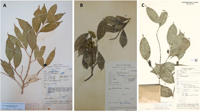
Figure 6.
Herbarium specimens of: (A) Dichapetalum gelanioides subsp. tuberculatum (SAN 57302) with 26 650 μg g−1 Ni and 257 μg g−1 Zn, (B) Dichapetalum gelanioides subsp. sumatranum (SAN 30460) with <5 μg g−1 Ni and 14 060 μg g−1 Zn, and (C) D. gelonioides subsp. tuberculatum specimen from Sumatra (which had 30 000 µg g−1 foliar Zn) (Baker et al.46) (all images are by A. van der Ent).
In Dichapetalum, Zn accumulation at extreme concentrations is not only limited to the leaves, but in other plant tissues as well, even though the distribution is uneven. The present study reveals as high as 8465 µg g−1 Zn concentrations in the phloem/bark of D. subsp. sumatranum. This exceptionally high Zn concentration in the phloem may be transported to the fruits and seeds. The mature fruit of D. subsp. sumatranum has high Zn concentrations of up to 2785 µg g−1. The high Zn concentration in the phloem and the fruits of D. subsp. sumatranum could have implications for Zn biofortification of edible crop plants. For crop plants, Zn concentrations decrease in the order of roots >shoots >fruits and seeds56. Notably, low Zn mobility in phloem limits the Zn concentrations in fruits and seeds, and these phloem-fed tissues rarely achieve Zn concentrations greater than 30–100 µg g−1 15. As a result, Zn concentrations are very low in grain, seed, fruit or tuber crops57–59. Consequently, Zn deficiency disorders are more prevalent in population with diets dominated by such crops60. Zinc is an essential trace element for all organisms, and particularly important for human nutrition57,58,61. The Institute of Medicine USA62 recommends a daily intake of 8–13 mg Zn, but up to 1/3 of the global population has inadequate dietary Zn intake because of limited access to Zn-rich foods, and consequently, Zn deficiency is a major health problem in developing countries63–66. Particularly, it is more problematic in Southeast Asia because of the consumption of diets low in Zn, mainly polished rice. Limited dietary Zn intake can be minimised by improving the available Zn in the diet, and biofortification of edible crops is a sustainable strategy especially in developing countries15,67–69. The target Zn concentrations are 28 µg g−1 in rice, and 38 µg g−1 in wheat grain and maize68. Zinc hyperaccumulators have the potential to advance biofortification, because insights into the biomolecular mechanisms of Zn acquisition may be applied to food crops15, and hyperaccumulator plant biomass may be used as a dietary supplement14. However, A. halleri and N. caerulescens are both temperate region plants with low biomass, and hence unsuited for cultivation in tropical regions where human Zn deficiency is prevalent. Hence, the discovery of a Zn hyperaccumulator plant species on ‘normal’ soils in Sabah provides a strong incentive to advance Zn biofortification measures in Southeast Asia. Insights gained from the mechanisms underlying the remarkable Zn translocation and accumulation especially in the phloem and phloem-fed tissues may be particulary useful. Evidence from transcriptome data suggests that Zn hyperaccumulator plant species have an enhanced capacity to protect cells from metal toxicity22,27. On the other hand, crop plants maintain low Zn concentration in the phloem sap as a strategy to avoid cellular toxicity58. Therefore, identifying the genes involved in the Zn hyperaccumulation in D. subsp. sumatranum could enable the breeding of genetically modified edible crops with enhanced abilities, especially translocation of Zn in the phloem. The increased Zn tolerance and mobility in phloem of these improved cultivars would therefore enrich the phloem-fed tissues (such as seeds/grains), and ultimately increase dietary Zn intake in Southeast Asia.
Another approach to increase dietary Zn intake in Southeast Asia may be to introduce the biomass of hyperaccumulators, such as D. subsp. sumatranum and D. subsp. pilosum, as a food supplement14. Considering a recommended intake of 8 mg Zn (Institute of Medicine USA62), an average 33% efficiency of human uptake70, and a leaf Zn concentration of 9000 µg g−1 in the leaves of D. subsp. sumatranum (Table 2), consuming 0.5 g dry material would meet one fifth of the daily requirements. As Zn in the leaves of Zn hyperaccumulators is likely bioavailable, human Zn uptake from these materials should not be an issue. However, there are major food safety concerns regarding the use of Zn hyperaccumulator plant species as food because A. halleri and N. caerulescens can also accumulate Cd in their leaves41. Cadmium is highly toxic to humans, and obviously consuming plant tissues with high bioavailable Cd is not recommended for human health71. Therefore, there is a need to test whether Dichapetalum-species occurring on ‘normal’ soils will accumulate Cd. Furthermore, many Dichapetalum-species are extremely toxic to livestock because they contain fluorinated compounds such as fluoroacetic acid and ω-fluorinated fatty acids72–76. Therefore, these fluorinated compounds will need to be detoxified before they are introduced as food supplements for human consumption, and toxicological assays are clearly required. Some Dichapetalum-species can also contain fluorine-free compounds, such as the triterpenoids group of dichapetalins that are being intensively investigated because of their strong cytotoxic activity, and potential application in anti-cancer drugs77–81.
Apart from the potential for agromining and biofortification applications, Dichapetalum-species can advance our understanding on both Ni and Zn hyperaccumulation. The mechanisms involved in the hyperaccumulation of trace elements are not fully understood, but the key processes include uptake by roots and xylem loading as well as sequestration in the leaf cells. Whether hyperaccumulation is primarily driven by root processes, or hypertolerance is determined by shoot processes remains inconclusive, at least in tropical hyperaccumulator plant species. Grafting experiments in which the rootstocks and shoot scions are reciprocally grafted from D. subsp. pilosum or D. subsp. sumatranum (Zn hyperaccumulating subsp.) and D. subsp. tuberculatum (Ni hyperaccumulating subsp.) may unravel the distinct roles of root and shoot processes in hyperaccumulation and hypertolerance. In addition, reciprocal dosing of Dichapetalum-species in which the Zn hyperaccumulating subsp. is dosed with Ni, and vice versa may provide more insights into the uptake, translocation, tolerance and accumulation of these trace elements. Furthermore, Ni and Zn stable isotope tracing experiments can provide useful information on both uptake and transfer mechanisms within the plant-soil systems. Therefore, Dichapetalum-species are an attractive model to study both Ni and Zn hyperaccumulation and hypertolerance in tropical hyperaccumulator plant species.
Materials and Methods
Herbarium XRF scanning
The Niton XL3t 980 analyser (Thermo-Fisher Scientific) uses a miniaturised X-ray tube (Ag anode; 6–50 kV, 0–200 µA max) as its excitation source. The X-ray tubes irradiates the sample with a stable source of high-energy X-rays, and fluorescent X-rays are detected, identified and quantified by the inbuilt Silicon Drift Detector (SDD) with ~185 eV, up to 60 000 cps, 4 µs shaping time. The instrument uses Compton normalisation for quantification, appropriate for the relatively low elemental concentrations found in plant material. A total of 590 dried plant samples originating from Sabah, Malaysia82 were used for the calibration. From each sample, a 6-mm diameter leaf disc (to match the XRF beam width) was extracted using a paper punch. A square of ~99.7% pure titanium (2 mm thick × 10 cm × 10 cm; Sigma-Aldrich 369489–90G) was used behind the specimens to provide a uniform background and block penetrating X-rays. XRF measurements were carried out in the ‘Soils Mode’ for 60 s. After scanning, the leaf samples were weighed, digested and analysed with Inductively Coupled Plasma Atomic Emission Spectroscopy (ICP-AES), following the procedures described below. Correction factors were derived by linear regression of XRF data against corresponding ICP-AES measurements.
In total, 91 herbarium species were measured at the herbarium of the Forest Research Centre in Sepilok, Sabah, Malaysia. This comprised two species: Dichapetalum grandifolium and Dichapetalum gelonioides (subsp. tuberculatum, pilosum, sumatranum). Each specimen was measured for 30 seconds in ‘Soil Mode’ and the raw XRF data was corrected using the regression formulas obtained from the calibration.
Collection and analysis of plant tissue and rhizosphere soil samples
Plant tissue samples (leaves, wood, bark, flowers) for bulk chemical analysis were collected in the natural habitats in Sabah, Malaysia: D. gelonioides subsp. tuberculatum (Mt Silam Forest Reserve), and D. subsp. pilosum and D. subsp. sumatranum (Sepilok-Kabili Forest Reserve) (Fig. 7). These samples were dried at 70 °C for five days in a drying oven and subsequently packed for transport to Australia and gamma irradiated at Steritech Pty. Ltd. in Brisbane following Australian Quarantine Regulations. The dried plant tissue samples were subsequently ground and ~300 mg material digested using 4 mL HNO3 (70%) and 1 mL H2O2 (30%) in a microwave oven (Milestone Start D) for a 45-minute programme and diluted to 30 mL with ultrapure water (Millipore 18.2 MΩ·cm at 25 °C). Further, bulk samples of D. gelonioides subsp. tuberculatum (leaves), D. subsp. sumatranum (leaves), D. subsp. sumatranum (stems), and D. subsp. pilosum (twigs and leaves) were collected. After oven-drying at 70 °C, subsamples of ~750 mg were ashed in a muffle furnace at 550 °C for 5 hrs and cooled to room temperature. Samples were weighed before and after ashing for gravimetric analysis. The ash was subsequently dissolved in 37% HCl (10 mL per sample), and analysed with ICP-AES as described below.
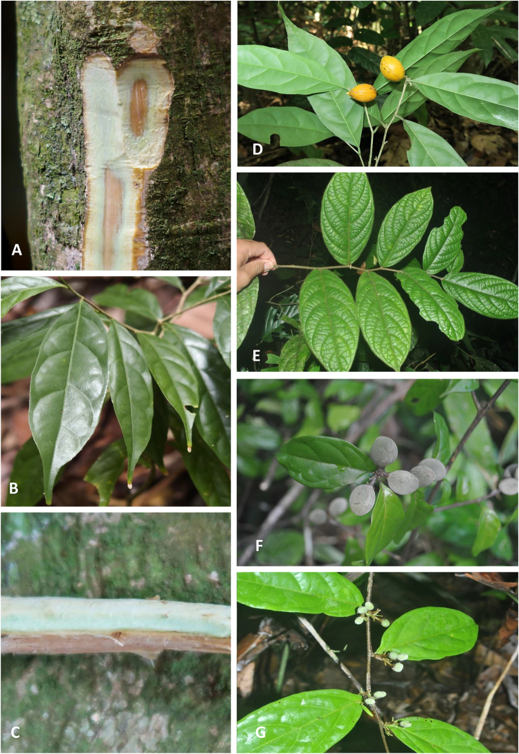
Figure 7.
The various D. gelonioides subspecies in the native habitat in Sabah: D. subsp. tuberculatum (A–D), D. subsp. pilosum (E) and D. subsp. sumatranum (F,G) (photos by Antony van der Ent and Sukaibin Sumail).
Rhizosphere soil samples were collected from near the roots of the Dichapetalum plants. Soil sub-samples (~300 mg) were digested using 9 mL 70% HNO3 and 3 mL 37% HCl per sample in a digestion microwave (Milestone Start D) for a program of 1.5 hours, and diluted to 45 mL with ultrapure water before analysis to obtain pseudo-total elemental concentrations. Soil pH was obtained in a 1 to 2.5 soil to water mixture after 2 hr shaking. Exchangeable trace elements were extracted in 0.1MSr(NO3)2 at a soil:solution ratio of 1:4 (10 gram soil with 40 mL solution) and 2 hr shaking time (adapted from Kukier & Chaney83). As a means of estimating potentially phytoavailable trace elements, the DTPA-extractant was used according to Becquer et al.84, which was adapted from the original method by Lindsay and Norvell85, with the following modifications: excluding triethanolamine (TEA), adjusted at pH 5.3, 5 g soil with 25 mL extractant, and extraction time of one hr. The plant digests and soil digests/extracts were analysed with ICP-AES (Varian Vista Pro II) for Ni, Co, Cr, Cu, Zn, Mn, Fe, Mg, Ca, Na, K, S and P; the Certified Reference Materials (CRMs) are given in Supplementary Table 2. Because of the low concentrations of our target elements in the Standard Reference Material Apple Leaves NIST 1515, we have included other CRMs (Standard Reference Material Tomato Leaves NIST 1573a and Standard Reference Material Spinach Leaves NIST 1570a) with relatively high Ni and Zn concentrations for improved accuracy (see Supplementary Table 2).
Collection and preparation of samples for Synchrotron X-ray Fluorescence Microscopy (XFM)
Plant tissue samples (roots, wood, leaves and phloem tissue) and rhizosphere soil samples were collected in the native rainforest habitat in Malaysian Borneo (Sepilok-Kabili Forest Reserve for D. subsp. sumatranum and D. subsp. pilosum and Mount Silam Forest Reserve for D. subsp. tuberculatum, in Sabah, Malaysia). Tissue samples intended for synchrotron analysis were excised with a razor blade, and immediately shock-frozen using a metal mirror technique in which the samples were pressed between a block of Cu-metal cooled by liquid N2 and a second Cu-metal block attached to a Teflon holder. This ensured extremely fast freezing of the plant tissue samples to prevent cellular damage by ice crystal formation. The samples were then wrapped in Al-foil, and transported in a cryogenic container (kept at <180 °C).
The samples required freeze-drying because they could not be kept frozen during the X-ray fluorescence microscopy (XFM) analysis. The samples were freeze-dried (using a Thermoline freeze-dryer) employing a long process of 4 days in which the temperature was stepwise increased, starting at −196 °C on a liquid N2 pre-cooled block (with the freeze-dryer set to −85 °C) until room temperature, to limit morphological changes of the tissues and elemental re-distribution.
Synchrotron X-ray Fluorescence Microscopy
The X-ray fluorescence microscopy (XFM) beamline employs an in-vacuum undulator to produce a brilliant X-ray beam. An Si(111) monochromator and a pair of Kirkpatrick-Baez mirrors delivers a monochromatic focused incident beam onto the specimen86. The Maia detector uses a large detector array to maximize detected signal and count-rates for efficient imaging. Maia enables high overall count-rates and uses an annular detector geometry, where the beam passes though the detector and strikes the sample at normal incidence87,88. This enables a large solid-angle (1.2 steradian) to be achieved in order to either maximize detected signal or to reduce the dose and potential damage to a specimen89. Maia is designed for event-mode data acquisition, where each detected X-ray event is recorded, tagged by detector number in the array, position in the scan and other metadata90. This approach eliminates readout delays and enables arbitrarily short pixel times (typically down to 0.1 ms) and large pixel count to be achieved for high definition imaging (typically 10–100 M pixels). The freeze-dried samples were mapped by sampling at intervals ranging from 2–10 μm and 0.5–5 ms transit time. The XRF event stream was analysed using the Dynamic Analysis method91,92 as implemented in GeoPIXE93.
Electronic supplementary material
Acknowledgements
We thank John Sugau, Jemson Miun (Forest Research Centre) and Sukaibin Sumail (Sabah Parks) for their support during the fieldwork in Malaysia. We acknowledge Ana Ocenar for undertaking herbarium XRF measurements of Dichapetalum specimens at the FRC Herbarium and we thank Gill Brown for support for the XRF scanning at the Queensland Herbarium. We thank the Sabah Forestry Department for granting permission to conduct research in the Silam FR and the Sepilok-Kabili FR, and the Sabah Biodiversity Council for research permits. This research was undertaken on the X-Ray Fluorescence Microscopy beamline of the Australian Synchrotron, ANSTO, Australia. We thank Hugh Harris (University of Adelaide), Martin de Jonge (ANSTO) and Rachel Mak (University of Sydney) for support with the synchrotron experiments. This work was supported by the Multi-modal Australian ScienceS Imaging and Visualisation Environment (MASSIVE). The French National Research Agency through the national “Investissements d’avenir” program (ANR-10-LABX-21, LABEX RESSOURCES21) and through the ANR-14-CE04–0005 Project “Agromine” is acknowledged for funding support to A. van der Ent and P.N. Nkrumah. A. van der Ent is the recipient of a Discovery Early Career Researcher Award (DE160100429) from the Australian Research Council. P.N. Nkrumah is the recipient of an Australian Government Research Training Program Scholarship and UQ Centennial Scholarship at The University of Queensland, Australia. We would like to thank the editor and the three anonymous reviewers for their constructive comments on an earlier version of this manuscript.
Author Contributions
P.N.N., G.E., P.D.E. and A.v.d.E. conducted the fieldwork and collected the samples. P.N.N. performed the laboratory analyses. A.v.d.E. and P.D.E. conducted the synchrotron XFM analysis. A.v.d.E. performed the light microscopy. P.N.N., G.E., P.D.E. and A.v.d.E. wrote the manuscript.
Competing Interests
The authors declare no competing interests.
Footnotes
Electronic supplementary material
Supplementary information accompanies this paper at 10.1038/s41598-018-26859-7.
Publisher's note: Springer Nature remains neutral with regard to jurisdictional claims in published maps and institutional affiliations.
References
- 1.Jaffre T, Brooks RR, Lee J, Reeves RD. Sebertia acuminata: a hyperaccumulator of nickel from New Caledonia. Science. 1976;193:579–580. doi: 10.1126/science.193.4253.579. [DOI] [PubMed] [Google Scholar]
- 2.Reeves RD. Tropical hyperaccumulators of metals and their potential for phytoextraction. Plant Soil. 2003;249:57–65. doi: 10.1023/A:1022572517197. [DOI] [Google Scholar]
- 3.van der Ent A, Baker AJM, Reeves RD, Pollard AJ, Schat H. Hyperaccumulators of metal and metalloid trace elements: facts and fiction. Plant Soil. 2013;362:319–334. doi: 10.1007/s11104-012-1287-3. [DOI] [Google Scholar]
- 4.Bani A, Echevarria G, Sulçe S, Morel JL. Improving the agronomy of Alyssum murale for extensive phytomining: a five-year field study. Int. J. Phytoremediation. 2015;17:117–127. doi: 10.1080/15226514.2013.862204. [DOI] [PubMed] [Google Scholar]
- 5.Chaney, R. L. et al. In Phytoremediation of Contaminated Soil and Water (CRC Press, 1999).
- 6.Chaney RL, et al. Improved understanding of hyperaccumulation yields commercial phytoextraction and phytomining technologies. J. Environ. Qual. 2007;36:1429–1443. doi: 10.2134/jeq2006.0514. [DOI] [PubMed] [Google Scholar]
- 7.Nkrumah PN, et al. Current status and challenges in developing nickel phytomining: an agronomic perspective. Plant Soil. 2016;406:55–69. doi: 10.1007/s11104-016-2859-4. [DOI] [Google Scholar]
- 8.van der Ent A, et al. Agromining: Farming for Metals in the Future? Environ. Sci. Technol. 2015;49:4773–4780. doi: 10.1021/es506031u. [DOI] [PubMed] [Google Scholar]
- 9.Chaney RL, Baklanov IA. Phytoremediation and Phytomining: Status and Promise. Adv. Bot. Res. 2017;83:189–221. doi: 10.1016/bs.abr.2016.12.006. [DOI] [Google Scholar]
- 10.Chaney RL, et al. Phytoremediation of soil metals. Curr. Opin. Biotechnol. 1997;8:279–284. doi: 10.1016/S0958-1669(97)80004-3. [DOI] [PubMed] [Google Scholar]
- 11.McGrath, S. P., Dunham, S. J. & Correll, R. L. In Phytoremediation of Contaminated Soil and Water (CRC Press, 1999).
- 12.McGrath SP, Zhao FJ, Lombi E. Plant and rhizosphere processes involved in phytoremediation of metal-contaminated soils. Plant Soil. 2001;232:207–214. doi: 10.1023/A:1010358708525. [DOI] [Google Scholar]
- 13.Schwartz C, Echevarria G, Morel JL. Phytoextraction of cadmium with Thlaspi caerulescens. Plant Soil. 2003;249:27–35. doi: 10.1023/A:1022584220411. [DOI] [Google Scholar]
- 14.Clemens S. How metal hyperaccumulating plants can advance Zn biofortification. Plant Soil. 2017;411:111–120. doi: 10.1007/s11104-016-2920-3. [DOI] [Google Scholar]
- 15.White, P. J. & Broadley, M. R. Physiological limits to zinc biofortification of edible crops. Front. Plant Sci. 2 (2011). [DOI] [PMC free article] [PubMed]
- 16.Boyd, R. S. The defense hypothesis of elemental hyperaccumulation: status, challenges and new directions. Plant Soil293 (2007).
- 17.Boyd RS. Plant defense using toxic inorganic ions: conceptual models of the defensive enhancement and joint effects hypotheses. Plant Science. 2012;195:88–95. doi: 10.1016/j.plantsci.2012.06.012. [DOI] [PubMed] [Google Scholar]
- 18.Broadhurst, C. et al. Interaction of nickel and manganese in accumulation and localization in leaves of the Ni hyperaccumulators Alyssum murale and Alyssum corsicum. Plant Soil314 (2009).
- 19.Broadhurst CL, Chaney RL, Angle JS, Erbe EF, Maugel TK. Nickel localization and response to increasing Ni soil levels in leaves of the Ni hyperaccumulator Alyssum murale. Plant Soil. 2004;265:225–242. doi: 10.1007/s11104-005-0974-8. [DOI] [Google Scholar]
- 20.Tappero, R. et al. Hyperaccumulator Alyssum murale relies on a different metal storage mechanism for cobalt than for nickel. New Phyt. 175 (2007). [DOI] [PubMed]
- 21.van der Ent A, et al. Nickel biopathways in tropical nickel hyperaccumulating trees from Sabah (Malaysia) Sci. Rep. 2017;7:41861. doi: 10.1038/srep41861. [DOI] [PMC free article] [PubMed] [Google Scholar]
- 22.Verbruggen N, Hermans C, Schat H. Molecular mechanisms of metal hyperaccumulation in plants. New Phyt. 2009;181:759–776. doi: 10.1111/j.1469-8137.2008.02748.x. [DOI] [PubMed] [Google Scholar]
- 23.van der Ent A, Erskine P, Sumail S. Ecology of nickel hyperaccumulator plants from ultramafic soils in Sabah (Malaysia) Chemoecol. 2015;25:243–259. doi: 10.1007/s00049-015-0192-7. [DOI] [Google Scholar]
- 24.van der Ent A, Repin R, Sugau J, Wong KM. Plant diversity and ecology of ultramafic outcrops in Sabah (Malaysia) Aust. J. Bot. 2015;63:204–215. doi: 10.1071/BT14214. [DOI] [Google Scholar]
- 25.Brooks RR, Wither ED. Nickel accumulation by Rinorea bengalensis (Wall.) O.K. J. Geochem. Explor. 1977;7:295–300. doi: 10.1016/0375-6742(77)90085-1. [DOI] [Google Scholar]
- 26.Pollard AJ, Reeves RD, Baker AJM. Facultative hyperaccumulation of heavy metals and metalloids. Plant Science. 2014;217–218:8–17. doi: 10.1016/j.plantsci.2013.11.011. [DOI] [PubMed] [Google Scholar]
- 27.Krämer U. Metal Hyperaccumulation in Plants. Annu. Rev. Plant Biol. 2010;61:517–534. doi: 10.1146/annurev-arplant-042809-112156. [DOI] [PubMed] [Google Scholar]
- 28.Baker AJM, Reeves RD, Hajar ASM. Heavy metal accumulation and tolerance in British populations of the metallophyte Thlaspi caerulescens J. & C. Presl (Brassicaceae) New Phyt. 1994;127:61–68. doi: 10.1111/j.1469-8137.1994.tb04259.x. [DOI] [PubMed] [Google Scholar]
- 29.Bert V, Bonnin I, Saumitou‐Laprade P, De Laguérie P, Petit D. Do Arabidopsis halleri from nonmetallicolous populations accumulate zinc and cadmium more effectively than those from metallicolous populations? New Phyt. 2002;155:47–57. doi: 10.1046/j.1469-8137.2002.00432.x. [DOI] [PubMed] [Google Scholar]
- 30.Bert, V., Macnair, M. R., De Laguerie, P., Saumitou‐Laprade, P. & Petit, D. Vol. 146, 225–233 (Cambridge, UK, 2000). [DOI] [PubMed]
- 31.Bert V, et al. Genetic basis of Cd tolerance and hyperaccumulation in Arabidopsis halleri. Plant Soil. 2003;249:9–18. doi: 10.1023/A:1022580325301. [DOI] [Google Scholar]
- 32.Reeves RD, Brooks RR. European species of Thlaspi L. (Cruciferae) as indicators of nickel and zinc. J. Geochem. Explor. 1983;18:275–283. doi: 10.1016/0375-6742(83)90073-0. [DOI] [Google Scholar]
- 33.Tang Y-T, et al. Lead, zinc, cadmium hyperaccumulation and growth stimulation in Arabis paniculata Franch. Environ. Exper. Bot. 2009;66:126–134. doi: 10.1016/j.envexpbot.2008.12.016. [DOI] [Google Scholar]
- 34.Peer, W. A. et al. Assessment of plants from the Brassicaceae family as genetic models for the study of nickel and zinc hyperaccumulation. New Phyt. 172 (2006). [DOI] [PubMed]
- 35.Assunção AGL, et al. Differential metal-specific tolerance and accumulation patterns among Thlaspi caerulescens populations originating from different soil types. New Phyt. 2003;159:411–419. doi: 10.1046/j.1469-8137.2003.00819.x. [DOI] [PubMed] [Google Scholar]
- 36.Assunção AGL, Schat H, Aarts MGM. Thlaspi caerulescens, an attractive model species to study heavy metal hyperaccumulation in plants. New Phyt. 2003;159:351–360. doi: 10.1046/j.1469-8137.2003.00820.x. [DOI] [PubMed] [Google Scholar]
- 37.Lasat, M. & Kochian, L. In Phytoremediation of Contaminated Soil and Water (CRC Press, 1999).
- 38.Milner MJ, Kochian LV. Investigating heavy-metal hyperaccumulation using Thlaspi caerulescens as a model system. Ann. Bot. 2008;102:3–13. doi: 10.1093/aob/mcn063. [DOI] [PMC free article] [PubMed] [Google Scholar]
- 39.Huguet S, et al. Cadmium speciation and localization in the hyperaccumulator Arabidopsis halleri. Environ. Exper. Bot. 2012;82:54–65. doi: 10.1016/j.envexpbot.2012.03.011. [DOI] [Google Scholar]
- 40.Küpper H, Lombi E, Zhao F-J, McGrath SP. Cellular compartmentation of cadmium and zinc in relation to other elements in the hyperaccumulator Arabidopsis halleri. Planta. 2000;212:75–84. doi: 10.1007/s004250000366. [DOI] [PubMed] [Google Scholar]
- 41.Reeves RD, Schwartz C, Morel JL, Edmondson J. Distribution and metal-accumulating behavior of Thlaspi caerulescens and associated metallophytes in France. Int. J. Phytoremediation. 2001;3:145–172. doi: 10.1080/15226510108500054. [DOI] [Google Scholar]
- 42.Schwartz C, et al. Testing of outstanding individuals of Thlaspi caerulescens for cadmium phytoextraction. Int. J. Phytoremediation. 2006;8:339–357. doi: 10.1080/15226510600992964. [DOI] [PubMed] [Google Scholar]
- 43.Escarré J, et al. Zinc and cadmium hyperaccumulation by Thlaspi caerulescens from metalliferous and nonmetalliferous sites in the Mediterranean area: implications for phytoremediation. New Phyt. 2000;145:429–437. doi: 10.1046/j.1469-8137.2000.00599.x. [DOI] [PubMed] [Google Scholar]
- 44.Stein, R. J. et al. Relationships between soil and leaf mineral composition are element-specific, environment-dependent and geographically structured in the emerging model Arabidopsis halleri. New Phyt. 213 (2017). [DOI] [PMC free article] [PubMed]
- 45.Punt W. Pollen morphology of the Dichapetalaceae with special reference to evolutionary trends and mutual relationships of pollen types. Rev. Palaeobot. Palynol. 1975;19:1–97. doi: 10.1016/0034-6667(75)90017-2. [DOI] [Google Scholar]
- 46.Baker, A., Proctor, J., Van Balgooy, M. & Reeves, R. Hyperaccumulation of nickel by the flora of the ultramafics of Palawan, Republic of the Philippines. The vegetation of ultramafic (serpentine) soils’. (Eds Baker, A. J. M., Proctor, J., Reeves, R. D.) pp, 291–304 (1992).
- 47.Datta S, Chaudhury K, Mukherjee PK. Hyperaccumulators from the serpentines of Andaman, India. Aust. J. Bot. 2015;63:243–251. doi: 10.1071/BT14244. [DOI] [Google Scholar]
- 48.Homer FA, Reeves RD, Brooks RR, Baker AJM. Characterization of the nickel-rich extract from the nickel hyperaccumulator Dichapetalum gelonioides. Phytochemistry. 1991;30:2141–2145. doi: 10.1016/0031-9422(91)83602-H. [DOI] [Google Scholar]
- 49.Baker AJM. Accumulators and excluders-strategies in the response of plants to heavy metals. J. Plant Nutr. 1981;3:643–654. doi: 10.1080/01904168109362867. [DOI] [Google Scholar]
- 50.Leenhouts PW. Dichapetalaceae. Flora Malesiana - Series 1, Spermatophyta. 1955;5:305–316. [Google Scholar]
- 51.Vaughan J, et al. Characterisation and hydrometallurgical processing of nickel from tropical agromined bio-ore. Hydrometallurgy. 2017;169:346–355. doi: 10.1016/j.hydromet.2017.01.012. [DOI] [Google Scholar]
- 52.Losfeld G, Escande V, Jaffré T, L’Huillier L, Grison C. The chemical exploitation of nickel phytoextraction: an environmental, ecologic and economic opportunity for New Caledonia. Chemosphere. 2012;89:907–910. doi: 10.1016/j.chemosphere.2012.05.004. [DOI] [PubMed] [Google Scholar]
- 53.Barbaroux R, et al. A new process for nickel ammonium disulfate production from ash of the hyperaccumulating plant Alyssum murale. Science of the Total Environment. 2012;423:111–119. doi: 10.1016/j.scitotenv.2012.01.063. [DOI] [PubMed] [Google Scholar]
- 54.Barbaroux R, Mercier G, Blais JF, Morel JL, Simonnot MO. A new method for obtaining nickel metal from the hyperaccumulator plant Alyssum murale. Separation and Purification Technology. 2011;83:57–65. doi: 10.1016/j.seppur.2011.09.009. [DOI] [Google Scholar]
- 55.van der Ent A, Cardace D, Tibbett M, Echevarria G. Ecological implications of pedogenesis and geochemistry of ultramafic soils in Kinabalu Park (Malaysia) Catena. 2018;160:154–119. doi: 10.1016/j.catena.2017.08.015. [DOI] [Google Scholar]
- 56.Broadley, M. R., White, P. J., Hammond, J. P., Zelko, I. & Lux, A. Vol. 173, 677–702 (Oxford, UK, 2007). [DOI] [PubMed]
- 57.White PJ, Broadley MR. Biofortifying crops with essential mineral elements. Trends Plant Sci. 2005;10:586–593. doi: 10.1016/j.tplants.2005.10.001. [DOI] [PubMed] [Google Scholar]
- 58.White, P. J. & Broadley, M. R. Vol. 182, 49–84 (Oxford, UK, 2009). [DOI] [PubMed]
- 59.Pfeiffer, W. H. & McClafferty, B. Vol. 47, S88–S105 (2007).
- 60.Graham RD, Welch RM, Bouis HE. Addressing micronutrient malnutrition through enhancing the nutritional quality of staple foods: Principles, perspectives and knowledge gaps. Adv. Agron. 2001;70:77–142. doi: 10.1016/S0065-2113(01)70004-1. [DOI] [Google Scholar]
- 61.Prasad AS. Discovery of human zinc deficiency: 50 years later. J. Trace Elem. Med. Biol. 2012;26:66–69. doi: 10.1016/j.jtemb.2012.04.004. [DOI] [PubMed] [Google Scholar]
- 62.Institute of Medicine USA. Dietary reference intakes for vitamin A, vitamin K, arsenic, boron, chromium, copper, iodine, iron, manganese, molybdenum, nickel, silicon, vanadium, and zinc: a report of the Panel on Micronutrients, Food and Nutrition Board, Institute of Medicine. (Washington, D.C.: National Academy Press, 2001).
- 63.Gibson R. Zinc deficiency and human health: etiology, health consequences, and future solutions. Plant Soil. 2012;361:291–299. doi: 10.1007/s11104-012-1209-4. [DOI] [Google Scholar]
- 64.Gibson RS. Zinc: the missing link in combating micronutrient malnutrition in developing countries. Proc. Nutr. Soc. 2006;65:51–60. doi: 10.1079/PNS2005474. [DOI] [PubMed] [Google Scholar]
- 65.Wessells KR, Brown KH. Estimating the global prevalence of zinc deficiency: results based on zinc availability in national food supplies and the prevalence of stunting. PLoS One. 2012;7:e50568. doi: 10.1371/journal.pone.0050568. [DOI] [PMC free article] [PubMed] [Google Scholar]
- 66.World Health, O. The world health report: reducing risks, promoting healthy life/World Health Organization. (Geneva: World Health Organization, 2002).
- 67.Bouis HE. Micronutrient fortification of plants through plant breeding: can it improve nutrition in man at low cost? Proc. Nutr. Soc. 2003;62:403–411. doi: 10.1079/PNS2003262. [DOI] [PubMed] [Google Scholar]
- 68.Bouis HE, Welch RM. Biofortification-a sustainable agricultural strategy for reducing micronutrient malnutrition in the global south. Crop Sci. 2010;50:S20–S32. doi: 10.2135/cropsci2009.09.0531. [DOI] [Google Scholar]
- 69.Hotz C. The potential to improve zinc status through biofortification of staple food crops with zinc. Food Nutr. Bull. 2009;30:S172–S178. doi: 10.1177/15648265090301S109. [DOI] [PubMed] [Google Scholar]
- 70.Roohani N, Hurrell R, Kelishadi R, Schulin R. Zinc and its importance for human health: an integrative review. J. Res. Med. Sci. 2013;18:144–157. [PMC free article] [PubMed] [Google Scholar]
- 71.Clemens, S., Aarts, M. G. M., Thomine, S. & Verbruggen, N. Plant science: the key to preventing slow cadmium poisoning. Trends Plant Sci. 18 (2012). [DOI] [PubMed]
- 72.Altschul, S. V. R. Drugs and foods from little-known plants: notes in Harvard University herbaria. (Cambridge, Mass.: Harvard University Press, 1973).
- 73.Esters, V. et al. Unusual amino acids and monofluoroacetate from Dichapetalum michelsonii (Umutambasha), a toxic plant from Rwanda. Planta Med. 79 (2013). [DOI] [PubMed]
- 74.Hall RJ. The distribution of organic fluorine in some toxic tropical plants. New Phyt. 1972;71:855–871. doi: 10.1111/j.1469-8137.1972.tb01965.x. [DOI] [Google Scholar]
- 75.O’Hagan D, et al. High levels of monofluoroacetate in Dichapetalum braunii. Phytochemistry. 1993;33:1043–1045. doi: 10.1016/0031-9422(93)85020-R. [DOI] [Google Scholar]
- 76.Vickery B, Vickery ML. Fluoride metabolism in Dichapetalum toxicarium. Phytochemistry. 1972;11:1905–1909. doi: 10.1016/S0031-9422(00)90151-1. [DOI] [Google Scholar]
- 77.Achenbach H, Asunka SA, Waibel R, Addae-Mensah I, Oppong IV. Dichapetalin A, a novel plant constituent from Dichapetalum madagascariense with potential antineoplastic activity. Nat. Prod. Lett. 1995;7:93–100. doi: 10.1080/10575639508043195. [DOI] [Google Scholar]
- 78.Addae-Mensah I, Waibel R, Asunka SA, Oppong IV, Achenbach H. The dichapetalins—A new class of triterpenoids. Phytochemistry. 1996;43:649–656. doi: 10.1016/0031-9422(96)00350-0. [DOI] [Google Scholar]
- 79.Fang L, et al. Cytotoxic constituents from the stem bark of Dichapetalum gelonioides collected in the Philippines. J. Nat. Prod. 2006;69:332–337. doi: 10.1021/np058083q. [DOI] [PMC free article] [PubMed] [Google Scholar]
- 80.Jing S-X, et al. Biologically active dichapetalins from Dichapetalum gelonioides. J. Nat. Prod. 2014;77:882–893. doi: 10.1021/np400971r. [DOI] [PubMed] [Google Scholar]
- 81.Long C, et al. Dichapetalins from Dichapetalum species and their cytotoxic properties. Phytochemistry. 2013;94:184–191. doi: 10.1016/j.phytochem.2013.03.023. [DOI] [PubMed] [Google Scholar]
- 82.van der Ent, A., Mulligan, D. R., Repin, R. & Erskine, P. D. Foliar elemental profiles in the ultramafic flora of Kinabalu Park (Sabah, Malaysia). Ecol. Res. In Press (2017).
- 83.Kukier U, Chaney RL. Amelioration of nickel phytotoxicity in muck and mineral soils. J. Environ. Qual. 2001;30:1949–1960. doi: 10.2134/jeq2001.1949. [DOI] [PubMed] [Google Scholar]
- 84.Becquer T, Bourdon E, Pétard J. Disponibilité du nickel le long d’une toposéquence de sols développés sur roches ultramafiques de Nouvelle-Calédonie. Comptes Rendus de l’Académie des Sciences. Série. 1995;2a(321):585–592. [Google Scholar]
- 85.Lindsay WL, Norvell WA. Development of a DTPA soil test for zinc, iron, manganese, and copper. Soil Sci. Soc. Am J. 1978;42:421–428. doi: 10.2136/sssaj1978.03615995004200030009x. [DOI] [Google Scholar]
- 86.Paterson, D. et al. In AIP Conference Proceedings. 219–222 (AIP).
- 87.Kirkham, R. et al. In AIP Conference Proceedings. 240–243 (AIP).
- 88.Siddons, D. et al. In Journal of Physics: Conference Series. 012001 (IOP Publishing).
- 89.Ryan CG, et al. Elemental X-ray imaging using the Maia detector array: The benefits and challenges of large solid-angle. Nucl. Instr. Meth. Phys. Res. A. 2010;619:37–43. doi: 10.1016/j.nima.2009.11.035. [DOI] [Google Scholar]
- 90.Ryan CG, et al. Maia x-ray fluorescence imaging: capturing detail in complex natural samples. Journal of Physics: Conference Series. 2014;499:012002. [Google Scholar]
- 91.Ryan CG. Quantitative trace element imaging using PIXE and the nuclear microprobe. International Journal of Imaging Systems and Technology. 2000;11:219–230. doi: 10.1002/ima.1007. [DOI] [Google Scholar]
- 92.Ryan CG, Jamieson DN. Dynamic analysis: on-line quantitative PIXE microanalysis and its use in overlap-resolved elemental mapping. Nucl. Instr. Meth. Phys. Res. B. 1993;77:203–214. doi: 10.1016/0168-583X(93)95545-G. [DOI] [Google Scholar]
- 93.Ryan CG, Cousens DR, Sie SH, Griffin WL. Quantitative analysis of PIXE spectra in geoscience applications. Nucl. Instr. Meth. Phys. Res. B. 1990;49:271–276. doi: 10.1016/0168-583X(90)90259-W. [DOI] [Google Scholar]
Associated Data
This section collects any data citations, data availability statements, or supplementary materials included in this article.


