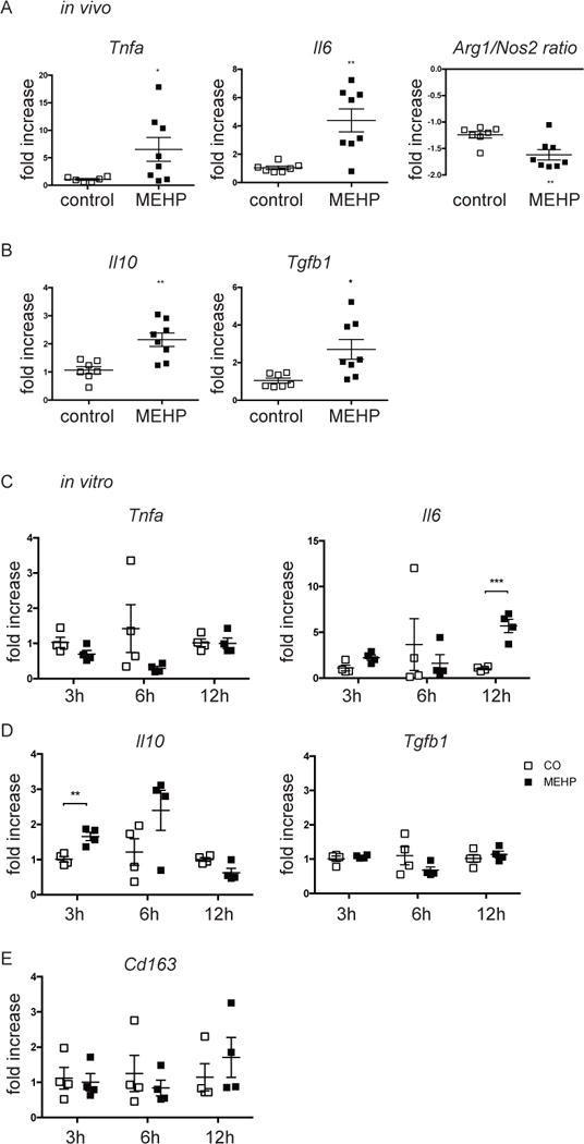
Figure 4. Infiltrating mononuclear immune cells are pro-inflammatory.

mRNA expression by (A-B) macrophages from animals 12 hours after exposure to MEHP or control or (C-E) testicular macrophages exposed to 200μM MEHP in vitro for 3, 6, or 12 hrs of (A,C) pro-inflammatory, (B,D) anti-inflammatory genes, or (E) Cd163, is presented as fold increase compared to gene expression of control macrophages. Data are means ± S.E.M. for n≥4. Statistical analysis was performed by the (A-B) unpaired two-tailed t-test and (C-E) the unpaired t-test and corrected for multiple comparisons using the Holm-Sidak method. *P<0.05, **P<0.01, ***P<0.001.
