Abstract
Background
The King-Devick (K-D) test of rapid number naming is a reliable visual performance measure that is a sensitive sideline indicator of concussion when time scores worsen (lengthen) from preseason baseline. Within cohorts of youth athletes <18 years old, baseline K-D times become faster with increasing age. We determined the relation of rapid number-naming time scores on the K-D test to electronic measurements of saccade performance during preseason baseline assessments in a collegiate ice hockey team cohort. Within this group of young adult athletes, we also sought to examine the potential role for player age in determining baseline scores.
Methods
Athletes from a collegiate ice hockey team received preseason baseline testing as part of an ongoing study of rapid rink-side performance measures for concussion. These included the K-D test (spiral-bound cards and tablet computer versions). Participants also performed a laboratory-based version of the K-D test with simultaneous infrared-based video-oculographic recordings using an Eye-Link 1000+. This allowed measurement of the temporal and spatial characteristics of eye movements, including saccadic velocity, duration, and intersaccadic interval (ISI).
Results
Among 13 male athletes, aged 18–23 years (mean 20.5 ± 1.6 years), prolongation of the ISI (a combined measure of saccade latency and fixation duration) was the measure most associated with slower baseline time scores for the EyeLink-paired K-D (mean 38.2 ± 6.2 seconds, r = 0.88 [95% CI 0.63–0.96], P = 0.0001), the K-D spiral-bound cards (36.6 ± 5.9 seconds, r = 0.60 [95% CI 0.08–0.87], P = 0.03), and K-D computerized tablet version (39.1 ± 5.4 seconds, r = 0.79 [95% CI 0.42–0.93], P = 0.001). In this cohort, older age was a predictor of longer (worse) K-D baseline time performance (age vs EyeLink-paired K-D: r = 0.70 [95% CI 0.24–0.90], P = 0.008; age vs K-D spiral-bound cards: r = 0.57 [95% CI 0.03–0.85], P = 0.04; age vs K-D tablet version: r = 0.59 [95% CI 0.06–0.86], P = 0.03) as well as prolonged ISI (r = 0.62 [95% CI 0.11–0.87], P = 0.02). Slower baseline K-D times were not associated with greater numbers of reported prior concussions.
Conclusions
Rapid number-naming performance using the K-D at preseason baseline in this small cohort of collegiate ice hockey players is best correlated with ISI among eye movement-recording measures. Baseline K-D scores notably worsened with increasing age, but not with numbers of prior concussions in this small cohort. While these findings require further investigation by larger studies of contact and non-contact sports athletes, they suggest that duration of contact sports exposure may influence preseason test performance.
Concussion is the mildest form of traumatic brain injury and is defined by a force to the head or body followed by any new neurological sign or symptom (1). A variety of symptoms may develop after a concussion, including those related to vision (2–7). Sports-related concussion is frequently underreported by high school and college-age athletes (6,8). Assessment of which athletes are vulnerable to continued injury following an event can be difficult given the potential for underreporting and lack of recognition.
Vision-based performance testing has been demonstrated to be a useful complement to sideline measures of balance, cognition, and symptoms. Rapid number naming using the King-Devick (K-D) test (9) has been administered in conjunction with components of the Sport Concussion Assessment Tool, 3rd Edition (SCAT3) (10) in collegiate and youth athletes (7). The K-D test requires saccades and other eye movements and is a useful measure for assessing the brain’s pathways related to vision (1,11). The K-D test is a reliable visual performance measure that is a sensitive sideline indicator of concussion when time scores worsen after an event, as compared to a preseason baseline (11). Within cohorts of youth athletes <18 years old, baseline K-D times also become faster with increasing age.
The purpose of our study was to examine the relation of rapid number-naming time scores on the K-D test to electronic, laboratory-based measurements of saccade performance in collegiate athletes. Within this cohort of young adult athletes, we also sought to explore the potential role for player age in determining baseline K-D scores.
METHODS
Study Participants
Student athletes from the New York University (NYU) men’s ice hockey team (n = 13, aged 18–23 years) were enrolled in this study during the preseason practice period. Study protocols were approved by the Institutional Review Board at the NYU School of Medicine, and informed consent was obtained from all study participants.
Participants underwent a series of tests, including K-D (9), a test of rapid number naming that uses spiral-bound cards or a computer-based tablet version, and components of the SCAT3 (10). SCAT3 components included the Symptom Evaluation questionnaire, Standardized Assessment of Concussion (SAC) (12), and a timed tandem gait test.
K-D Test of Rapid Number Naming
We requested that participants complete the K-D, a vision-based rapid number test in 3 separate platforms (in a randomized order). The first platform was a spiral-bound booklet version of the K-D; the second was the K-D on a computerized tablet, and the third was a computer digitized version of the K-D linked with formal eye movement recordings (EyeLink 1000+ infrared-based video-oculography; SR Research, Mississauga, Canada). Saccadic eye movements were analyzed and the inter-saccadic interval (ISI) was calculated. The ISI is a measure of time between saccades and represents a combined measure of saccade latency and fixation duration (13).
K-D Test Platform 1: Spiral-Bound Version
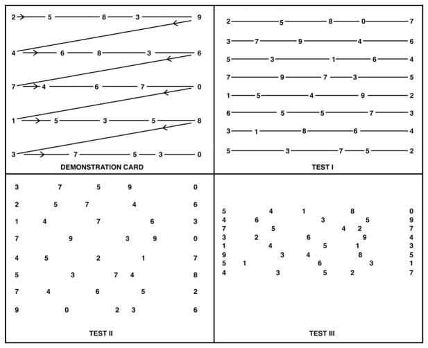
Participants were instructed to perform the K-D test in the spiral-bound booklet version (Fig. 1). Booklets were held at a comfortable distance and timing of the test began when the subject read aloud the numbers from left to right on each test card. Participants were instructed to read as quickly as they could without making any errors. Reading times were measured for each card and errors for each test were recorded by the test administrator. Each participant completed the K-D twice (Trial 1 and Trial 2). The best time (shortest time) of the 2 trials was recorded.
FIG. 1.
Test card formats for the King-Devick (K-D) test of rapid number naming. To perform the King-Devick test, participants are asked to read the numbers on each card from left to right as quickly as possible, but without making any errors. Following completion of the demonstration card (upper left), subjects are then asked to read each of the 3 test cards in the same manner. The times required to complete each card are recorded in seconds using a stopwatch. The sum of the 3 test card time scores constitutes the summary score for the entire test, the King-Devick time score. Numbers of errors made in reading the test cards are also recorded; misspeaks on numbers are recorded as errors only if the subject does not immediately correct the mistake before going on to the next number.
K-D Test Platform 2: Tablet Version
The K-D test was also administered on an iPad. Similar to the spiral-bound version of the K-D test, participants were instructed to hold the iPad at a comfortable distance and read the numbers aloud quickly from left to right. The test administrator was responsible for changing the screen to the next K-D card when the participant had completed reading the numbers. The time for each test card was recorded immediately after the administrator tapped the screen, bringing the participant to the next test card. After completing each card, the administrator tapped the screen to record the time. A total of 3 time scores were recorded and then summed for the total time. The participant was allowed to pause between test cards, starting the next card only after notifying the administrator when ready to begin the next card. In addition, the test administrator recorded errors for each test card on a separate form.
For the iPad platform, there were 2 versions available. Version 1 was identical to the numbers of the spiral version. Version 2 utilized a different sequence of numbers. Participants were randomized to take either Version 1 or 2 of the K-D test on the iPad. Similar to the spiral-bound booklet version of the K-D test, participants took the test on the iPad twice (Trial 1 and Trial 2). The best time (shortest time) of the 2 trials was recorded.
K-D Test Platform 3: Computerized Eye Link 1000+ Eye Movement Recording Version
Participants performed a laboratory-based version of the K-D test with simultaneous infrared-based video-oculographic recordings using the EyeLink 1000+. All participants completed a 13-point spatial calibration procedure and 1 trial of reading 3 computer-generated K-D test cards while undergoing simultaneous eye movement recording with infrared-based video-oculography (Eye link 1000+; SR Research). The system recorded both eyes with a sampling frequency of 500 Hz and a spatial accuracy of approximately 0.5°. Eye movement data were analyzed off-line using custom Matlab software. Data between card presentations were automatically identified for exclusion and manually confirmed, and data within 100 ms of a blink were automatically eliminated. The total reading times and error rates were recorded. Additional details, including full methodologies, recording procedures, and KD eye movement analysis techniques, have been published (14).
Sport Concussion Assessment Tool—Third Edition Component Testing
The SCAT3 is a sideline tool that assesses symptoms, cognition, and balance. The test battery combines the Standardized Assessment of Concussion (SAC) (12) and a modified Balance Error Scoring System (BESS) or timed tandem gait test (10,15).
Standardized Assessment of Concussion
The SAC is a brief cognitive battery that captures the domains of orientation, immediate memory, concentration, and delayed recall. A maximum total score of 30 is generated by adding the 4 subscores from each section. A worsening SAC score of 2–4 points from baseline was used to determine a clinically meaningful change based on recent guidelines, and this threshold was used to examine a worsening SAC score dichotomously (10). The SCAT3 Symptom Checklist, an evaluation of 22 symptoms associated with concussion, was also administered to all participants (10).
Timed Tandem Gait Test
The timed tandem gait (TTG) test, a balance component of the SCAT3, was recorded for each participant. The TTG was chosen over the BESS due to evidence suggesting that this is less impacted by exercise and fatigue than static balance tasks (16). To perform the TTG, participants were instructed to walk along a 38-mm-wide × 3-m-long sports tape. Athletes placed one foot in front of the other, without shoes or skates, along the line as quickly and accurately as possible. The best time of 4 trials back and forth along the tape was recorded as the official score.
Statistical Analyses
Statistical analyses were performed using Stata 14.2 (Stata-Corp, College Station, TX). Pearson linear correlations with 95% confidence intervals (CI) were calculated to examine the relation of age with preseason baseline test scores, after examining the distributions of measures in this small cohort to fit assumptions for normality (skewness < |1.0|, median nearly identical to mean summary measures, and results of the Shapiro-Wilk test [P values >0.05]). The linear correlations were also calculated to examine the relation of vision-based test scores to SAC and TTG in this cohort. Normal Q-Q plots, leverage plots and residual plots were examined to identify possible influential observations. Regression models were developed to predict the effect of age on the observed K-D measurements. Adjusted models were calculated when an influential point was identified with Cook’s distance; the point was then removed and the model was recalculated.
RESULTS
Among 13 male athletes (mean age 20.5 ± 1.6 years), ISI prolongation was the quantified eye movement parameter most associated with slower baseline time scores for all the K-D test platforms. These included the EyeLink-paired K-D (mean 38.2 ± 6.2 seconds, r = 0.88 [95% CI 0.63–0.96], P = 0.0001), the K-D spiral-bound cards (36.6 ± 5.9 seconds, r = 0.60 [95% CI 0.08–0.87], P = 0.03) and the K-D tablet version (39.1 ± 5.4 seconds, r = 0.79 [95% CI 0.42–0.93], P = 0.001).
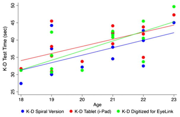
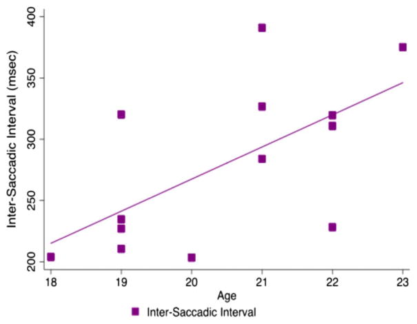
In exploring the potential relation of age with K-D scores, older age in this cohort was a predictor of longer (worse) K-D scores from preseason baseline time across all 3 K-D platforms (age vs EyeLink-paired K-D: r = 0.70 [95% CI 0.24–0.90], P = 0.008; age vs K-D spiral-bound cards: r = 0.57 [95% CI 0.03–0.85], P = 0.04; age vs K-D tablet version: r = 0.59 [95% CI 0.06–0.86], P = 0.03, Fig. 2). Older age was also associated with a prolonged ISI across time (r = 0.62 [95% CI 0.11–0.87], P = 0.02, Fig. 3). Slower baseline K-D times were not associated with greater numbers of reported prior concussions. In addition, the SAC and TTG scores were not associated with worse K-D scores (SAC vs K-D tablet version: r = 0.30 [95% CI −0.30–0.73], P = 0.32; TTG vs K-D tablet version: r = 0.10 [95% CI −0.48–0.62] P = 0.74), or with increasing or decreasing age (age vs SAC: r = −0.11 [95% CI −0.62–0.47], P = 0.72; age vs TTG: r = 0.29 [95% CI −0.31–0.73], P = 0.33).
FIG. 2.
Scatterplot of King-Devick (K-D) test times vs athlete age indicates that in this cohort of participants aged 18–23 years, there is a prolongation of K-D test time with increasing age. This is demonstrated across all the 3 test platforms, including K-D spiral (blue line), K-D computerized tablet/iPad (red line) and K-D digitized for the EyeLink infrared-based video-oculography (green line).
FIG. 3.
Scatterplot of intersaccadic interval (ISI) vs age demonstrating that with increasing age, the ISI also increases.
Linear regression models with unadjusted and adjusted P values (adjusted for possible influential points/outliers) demonstrated that with increasing age, K-D scores also increased regardless of the testing platform used (unadjusted models: age vs EyeLink-paired K-D: P = 0.01; age vs K-D spiral-bound cards: P = 0.04; age vs K-D tablet version: P = 0.03; adjusted models: age vs EyeLink-paired K-D: P = 0.001; age vs K-D spiral-bound cards: P = 0.003; age vs K-D tablet version: P = 0.003). Thus, removal of the potential influential points did not modify the significance of the results.
DISCUSSION
Results of this investigation of a small cohort of collegiate ice hockey players suggests that, during preseason baseline testing of the vision-based K-D, the ISI was the quantified eye movement parameter most strongly associated with rapid number-naming test times. In addition, our study generated the hypothesis that increasing age, even within the limits of a collegiate cohort, may be associated with prolonged K-D test times and ISI.
This is the first study to examine ocular motor behavior during performance of a digitized version of the K-D test in a preseason baseline for a collegiate athlete cohort. The ISI has previously been shown to have the best correlation with K-D times among healthy volunteers, and in chronic concussion during eye movement recordings (14,17). The K-D test is useful in identifying athletes with concussion in contact sports across all age groups (7,18–20). This test can also be easily performed by nonmedical personnel, including parents, making it a practical performance measure to complement clinical judgment and existing concussion protocols on the sidelines (21).
It also has been demonstrated that athletes have abnormal saccade measurements following concussion (22,23). Tests that assess saccadic eye movements enable the analysis of a number of pathways throughout the brain including attention, motor planning, and visual-spatial integration (24). The ISI correlated with the K-D test and was useful for studying concussion. Saccadic eye movements involve the integration of information throughout several areas of the brain including the frontal eye fields, dorsolateral prefrontal cortex, intraparietal sulcus, supplementary eye fields, and deep structures of the brainstem (24,25). In concussion, pathways involved in the cognitive control of vision are vulnerable to injury based on diffusion tensor imaging studies (26,27). Injury to the fronto-parietal circuits have been demonstrated and can impair eye movement behavior (28). With concussion, K-D test times have been shown to worsen (prolonged time) from a pre-season baseline score (7,19,20).
Baseline KD scores and ISI worsened with increasing age but not with number of prior concussions in this small cohort. This suggests that duration of contact sport exposure history may influence preseason baseline rapid number-naming performance. In contrast, in a younger athletic cohort (aged 5–17 years), K-D scores showed improvement (faster times) with increasing age (7). This may be due to maturation of grey matter and white matter tracts during childhood development (29). The frontal lobe circuits which are integral in eye movement tasks begin to stabilize in adolescence, suggesting mechanisms that may contribute to improved KD scores (29).
Longer durations of contact sport exposure have been associated with greater degrees of cognitive impairment (30). Among 18 boxers (age 25–60 years) who underwent neuropsychological testing, duration of contact sport exposure based on number of professional fights and age correlated with worsening cognitive indices as opposed to the number of knock-outs or episodes of amnesia (30). In a study of active American professional football players, cognitive function was worse in older players with the apolipoprotein E4 (APOE) allele compared to players without this allele, or less experienced (younger) players of any genotype (31). This association was present independently of the numbers of concussions reported. This study suggested that cognitive impairment in professional athletes may be influenced by age and APOE genotype, in addition to duration of exposure to contact sports.
Our study of the relation of baseline rapid number-naming time scores with eye movement recording measurements in a collegiate athlete cohort suggests a potential role for athlete age at the time of testing as a factor in determining K-D scores. We recognize that this finding must be confirmed in a larger study cohort of contact and noncontact sport athletes, and also must be examined in the context of differences between males and females, types of sport, age at first contact sport participation, and other relevant factors. As noted in several recent studies, the prolongation of the ISI is the eye movement recording measurement that is, most strongly related to K-D test times among individuals with a history of concussion (14,17). Important next steps for the investigation of vision-based rapid sideline tests and their eye movement underpinnings as recorded in the laboratory will include: 1) further evaluation of a rapid picture-naming test platform which captures visual and cognitive pathways that are additive to rapid number naming, 2) determination of the role for age in the developmental performance of rapid number and picture naming across the youth age groups, and within additional collegiate cohorts engaged in contact sports, and 3) examination of how vision-based rapid sideline tests can reflect both concussion history and symptom recovery in the context of acute injury.
We have demonstrated that the ISI correlates well with K-D scores, and that increasing age may be a marker for duration of contact sports exposure. The worse K-D preseason baseline scores with increasing age in our collegiate cohort also supports further examination of the clinical significance of subconcussive events.
Footnotes
S. L. Galetta and L. J. Balcer have received honoraria for consulting from Biogen. The remaining authors report no conflicts of interest.
STATEMENT OF AUTHORSHIP
Category 1: a. Conception and design: L. Hasanaj, J. C. Rucker, S. L. Galetta, and L. J. Balcer; b. Acquisition of data: L. Hasanaj, S. P. Thawani, N. Webb, J. D. Drattell, L. Serrano, R. C. Nolan, J. Raynowska, T. E. Hudson, J. -R. Rizzo, and W. Dai; c. Analysis and interpretation of data: L. Hasanaj, S. P. Thawani, T. E. Hudson, W. Dai, J. -R. Rizzo, J. C. Rucker, S. L. Galetta, and L. J. Balcer.
Category 2: a. Drafting the manuscript: L. Hasanaj, S. P. Thawani, S. L. Galetta, L. J. Balcer; b. Revising it for intellectual content: L. Hasanaj, S. P. Thawani, N. Webb, J. D. Drattell, L. Serrano, R. C. Nolan, J. Raynowska, T. E. Hudson, J. -R. Rizzo, W. Dai, and S. L. Galetta.
Category 3: a. Final approval of the completed manuscript: L. Hasanaj, S. P. Thawani, N. Webb, J. D. Drattell, L. Serrano, R. C. Nolan, J. Raynowska, T. E. Hudson, J. -R. Rizzo, W. Dai, and S. L. Galetta.
References
- 1.Ventura RE, Balcer LJ, Galetta SL. The neuro-ophthalmology of head trauma. Lancet Neurol. 2014;13:1006–1016. doi: 10.1016/S1474-4422(14)70111-5. [DOI] [PubMed] [Google Scholar]
- 2.Guskiewicz KM, Marshall SW, Bailes J, McCrea M, Harding HP, Jr, Matthews A, Mihalik JR, Cantu RC. Recurrent concussion and risk of depression in retired professional football players. Med Sci Sports Exerc. 2007;39:903–909. doi: 10.1249/mss.0b013e3180383da5. [DOI] [PubMed] [Google Scholar]
- 3.Guskiewicz KM, McCrea M, Marshall SW, Cantu RC, Randolph C, Barr W, Onate JA, Kelly JP. Cumulative effects associated with recurrent concussion in collegiate football players: the NCAA Concussion Study. JAMA. 2003;290:2549–2555. doi: 10.1001/jama.290.19.2549. [DOI] [PubMed] [Google Scholar]
- 4.McCrory P, Meeuwisse WH, Aubry M, Cantu RC, Dvořák J, Echemendia RJ, Engebretsen L, Johnston K, Kutcher JS, Raftery M, Sills A, Benson BW, Davis GA, Ellenbogen R, Guskiewicz KM, Herring SA, Iverson GL, Jordan BD, Kissick J, McCrea M, McIntosh AS, Maddocks D, Makdissi M, Purcell L, Putukian M, Schneider K, Tator CH, Turner M. Consensus statement on concussion in sport: the 4th International Conference on Concussion in Sport, Zurich, November 2012. J Athl Train. 2013;48:554–575. doi: 10.4085/1062-6050-48.4.05. [DOI] [PMC free article] [PubMed] [Google Scholar]
- 5.McKee AC, Stern RA, Nowinski CJ, Stein TD, Alvarez VE, Daneshvar DH, Lee HS, Wojtowicz SM, Hall G, Baugh CM, Riley DO, Kubilus CA, Cormier KA, Jacobs MA, Martin BR, Abraham CR, Ikezu T, Reichard RR, Wolozin BL, Budson AE, Goldstein LE, Kowall NW, Cantu RC. The spectrum of disease in chronic traumatic encephalopathy. Brain. 2013;136:43–64. doi: 10.1093/brain/aws307. [DOI] [PMC free article] [PubMed] [Google Scholar]
- 6.Torres DM, Galetta KM, Phillips HW, Dziemianowicz EM, Wilson JA, Dorman ES, Laudano E, Galetta SL, Balcer LJ. Sports-related concussion: anonymous survey of a collegiate cohort. Neurol Clin Pract. 2013;3:279–287. doi: 10.1212/CPJ.0b013e3182a1ba22. [DOI] [PMC free article] [PubMed] [Google Scholar]
- 7.Galetta KM, Morganroth J, Moehringer N, Mueller B, Hasanaj L, Webb N, Civitano C, Cardone DA, Silverio A, Galetta SL, Balcer LJ. Adding vision to concussion testing: a prospective study of sideline testing in youth and collegiate athletes. J Neuroophthalmol. 2015;35:235–241. doi: 10.1097/WNO.0000000000000226. [DOI] [PubMed] [Google Scholar]
- 8.Register-Mihalik JK, Guskiewicz KM, McLeod TC, Linnan LA, Mueller FO, Marshall SW. Knowledge, attitude, and concussion-reporting behaviors among high school athletes: a preliminary study. J Athl Train. 2013;48:645–653. doi: 10.4085/1062-6050-48.3.20. [DOI] [PMC free article] [PubMed] [Google Scholar]
- 9.Oride MK, Marutani JK, Rouse MW, DeLand PN. Reliability study of the Pierce and King-Devick saccade tests. Am J Optom Physiol Opt. 1986;63:419–424. doi: 10.1097/00006324-198606000-00005. [DOI] [PubMed] [Google Scholar]
- 10.Guskiewicz KM, Register-Mihalik J, McCrory P, McCrea M, Johnston K, Makdissi M, Dvorák J, Davis G, Meeuwisse W. Evidence-based approach to revising the SCAT2: introducing the SCAT3. Br J Sports Med. 2013;47:289–293. doi: 10.1136/bjsports-2013-092225. [DOI] [PubMed] [Google Scholar]
- 11.Galetta KM, Liu M, Leong DF, Ventura RE, Galetta SL, Balcer LJ. The King-Devick test of rapid number naming for concussion detection: meta-analysis and systematic review of the literature. Concussion. doi: 10.2217/cnc.15.8. [published online September 10, 2015] Available at: https://doi.org/10.2217/cnc.15.8. [DOI] [PMC free article] [PubMed]
- 12.McCrea M, Kelly JP, Kluge J, Bartolic E, Finn G, Baxter B. Standardized assessment of concussion in football players. Neurology. 1997;48:586–588. doi: 10.1212/wnl.48.3.586. [DOI] [PubMed] [Google Scholar]
- 13.Roos JC, Calandrini DM, Carpenter RH. A single mechanism for the timing of spontaneous and evoked saccades. Exp Brain Res. 2008;187:283–293. doi: 10.1007/s00221-008-1304-1. [DOI] [PubMed] [Google Scholar]
- 14.Rizzo JR, Hudson TE, Dai W, Desai N, Yousefi A, Palsana D, Selesnick I, Balcer LJ, Galetta SL, Rucker JC. Objectifying eye movements during rapid number naming: methodology for assessment of normative data for the King-Devick test. J Neurol Sci. 2016;362:232–239. doi: 10.1016/j.jns.2016.01.045. [DOI] [PMC free article] [PubMed] [Google Scholar]
- 15.Furman GR, Lin CC, Bellanca JL, Marchetti GF, Collins MW, Whitney SL. Comparison of the balance accelerometer measure and balance error scoring system in adolescent concussions in sports. Am J Sports Med. 2013;41:1404–1410. doi: 10.1177/0363546513484446. [DOI] [PMC free article] [PubMed] [Google Scholar]
- 16.Schneiders AG, Sullivan SJ, Handcock P, Gray A, McCrory PR. Sports concussion assessment: the effect of exercise on dynamic and static balance. Scand J Med Sci Sports. 2012;22:85–90. doi: 10.1111/j.1600-0838.2010.01141.x. [DOI] [PubMed] [Google Scholar]
- 17.Rizzo JR, Hudson T, Dai W, Birkemeier J, Pasculli R, Selesnick I, Balcer L, Galetta S, Rucker J. Rapid number naming in chronic concussion: eye movements in the King-Devick test. Ann Clin Translat Neurol. 2016;3:801–811. doi: 10.1002/acn3.345. [DOI] [PMC free article] [PubMed] [Google Scholar]
- 18.Galetta KM, Barrett J, Allen M, Madda F, Delicata D, Tennant AT, Branas CC, Maguire MG, Messner LV, Devick S, Galetta SL, Balcer LJ. The King-Devick test as a determinant of head trauma and concussion in boxers and MMA fighters. Neurology. 2011;76:1456–1462. doi: 10.1212/WNL.0b013e31821184c9. [DOI] [PMC free article] [PubMed] [Google Scholar]
- 19.Galetta KM, Brandes LE, Maki K, Dziemianowicz MS, Laudano E, Allen M, Lawler K, Sennett B, Wiebe D, Devick S, Messner LV, Galetta SL, Balcer LJ. The King-Devick test and sports-related concussion: study of a rapid visual screening tool in a collegiate cohort. J Neurol Sci. 2011;309:34–39. doi: 10.1016/j.jns.2011.07.039. [DOI] [PubMed] [Google Scholar]
- 20.Galetta MS, Galetta KM, McCrossin J, Wilson JA, Moster S, Galetta SL, Balcer LJ, Dorshimer GW, Master CL. Saccades and memory: baseline associations of the King-Devick and SCAT2 SAC tests in professional ice hockey players. J Neurol Sci. 2013;328:28–31. doi: 10.1016/j.jns.2013.02.008. [DOI] [PubMed] [Google Scholar]
- 21.Leong DF, Balcer LJ, Galetta SL, Liu Z, Master CL. The King-Devick test as a concussion screening tool administered by sports parents. J Sports Med Phys Fitness. 2014;54:70–77. [PubMed] [Google Scholar]
- 22.Heitger MH, Jones RD, Macleod AD, Snell DL, Frampton CM, Anderson TJ. Impaired eye movements in post-concussion syndrome indicate suboptimal brain function beyond the influence of depression, malingering or intellectual ability. Brain. 2009;132(pt 10):2850–2870. doi: 10.1093/brain/awp181. [DOI] [PubMed] [Google Scholar]
- 23.Heitger MH, Jones RD, Anderson TJ. A new approach to predicting postconcussion syndrome after mild traumatic brain injury based upon eye movement function. Conf Proc IEEE Eng Med Biol Soc. 2008;2008:3570–3573. doi: 10.1109/IEMBS.2008.4649977. [DOI] [PubMed] [Google Scholar]
- 24.DeSouza JF, Menon RS, Everling S. Preparatory set associated with pro-saccades and anti-saccades in humans investigated with event-related FMRI. J Neurophysiol. 2003;89:1016–1023. doi: 10.1152/jn.00562.2002. [DOI] [PubMed] [Google Scholar]
- 25.Pierrot-Deseilligny C, Milea D, Muri RM. Eye movement control by the cerebral cortex. Curr Opin Neurol. 2004;17:17–25. doi: 10.1097/00019052-200402000-00005. [DOI] [PubMed] [Google Scholar]
- 26.Lipton ML, Kim N, Park YK, Hulkower MB, Gardin TM, Shifteh K, Kim M, Zimmerman ME, Lipton RB, Branch CA. Robust detection of traumatic axonal injury in individual mild traumatic brain injury patients: intersubject variation, change over time and bidirectional changes in anisotropy. Brain Imaging Behav. 2012;6:329–342. doi: 10.1007/s11682-012-9175-2. [DOI] [PubMed] [Google Scholar]
- 27.Maruta J, Lee SW, Jacobs EF, Ghajar J. A unified science of concussion. Ann N Y Acad Sci. 2010;1208:58–66. doi: 10.1111/j.1749-6632.2010.05695.x. [DOI] [PMC free article] [PubMed] [Google Scholar]
- 28.Gentry LR, Godersky JC, Thompson B. MR imaging of head trauma: review of the distribution and radiopathologic features of traumatic lesions. AJR Am J Roentgenol. 1988;150:663–672. doi: 10.2214/ajr.150.3.663. [DOI] [PubMed] [Google Scholar]
- 29.Luna B, Velanova K, Geier CF. Development of eye-movement control. Brain Cogn. 2008;68:293–308. doi: 10.1016/j.bandc.2008.08.019. [DOI] [PMC free article] [PubMed] [Google Scholar]
- 30.Casson IR, Siegel O, Sham R, Campbell EA, Tarlau M, DiDomenico A. Brain damage in modern boxers. JAMA. 1984;251:2663–26677. [PubMed] [Google Scholar]
- 31.Kutner KC, Erlanger DM, Tsai J, Jordan B, Relkin NR. Lower cognitive performance of older football players possessing apolipoprotein E epsilon4. Neurosurgery. 2000;47:651–657. doi: 10.1097/00006123-200009000-00026. [DOI] [PubMed] [Google Scholar]


