

Abstract
Rationale:
Pheochromocytomas are rare catecholamine-secreting tumors arising from adrenomedullary chromaffin cells, usually causing hypertension, palpitation and headache. However, pheochromocytoma crisis, on the contrary, might present with hypotension, multiple organ dysfunction or even mimicking other diseases, leaving physician with diagnostic difficulties. In this study, we present a case featured hypotension, shock and multiple organ dysfunction syndrome on admission, which nearly lead us to miss the diagnosis of pheochromocytoma.
Patient concerns:
A 14-year-old female student presented with cough, hemoptysis and dyspnea for one week was reported.
Diagnoses:
The laboratory test showed significantly increase in plasma norepinephrine and 24-hour urine norepinephrine, the enhanced CT of bilateral adrenal gland showed two round-like masses (left: 4 × 5 × 3 cm; right: 6 × 4 × 3 cm) with soft tissue density in each adrenal gland. The post-surgical pathology confirmed the diagnosis of pheochromocytoma.
Interventions:
The resection of bilateral adrenal tumors was conducted after the preoperative medical treatment of phenoxybenzamine for two weeks.
Outcomes:
The patient underwent follow-up for a year and a half and showed no signs of recurrence.
Lessons:
The diagnosis and treatment process of the patient in this study indicates us that when we meet a patient with hypotension and multiple organ dysfunctions in a relatively short time, the suspicion of pheochromocytoma should not be missed.
Keywords: case report, emergency, hypotension, liver dysfunction, pheochromocytoma crisis
1. Introduction
According to the latest clinical practice guideline of pheochromocytoma and paraganglioma, a pheochromocytoma is defined as a tumor deriving from adrenomedullary chromaffin cells that produces one or more catecholamines: epinephrine, norepinephrine, and dopamine.[1] Pheochromocytoma crisis (PCC) is an endocrine emergency associated with significant mortality, with a prevalence of 7% to 18% in patients with pheochromocytoma.[2,3] It is defined as the acute severe presentation of catecholamine-induced hemodynamic instability causing end-organ damage or dysfunction.[4] In this study, we present a patient with PCC featured hypotension, hemoptysis ,and abnormal liver function, whose initial clinical, laboratory, and radiography performance nearly lead us to exclude pheochromocytoma.
2. Case report
This is a case report approved by National clinical trials agencies, Sichuan University of West China Hospital GCP Center and has obtained patient's informed consent. A 14-year-old female student was presented to our hospital, complaining of cough, hemoptysis, and dyspnea for 1 week, getting worsening and accompanying with pale face, profuse sweating, palpitation, chest pain, nausea, and vomiting for half a day.
On physical examination, the patient was clear in consciousness, but found to have mental fatigue, pale face, and cyanotic lips. Her blood pressure (BP) was 72/50 mmHg, heart rate (HR) 126 bpm, respiratory rate (RR) 35 cpm, body temperature 35.9°C, and oxygen saturation 82%. Her extremities were wet and cold and marbling-like lines could be seen on the skin of her lower limbs.
Initial laboratory tests demonstrated leukocytosis (WBC 22.47 × 10^9/L, NEUT% 91.5%) and significantly increased serum procalcitonin of 3 ng/mL (normal value is less than 0.046 ng/mL). The blood gas analysis revealed hypoxic respiratory failure (pH 7.48, PaO2 54 mmHg, PaCO2 26 mmHg, HCO3– 18.9 mmol/L). The chest computed tomography (CT) and screening tests for serum antibodies both showed signs of infection, with Moraxella catarrhalis cultured in the sputum.
Her liver and renal function was also abnormal (alanine transaminase [ALT] 145 U/L, glutamic-oxaloacetic transaminase 156 U/L, creatinine 126 umol/L). The myocardial markers in plasma increased. The electrocardiogram showed a sinus tachycardia, ST segment elevation and high voltage of left ventricle. The echocardiography showed enlargement of left atrium and ventricle with thickened left ventricular wall. The apex had a “ballooning sign,” and the systolic left ventricular function was impaired (ejection fraction 39%).
On administration, the diagnosis was considered as “pneumonia, septic shock, and multiple organ dysfunction syndrome.” After treating with antibiotic, fresh frozen plasma, furosemide, cedilanid, and methylprednisolone, the patient's condition was getting better.
However, she often complained of paroxysmal palpitation, chest distress, and headache. The patient denied the history of nephritis, her laboratory test and radiography showed no evidence of parenchymal renal disease and renal artery stenosis. The blood levels of thyroid hormone, catecholamine, rennin-angiotensin-aldosterone system and plasma cortical reported no abnormity.
On the 13th day after administration, the patient complained of fulgurating pounding headache, palpitation, chest distress, pallor, and profuse sweating. The BP was 182/138 mmHg, the HR was 128 bpm. Given the suspicion of pheochromocytoma, the tests of plasma and urine catecholamines were performed, which showed significantly increased plasma norepinephrine of 12618 ng/L (normal value:174–357 ng/L) and 24-hour urine norepinephrine of 2254 ng/24-hour urine (16.3–41.5 ug/24 hours urine), lightly increased plasma epinephrine of 116 ng/L (60–104 ng/L) and decreased urine epinephrine of 2.46 ng/24-hours urine (normal value: 7.5–21.9 ug/24 hours urine). Further the enhanced CT of bilateral adrenal gland were performed and the images showed two round-like masses (left: 4 × 5 × 3 cm; right: 6 × 4 × 3 cm) with soft tissue density in each adrenal gland (Fig. 1).
Figure 1.
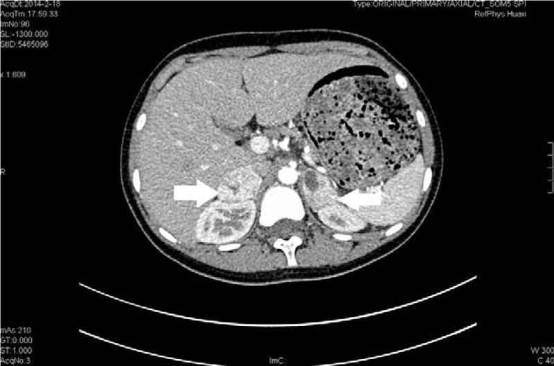
Enhanced CT of bilateral adrenal glands, showing 2 round-like masses (left: 4 × 5 × 3 cm; right: 6 × 4 × 3 cm) with soft tissue density in each adrenal gland, which were irregularly reinforced in arterial phase.(white arrowhead). CT = computed tomography.
The phenoxybenzamine was administrated for 2 weeks to control blood pressure, and then the resection of bilateral adrenal tumors was conducted. Pathological examination confirmed the diagnosis of bilateral adrenal pheochromocytoma. The patient underwent follow-up for 1.5 years and showed no signs of recurrence.
3. Discussion
The typical manifestations of pheochromocytoma include hypertension, forceful heartbeat, pallor, tremor, headache, and diaphoresis. However, the clinical manifestations of the present case featured hypotension, shock, and multiple organ dysfunction syndrome on admission, which nearly led us to miss the diagnosis of pheochromocytoma.
However, in the process of treatment, the patient always complained of paroxysmal palpitation, chest distress and headache. And her echocardiography and electrocardiogram showed the enlargement of left ventricle with thickened left ventricular wall. These proofs prompted us to doubt the possibility of hypertension, which was concealed by the state of circulatory failure before. Finally the ambulatory blood pressure monitoring (ABPM) results confirmed the presence of hypertension. Hypertension in children and adolescent is defined as systolic blood pressure (SBP) and/or diastolic blood pressure (DBP), persistently above the 95th percentile for sex, age, and height on three or more occasions.[5,6] All pediatric hypertension is recommended to screen for secondary cause, among which renal (parenchymal and vascular) diseases are the largest categories, accounting for 70% to 90% for all cases. During the process of screening, the renal diseases were preliminarily ruled out. However, the patient lately had an episode of hyperadrenergic spells (headache, palpitation, pallor, and sweating) with the laboratory test and radiography supported the diagnosis of pheochromocytoma.
Actually, the dramatic emergency in the present case was proved to be PCC. According to Newell's report, severe PCC could include severe hypertension or hypotension, multi-organ system failure, high fever, and encephalopathy.[7] A PCC without sustained hypotension is classed as “type A crisis,” while a PCC with sustained hypotension is classed as “type B crisis,” which is often presented with shock and multi-organ dysfunction.
Cardiovascular manifestations are the most common presentations of pheochromocytoma crisis, including cardiomyopathy, myocardial infarction, arrhythmia, and cardiogenic shock.[8] In our case, the patient presented with shock, ischaemic changes on ECG, and raised troponin. As she had had symptoms of upper respiratory tract infection, it was hard to distinguish the catecholamine cardiomyopathy from the viral myocarditis. Fortunately, the images of echocardiography showed the sign of transient left ventricular apical ballooning, which was the typical feature of stress-related cardiomyopathy (tako-tsubo or tako-tsubo like cardiomyopathy).[9] Though the causes of catecholamine cardiomyopathy and tako-tsubo are different, the common mechanism of these injuries is catecholamine-mediated, and most cases result in complete or partial reversal after certain treatment.
Besides circulation system, respiratory system can be also dysfunctional in PCC. Pulmonary edema, massive hemoptysis can be the main manifestations.[4] In the present case, the patient visited to the hospital with the main subject of cough and hemoptysis. Hemoptysis as a presenting symptom of pheochromocytoma was thought to be closely associated with fluctuations in blood pressure.[10–12] The long-term stimulation of catecholamine had weakened the ejection function of heart. So the severe paroxysmal hypertension could easily result in acute left heart failure and pulmonary edema.
In the present case, the patient had abnormal liver function with an ALT of 145 IU/L, increasing more than 3 folds. She denied the history of any chronic liver disease. The laboratory tests helped to exclude any viral or autoimmune liver disease. Eun et al[13] reported a case of pheochromocytoma with abnormal liver function in 2014. In this case, the ALT was 317 IU/L, with normal total bilirubin. The abnormal liver function was thought to be associated with repeated overproduction of hepatocytes, which was secondary to the increased resistance of liver arterioles and veins, and decreased blood flow and oxygen, led by overstimulation of norepinephrine on α-adrenalin receptors.[14]
4. Conclusion
We presented a case of PCC featured clinically hypotension, catecholamine-induced cardiomyopathy, hemoptysis, and abnormal liver function. All the symptoms are not the common manifestations of pheochromocytosis that could be frequently unrecognized by clinicians. It indicates us that when we meet a patient with hypotension and multiple organ dysfunctions in a relatively short time, the suspicion of pheochromocytoma should not be missed.
Author contributions
Conceptualization: Renhua Wu.
Data curation: Shishi Xu.
Investigation: Fang Zhang.
Methodology: Nanwei Tong.
Project administration: Lizhi Tang.
Resources: Yuwei Zhang.
Supervision: Nanwei Tong, Yuwei Zhang.
Validation: Fang Zhang.
Writing – original draft: Renhua Wu.
Writing – review & editing: Xinlei Chen.
Footnotes
Abbreviations: ABPM = ambulatory blood pressure monitoring, BP = blood pressure, CT = computed tomography, DBP = diastolic blood pressure, HR = heart rate, PCC = pheochromocytoma crisis, RR = respiratory rate, SBP = systolic blood pressure.
The authors declare no conflicts of interest.
References
- [1].Lenders JW, Duh QY, Eisenhofer G, et al. Endocrine Society. Pheochromocytoma and paraganglioma: an endocrine society clinical practice guideline. J Clin Endocrinol Metab 2014;99:1915–42. [DOI] [PubMed] [Google Scholar]
- [2].Scholten A, Cisco RM, Vriens MR, et al. Pheochromocytoma crisis is not a surgical emergency. J Clin Endocrinol Metab 2013;98:581–91. [DOI] [PubMed] [Google Scholar]
- [3].Guerrero MA, Schreinemakers JM, Vriens MR, et al. Clinical spectrum of pheochromocytoma. J Am Coll Surg 2009;209:727–32. [DOI] [PubMed] [Google Scholar]
- [4].Whitelaw BC, Prague JK, Mustafa OG, et al. Phaeochromocytoma crisis. Clin Endocrinol (Oxf) 2014;80:13–22. [DOI] [PubMed] [Google Scholar]
- [5].Lurbe E, Cifkova R, Cruickshank JK, et al. European Society of Hypertension. Management of high blood pressure in children and adolescents: recommendations of the European Society of Hypertension. J Hypertens 2009;27:1719–42. [DOI] [PubMed] [Google Scholar]
- [6].Julie R, Ingelfinger MD. The child or adolescent with elevated blood pressure. N Engl J Med 2014;370:2316–25. [DOI] [PubMed] [Google Scholar]
- [7].Newell KA, Prinz RA, Pickleman J, et al. Pheochromocytoma multisystem crisis. A surgical emergency. Arch Surg 1988;123:956–9. [DOI] [PubMed] [Google Scholar]
- [8].Prejbisz A, Lenders JW, Eisenhofer G, et al. Cardiovascular manifestations of phaeochromocytoma. J Hypertens 2011;29:2049–60. [DOI] [PubMed] [Google Scholar]
- [9].Gravina M, Casavecchia G, D’Alonzo N, et al. Pheochromocytoma mimicking Takotsubo cardiomyopathy and hypertrophic cardiomyopathy: a cardiac magnetic resonance study. Am J Emerg Med 2017;35:353–5. [DOI] [PubMed] [Google Scholar]
- [10].Yoshida T, Ishihara H. Pheochromocytoma presenting as massive hemoptysis and acute respiratory failure. Am J Emerg Med 2009;27:626.e3–4. [DOI] [PubMed] [Google Scholar]
- [11].Park M, Hryniewicz K, Setaro JF. Pheochromocytoma presenting with myocardial infarction, cardiomyopathy, renal failure, pulmonary hemorrhage, and cyclic hypotension: case report and review of unusual presentations of pheochromocytoma. J Clin Hypertens (Greenwich) 2009;11:74–80. [DOI] [PMC free article] [PubMed] [Google Scholar]
- [12].Frymoyer PA, Anderson GH, Jr, Blair DC. Hemoptysis as a presenting symptom of pheochromocytoma. J Clin Hypertens 1986;2:65–7. [PubMed] [Google Scholar]
- [13].Eun CR, Ahn JH, Seo JA, et al. Pheochromocytoma with markedly abnormal liver function tests and severe leukocytosis. Endocrinol Metab (Seoul) 2014;29:83–90. [DOI] [PMC free article] [PubMed] [Google Scholar]
- [14].Wang P, Tait SM, Chaudry IH. Sustained elevation of norepinephrine depresses hepatocellular function. Biochim Biophys Acta 2000;1535:36–44. [DOI] [PubMed] [Google Scholar]