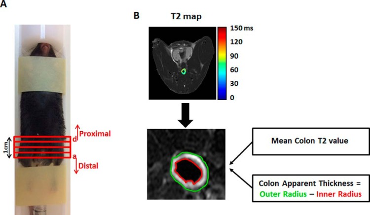
Figure 1.
In vivo mouse magnetic resonance imaging (MRI) colonoscopy experiment. Mice were set up in a dedicated MRI animal holder (A). The length of the scanned colon was ∼1 cm. Histological sections were taken from 4 regions [in red, sections (a)–(d)] of this area. The most distal portion of the colon is defined as (a) and the most proximal portion as (d). T2 maps of the colon, calculated on a pixel-by-pixel basis, with selection of two regions of interest, yellow and red, representing the outer and inner colon radii, respectively (B). Mean T2 value and apparent thickness were calculated for each section of the colon.

