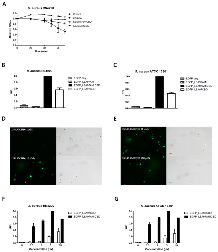
Figure 5.
Determination of the role of the LysSA97 amidase domain. (A) Activity comparison of LysSA97 and LSA97CHAPCBD against S. aureus RN4220. Equimolar concentrations (2.0 μM) of purified enzymes from the full length and truncation constructs were added to a 1 ml suspension of intact S. aureus RN4220, cells and the relative decrease in turbidity was monitored. Relative cell binding activities of 10 μM of EGFP_LSA97AMI, EGFP_LSA97AMICBD, and EGFP_LSA97CBD with (B) S. aureus RN4220 and (C) S. aureus ATCC 13301. Optical and florescence images of S. aureus RN4220 after the addition of (D) EGFP_LSA97CBD and (E) EGFP_LSA97AMICBD at 1 μM (top) and 10 μM (bottom). At different concentrations, the relative cell binding activities of EGFP_LSA97CBD and EGFP_LSA97AMICBD were measured with (F) S. aureus RN4220 and (G) S. aureus ATCC 13301. The data shown are the mean values from three independent measurements and the error bars represent the standard deviations. The scale bar represents 2 μm.

