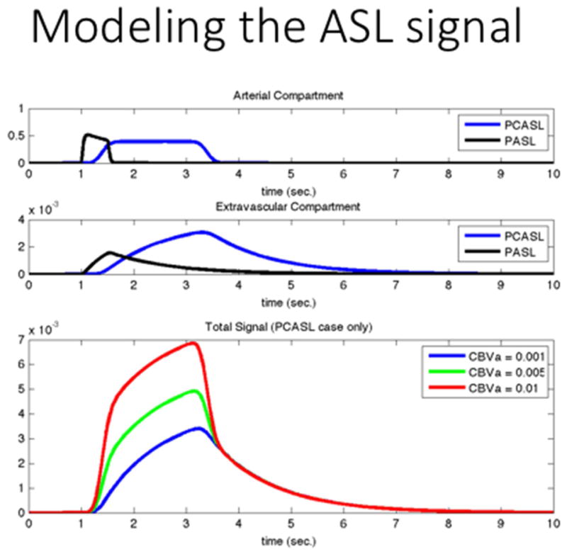
Figure 8.

ASL signal models capture the concentration of the label in a voxel as they move through the arterial compartment into the extravascular compartment, using the two-compartment model (without the pass-thru artery) in the middle panel of figure 7. Generally, the observed signal is the sum of the two compartments, unless the arterial compartment is suppressed during the acquisition.
