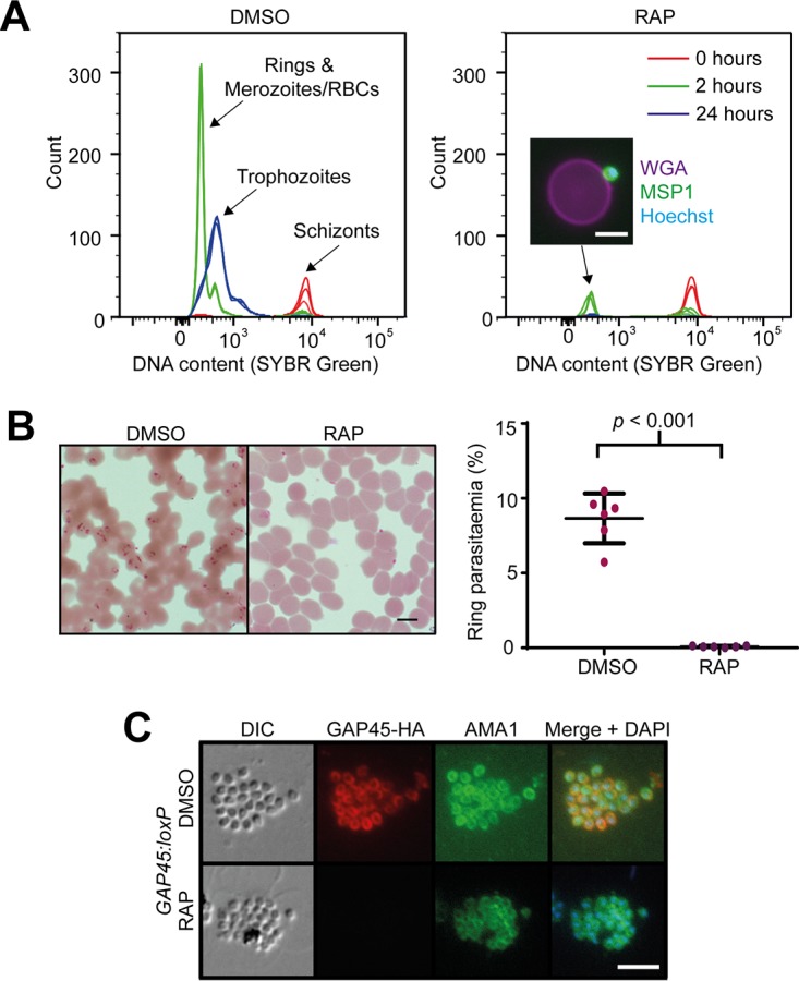
FIG 5 .

GAP45 is required for invasion but not for microneme discharge. (A) Flow cytometry-based invasion assay showing failure of ΔGAP45 parasites to develop from schizonts (t = 0 h) through rings (t = 2 h) to trophozoites (t = 24 h). The inset image shows a merozoite, stained with anti-MSP1 antibody and an Alexa 488 secondary antibody (green) and Hoechst stain (blue), bound to an uninfected RBC stained with wheat germ agglutinin (WGA) conjugated to Alexa 647 (magenta). Bar, 5 µm. Comparable results were observed in five additional flow cytometry-based invasion assays. (B) Giemsa-stained thin films showing the presence of ring-stage parasites in the invaded mock-treated (DMSO) GAP45:loxP parasites and absence of rings in the RAP-treated (ΔGAP45) GAP45:loxP parasites. Bar, 5 µm. This was observed in more than 10 experiments. Quantification of rings by manual counting of Giemsa-stained films in one representative experiment is shown (right). (C) IFA showing relocalization of microneme protein AMA1 to the merozoite periphery in both mock-treated and ΔGAP45 parasites. Bar, 5 µm.
