Fig. 6.
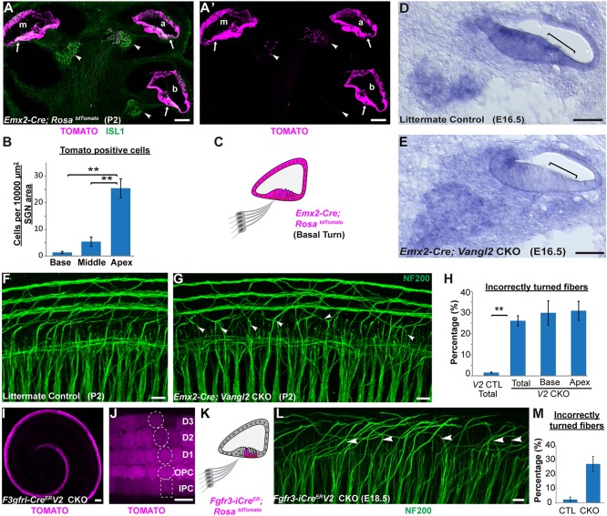
Vangl2 acts non-autonomously to guide type II SGN peripheral axon turning. (A,A′) Cross-section of an Emx2-Cre; RosatdTomato cochlea at P2 shows Cre-mediated recombination in the apical (a), middle (m) and basal (b) turns of the cochlear duct and organ of Corti (arrows). Limited recombination overlaps with ISL1-immunopositive neurons of the SGN (arrowheads) in a gradient along the length of the cochlea and is more frequent in the apical turn. (B) Quantification of tdTomato activation in the SGN shows a significant decrease from cochlear apex to base. (C) Schematic representation of Emx2-Cre-mediated recombination in the cochlear base. (D,E) In situ hybridization demonstrates reduction of Vangl2 mRNA in the Vangl2 CKO organ of Corti (square brackets) but not in the SGN. (F,G) Turning errors (arrowheads) are frequent in the basal turn of Emx2-Cre; Vangl2 CKOs. (H) Quantification of turning errors throughout the total length of Vangl2 CKO (n=5) cochlea relative to littermate controls (n=4), and comparison of turning errors between type II SGNs innervating the base versus apex. (I,J) In Fgfr3-iCreER; RosatdTomato cochlea at E18.5, recombination is restricted to supporting cells in the organ of Corti following tamoxifen induction initiated at E14.5. Dashed outlines indicate relative position of each supporting cell type. (K) Schematic representation of Fgfr3-iCreER-mediated recombination in the organ of Corti. (L) Incorrectly turned type II fibers (arrowheads) are readily detected in Fgfr3-iCreER; Vangl2 CKOs. (M) Quantification of turning errors in the middle turn of the Fgfr3-iCreER; Vangl2 CKO cochlea (n=3) relative to littermate controls (n=3). Data are mean±s.e.m. Asterisks show significant differences between genotypes using Student's t-test (**P<0.01). Scale bars: 100 µm in A,A′,I; 50 µm in D,E; 10 µm in F,G,J,L.

