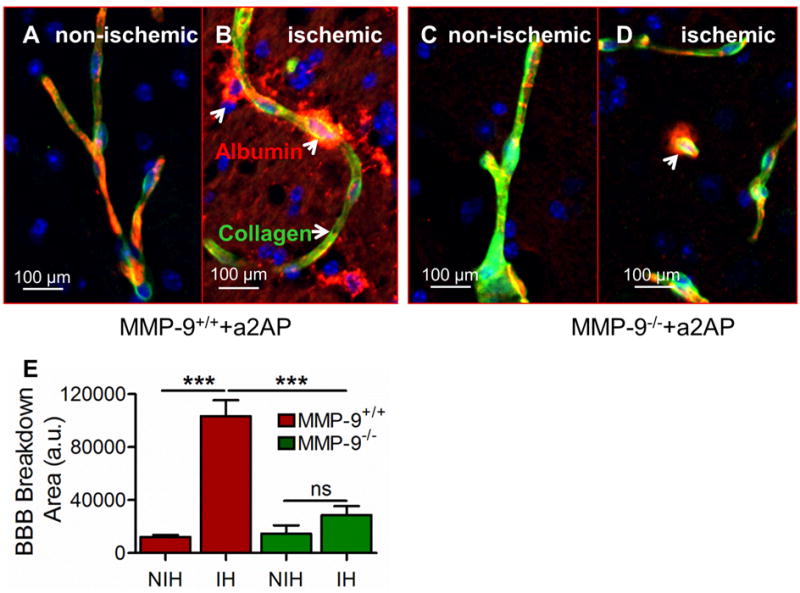
Figure 4. Effect of increased bloodα2-antiplasmin levels on BBB breakdown in MMP-9−/− vs MMP-9+/+ mice.

BBB breakdown was examined by leakage of albumin from intravascular to extravascular brain parenchymal tissue in MMP-9−/− and MMP-9+/+ mice supplemented with α2-antiplasmin. (A and B) Representative, merged 20X magnification (Scale bar = 100 μm) images of albumin leakage (red, arrows) outside blood vessels (type IV collagen; green) showing BBB breakdown (white arrows) in the ischemic vs. the non-ischemic hemisphere in MMP-9+/+ mice. Yellow color shows overlapping expression of collagen and albumin. (C and D) BBB breakdown in ischemic vs non-ischemic hemisphere in MMP-9−/− mice (E) The bar graph shows the average albumin positive area (arbitrary units, a.u.) in the ischemic hemisphere (IH) and the non-ischemic (NIH) hemisphere in MMP-9+/+ and MMP-9−/− mice, quantified in at least 8-10 representative images from each hemisphere. N=4-6, ***p<0.001, ns- non significant.
