Abstract
Aims
Plating displaced proximal humeral fractures is associated with a high rate of screw perforation. Dynamization of the proximal screws might prevent these complications. The aim of this study was to develop and evaluate a new gliding screw concept for plating proximal humeral fractures biomechanically.
Methods
Eight pairs of three-part humeral fractures were randomly assigned for pairwise instrumentation using either a prototype gliding plate or a standard PHILOS plate, and four pairs were fixed using the gliding plate with bone cement augmentation of its proximal screws. The specimens were cyclically tested under progressively increasing loading until perforation of a screw. Telescoping of a screw, varus tilting and screw migration were recorded using optical motion tracking.
Results
Mean initial stiffness (N/mm) was 581.3 (sd 239.7) for the gliding plate, 631.5 (sd 160.0) for the PHILOS and 440.2 (sd 97.6) for the gliding augmented plate without significant differences between the groups (p = 0.11). Mean varus tilting (°) after 7500 cycles was comparable between the gliding plate (2.6; sd 1.9), PHILOS (1.2; sd 0.6) and gliding augmented plate (1.7; sd 0.9) (p = 0.10). Similarly, mean screw migration(mm) after 7500 cycles was similar between the gliding plate (3.02; sd 2.85), PHILOS (1.30; sd 0.44) and gliding augmented plate (2.83; sd 1.18) (p = 0.13). Mean number of cycles until failure with 5° varus tilting were 12702 (sd 3687) for the gliding plate, 13948 (sd 1295) for PHILOS and 13189 (sd 2647) for the gliding augmented plate without significant differences between the groups (p = 0.66).
Conclusion
Biomechanically, plate fixation using a new gliding screw technology did not show considerable advantages in comparison with fixation using a standard PHILOS plate. Based on the finding of telescoping of screws, however, it may represent a valid approach for further investigations into how to avoid the cut-out of screws.
Cite this article: Y. P. Acklin, I. Zderic, J. A. Inzana, S. Grechenig, R. Schwyn, R. G. Richards, B. Gueorguiev. Biomechanical evaluation of a new gliding screw concept for the fixation of proximal humeral fractures. Bone Joint Res 2018;7:422–429. DOI: 10.1302/2046-3758.76.BJR-2017-0356.R1.
Keywords: Proximal humeral fracture, Dynamic fixation, Gliding humerus plate, PHILOS
Article focus
This study’s aim was to develop and test biomechanically a prototype plate for fixation of proximal humerus fractures which integrates a new gliding screw concept enabling dynamic compression in the fracture gap.
Key messages
Although from biomechanical perspective plate fixation with a new gliding screw concept did not show considerable advantages over PHILOS plating, based on the initiation of screw telescoping it may represent a valid alternative to the latter, especially in terms of cut-out prevention.
Strengths and limitations
This study presents a novel concept for improvement of plate fixation in the treatment of proximal humeral fractures together with new insights into the biomechanics of such fixed fractures, which can be used in the development of the next generation implants. The main strength of this study is the application of a reliable tracking system for precise analysis of interfragmentary and inter-implant movements.
The main limitation of this study is the low number of used specimens, restricting generalized clinical application. In addition, the specimens were preserved with Thiel’s method, reported to modify the biomechanical bone properties. Moreover, the biomechanical testing addressed loading in an idealized setting, neglecting such clinical aspects as muscle traction, soft tissue interference and compliance. In contrast to some previous biomechanical studies, the uniaxial loading was applied with the humeral axis kept at constant inclination, thus compromising to some extend the loading environment.
Introduction
Proximal humeral fractures are common. Their incidence in elderly patients with osteoporotic bone is increasing.1,2 Minimally displaced fractures show good results with conservative treatment.3-5 In severely displaced fractures, internal fixation using a plate or a nail, which provides sufficient biomechanical stability, has become widely accepted.6-9 However, it is still associated with a high complication and reoperation rate.10-12 One severe adverse effect is migration of screws with perforation of the cortex. This occurs in 8 to 11% of the cases, with a glenoid damage in up to 56% of them.13 There is a consensus in the literature that less rigid fixation is required, particularly for osteoporotic fractures, in order to reduce stresses at the bone-implant interface and prevent migration of the screws without compromising the stability of the construct.14-17 Although some semi-rigid or elastic implants have recently been used for fixation of proximal humeral fractures, no studies have investigated means of preventing screw cut-out in poor bone quality when using dynamic plating.9,18-22 Recently, a new prototype gliding plate, intended to enable dynamic compression of the fracture, was developed for fixation at the proximal humerus. Its design is adapted from the PHILOS plate (DePuy Synthes, West Chester, Pennsylvania), replacing the proximal locking holes with four short barrels for insertion of 3.5 mm gliding screws. The axes of the barrels are parallel to each other in order to enhance gliding of the screws. The construct allows anchorage in locations with high subchondral bone mineral density (BMD) and the blunt tips of the screws might help to reduce perforation. Cement can be added around the tips of the screws by injection into the predrilled bone holes.
The aim of this study was to evaluate the new concept for plating of three-part proximal humeral fractures biomechanically – with and without added bone cement – and compare it with the well-established PHILOS plate fixation. The hypothesis was that the gliding of the four proximal screws would prevent perforation of the joint better, and provide similar resistance to varus collapse, compared with PHILOS fixation; and that augmentation with cement would enhance additional resistance to varus collapse.
Materials and Methods
First, a pilot study, based on finite element analysis (FEA), was conducted to determine the optimal inclination of the four proximal gliding screws of the prototype plate which would be required to minimize stresses in the bone. For that purpose, three fresh-frozen (-20°C) human cadaveric humeri were scanned with high-resolution peripheral quantitative CT (HR-pQCT) using XtremeCT (Scanco Medical AG, Brüttisellen, Switzerland).
Implant placement, virtual osteotomies, meshing and material property assignments were performed using ScanIP software (Simpleware Ltd., Exeter, UK). Two osteotomies, simulating a three-part proximal humeral fracture without medial support according to Neer,23 were created in each specimen prior to scanning (Fig. 1a).24 The BMD, evaluated in the whole humeral head from the CT data, was 123 mg hydroxyapatite (HA)/cm3 for bone 1, 114 mg HA/cm3 for bone 2 and 100 mg HA/cm3 for bone 3. The Young’s modulus was assigned element-wise according to the BMD-modulus relationship used by Dragomir-Daescu et al:25 E = 14664ρ1.49 MPa, where E is Young’s modulus and ρ is the element’s apparent BMD obtained from the HR-pQCT scan. Poisson’s ratio was set to 0.3. All screws and both plate types were assigned a linear elastic Young’s modulus of 186.4 GPa with 0.3 as Poisson’s ratio. The three fracture fragments of each specimen were anatomically reduced and fixed virtually with either the gliding plate or PHILOS plate. The four most proximal screw holes of the latter were occupied with locking screws to ensure a similar configuration in the two plate constructs. The contact at the bone-screw interface was modelled as tied. The interaction between the plate and screws was modelled as surface-to-surface contact with a coefficient of friction of 0.1.
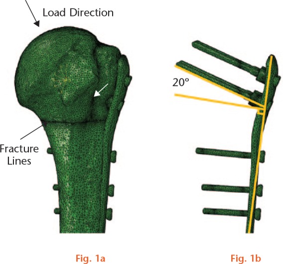
Examples of the finite element model for evaluation of the principal compressive bone strains at the tip of the four proximal screws of the prototype plate: a) a virtually instrumented specimen after anatomical reduction of the fragments. The red arrows denote the fracture lines and the yellow arrow the direction of loading as adapted from Bergman et al;24 b) prototype plate with 20° screw angle configuration.
The FEA models were solved using Abaqus software v6.13 (Simulia; Dassault Systèmes, Waltham, Massachusetts). A compressive load of 250 N was applied to the articular surface of each model, with the humeral axis inclined at 25° adduction (Fig. 1a). The loaded surface area was defined by projecting a vertically aligned cylinder of 30 mm diameter, whose axis crossed the centre of the humeral head on its surface. All translations perpendicular to the applied load and rotations around the compressive axis were constrained. Distally, a cardan joint coupling to the ground was simulated by connecting the shaft nodes, located in the 5 mm most distal part, to a control node located 200 mm distally, as measured from the most proximal aspect of the humeral head along the axis of the shaft. All translations of this control node and the rotation around the humeral shaft axis were constrained. The peak principal compressive strains at the tip of the four proximal screws of both implant systems were calculated for the standard screw angles of the PHILOS plate as well as for 20° (Fig. 1b) and 30°angles of the gliding plate. The results consistently indicated lower peak values for the gliding plate with 30° versus 20° screw angle as well as versus the PHILOS screw angles (Fig. 2). Based on these results, gliding plate prototypes were produced from stainless steel 316 L (DIN 1.4441) with a 30° inclination of the four screws (Fig. 3).
Fig. 2.
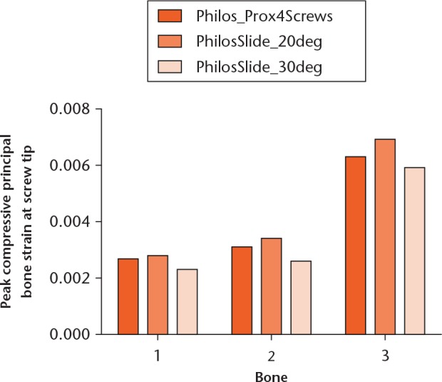
Graph showing peak principal compressive bone strains at the tip of the four proximal screws for the standard screw angles of the PHILOS plate (Philos_Prox4Screws) as well as for 20° and 30° angles of the gliding plate (PhilosSlide_20deg and PhilosSlide_30deg, respectively). The 30° screw angle configuration proved to be the most advantageous.
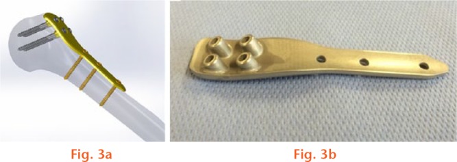
a) Gliding plate computer model; b) photograph of the back side of the gliding plate showing the four parallel aligned barrels.
A total of 12 paired human cadaveric proximal humeri, preserved with Thiel’s method,26 were used in the main study. Donors gave their informed consent within the donation of anatomical gift statement during their lifetime. The specimens were stripped of all soft tissue. The mean BMD of the humeral heads measured using clinical CT (SOMATOM Emotion 6, Siemens AG, Forchheim, Germany), was 87.0 mg HA/cm3 (sd 56.8).
Two osteotomies, simulating a three-part fracture with no medial support according to Neer,23 were created using a 1 mm oscillating saw blade. The three-part fracture represented a standard proximal humeral fracture consisting of a shaft, head and greater tubercle fragment. Eight paired specimens were assigned to two groups with an equal left and right number of bones for instrumentation with either the prototype gliding plate or PHILOS plate. The other four pairs were assigned for instrumentation using the gliding plate with bone cement augmentation at the tip of the four proximal screws.
Instrumentation was performed after reduction of the fracture. All plates were fixed distally to the humeral shaft with three 3.5 mm cortical screws. The plates in group 1 (gliding plate) were fixed proximally using four 3.5 mm gliding screws. In group 2 (PHILOS) the PHILOS aiming block was used for the proximal instrumentation of six 3.5 mm locking screws engaging the six most proximal screw holes. In group 3 (gliding augmented plate) the plate was fixed proximally with four 3.5 mm gliding screws which were augmented with 1 ml polymethylmethacrylate (PMMA)-based bone cement (Traumacem, DePuy Synthes, Zuchwil, Switzerland). The cement was prepared and 1 ml of it injected at the tip of the screw before its insertion, according to Röderer et al,27 using a standard vertebroplasty syringe and needle of DePuy Synthes. The length of each screw was defined using a depth gauge. Whereas the PHILOS plate and all screws were made of the titanium-based alloy Ti-6Al-7Nb (TAN) as provided by DePuy Synthes, the gliding plate prototypes were made of stainless steel (DIN 1.4441 – 316 L medical). Each specimen was distally cut at a length of 150 mm, and the distal 65 mm were embedded in PMMA (SCS-Beracryl, Suter-Kunststoffe AG, Fraubrunnen, Switzerland). Four retro-reflective marker sets were attached to the humeral head, the shaft, the most proximal screw (number one) and the plate of each specimen for optical motion tracking during testing.
Biomechanical testing was performed on a servo-hydraulic test system (Bionix 858.20, MTS Systems, Eden Prairie, Minnesota) equipped with a 4 kN/100 Nm load cell. The setup is shown in Figure 4. Each specimen was tested in 30° adduction in correspondence with the humeral loading angle as measured in vivo by Bergmann et al.24 For that purpose, the distally embedded humerus was fixed to the base of the machine using an angulated holder. The humeral head was loaded in axial compression along the machine axis using a spherically shaped PMMA shell cup attached to the load cell, which itself was interconnected to the machine actuator.
Fig. 4.
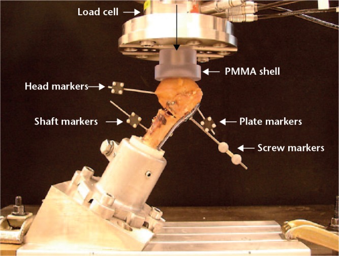
Test setup with a humerus instrumented with a PHILOS plate, equipped with four retro-reflective markers for optical motion tracking and mounted for biomechanical testing. The vertical arrow indicates the direction of loading (PMMA, polymethylmethacrylate).
Progressively increasing cyclical compression loading was applied with a physiological profile of each cycle at a rate of 2 Hz. While the valley load was kept at 20 N constant, the peak load, starting at 100 N, increased at a rate of 0.05 N/cycle until indication of a screw perforation through the humeral head.
Axial displacement (mm) and axial load (N) were acquired at a rate of 128 Hz. Based on these, initial stiffness of the bone-implant construct (N/mm) was calculated from the ascending linear slope of the load-displacement curve between 30 N and 90 N compression in the third loading cycle to exclude settling effects at the beginning of the cyclical test. Anteroposterior radiographs were taken for radiological assessment under peak load using a triggered C-arm at the beginning of the cyclical test and then at timed intervals every 250 cycles. Relative movements of the humeral head with respect to the shaft were investigated together with those between the most proximal screw and the plate in all six degrees of freedom using three-dimensional (3D) motion tracking analysis with five Qualisys ProReflex MCU digital cameras (Qualisys AB, Gothenburg, Sweden). For this purpose, the coordinates of the markers were continuously recorded throughout the cyclical test at a rate of 100 Hz. Based on the motion tracking data, varus tilting was calculated from the rotational movements of the humeral head in the coronal plane with respect to the shaft. Moreover, the number of cycles to mechanical failure with the corresponding peak load at failure, arbitrarily defined as 5° varus collapse, were derived from the magnitude series of the varus tilting over time. Furthermore, screw migration was calculated as the magnitude of the relative 3D translational movement of the humeral head and the tip of the most proximal screw. This screw was, as indicated, marked with a retro-reflective marker for tracking of its movement. Moreover, screw telescoping, defined as movement of the most proximal screw along its axis relative to the plate, was calculated to evaluate the performance of the gliding mechanism of the prototype plate. Finally, the number of cycles to cut-out was radiologically defined with the corresponding peak load at cut-out from the radiographs taken with the triggered C-arm. The outcomes varus tilting, screw migration and screw telescoping were derived after 7500 cycles under peak loading in relation to their values at cycle 1 under peak loading. The 7500 cycles represented the highest number when all specimens had not yet failed by cut-out and therefore were considered as an appropriate time point for evaluation.
Statistical analysis
Statistical analysis was performed using SPSS software package (IBM SPSS Statistics, V23; IBM, Armonk, New York). Data were screened for normal distribution with the Shapiro-Wilk test. Differences between the paired treatment groups were investigated using the Wilcoxon-Signed Rank test. Kruskal-Wallis test was used to detect differences between the non-paired groups. The level of significance was set to 0.05 for all statistical tests.
Results
Mean BMD in the three groups was 81.3 mg HA/cm3 (sd 52.3) for the gliding plate, 85.6 mg HA/cm3 (sd 52.8) for the PHILOS and 88.4 mg HA/cm3 (sd 74.3) for the gliding augmented plate, with homogeneous distribution among the groups, p = 0.83.
The mean initial stiffness was 581.3 N/mm (sd 239.7) for the gliding plate, 631.5 N/mm (sd 160.0) for the PHILOS and 440.2 N/mm (sd 97.6) for the gliding augmented plate, with no significant differences between the groups, p = 0.11.
Telescoping of the most proximal screw after 7500 cycles in the two groups with prototype plates was 0.37 mm (sd 0.31) for the gliding plate and 0.89 mm (sd 0.76) for the gliding augmented plate, with no significant difference between them, p = 0.24. The PHILOS plate group was excluded from this evaluation because of its locking screws with no telescoping.
The mean varus tilting after 7500 cycles was 2.6°(sd 1.9°) for the gliding plate, 1.2° (sd 0.6°) for the PHILOS and 1.7° (sd 0.9°) for the gliding augmented plate, with no significant differences between the groups, p = 0.10.
The mean screw migration was 3.02 mm (sd 2.85) for the gliding plate, 1.30 mm (sd 0.44) for the PHILOS and 2.83 mm (sd 1.18) for the gliding augmented plate, with no significant differences between the groups, p = 0.13.
The mean number of cycles to mechanical failure and corresponding load at failure in the three groups was 12702 (sd 3687) and 735.1 N (sd 184.3) for the gliding plate, 13948 (sd 1295) and 797.4 N (sd 64.8) for the PHILOS, and 13189 (sd 2647) and 759.5 N (sd 132.3) for the gliding augmented plate, with no significant differences between the groups, p = 0.66 (Fig. 5).
Fig. 5.
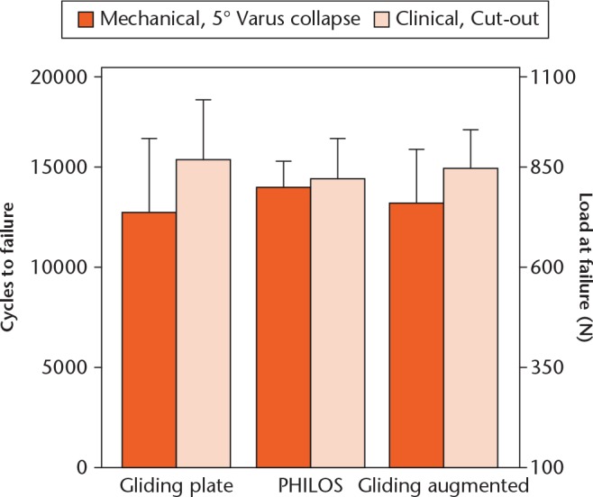
Cycles to mechanical and clinical failure and corresponding load at failure in the three groups (mean, sd).
The number of cycles to cut-out and corresponding load at cut-out in the three groups was 15344 (sd 3056) and 867.2 N (sd 152.8) for the gliding plate, 14406 (sd 1964) and 820.3 N (sd 98.2) for the PHILOS, and 14906 (sd 2922) and 845.3 (sd 96.1) for the gliding augmented plate, with no significant differences between the groups, p = 0.74 (Fig. 5).
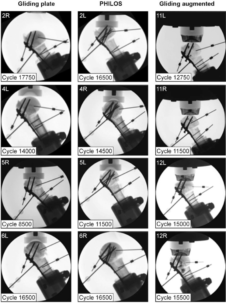
Examples of radiographs of the 12 humeri from the three groups with radiologically identified cut-out of the most proximal screws are shown in Figure 6.
Fig. 6.
Examples of radiographs of 12 humeri from the three groups with radiologically identified cut-out of the most proximal screw and respective number of cycles to cut-out.
Discussion
This study compared a new gliding screw concept versus conventional locking plate fixation of proximal humeral fractures. Although the gliding mechanism showed that dynamic fixation of the humeral head fragment is possible, it did not seem to be effective enough to reduce considerably screw perforation or varus tilting under the test conditions. On the other hand, PHILOS plating did not outperform the gliding plate fixation in any aspect, highlighting the potential of the gliding screw approach. However, some points need to be addressed in order to understand and further improve the gliding screw concept.
In its current version, the gliding plate included four proximal screws, whereas six were used for PHILOS plating. In addition, the angle of the proximal screw in the PHILOS plate was smaller than its 30° angle in the gliding plate. Although the 30° angle was found to be beneficial in FEA simulation, the influence of a varying number of gliding screws and/or changes in their inclination on the construct stability remains unknown for in vitro and in vivo loading scenarios.
Although augmentation with bone cement might increase the fixation strength when plating these fractures,27-29 in the present study its addition did not result in superiority over the other two fixation constructs. This could be ascribed to the technique of augmentation using the injection of cement into the bone holes prior to insertion of the screws, which can be considered restrictedly effective. We anticipate that more targeted injection of cement using cannulated screws with perforations around their tip would enhance anchorage and decrease screw migration.
An advantage of the gliding plate is that its screws allow optimal anchorage in the humeral head by targeting zones with the highest subchondral BMD, in the cranial and posterior subchondral bone.30,31 Our specimens included osteoporotic bone with low BMD compared with that of a normal lumbar vertebral body, with a mean BMD of 1.03 g/cm3.32 This can be explained by the specimens’ pretreatment with the Thiel Method.33
The blunt tips of the screws might reduce the risk of perforation. The screws were inclined at 30° in order to be aligned towards the main trajectory of the force acting on the humeral head, as reported by Bergmann et al24 who measured joint contact forces in six degrees of freedom using shoulder prostheses with telemetric data transmission. In their study the forces and moments were recorded during activities including abduction, flexion, extension, lifting a coffee pot, nailing, steering, walking with two crutches, lifting a weight and combing hair. The directions of the peak forces proved to be similar for the diverse activities.24,34
A previous study, that seems to be most related to the current work with regard to the development of implants, builds on a semi-rigid implant, the Humerusblock NG (Synthes Innovation Workshop, Salzburg, Austria), with modified tips of the pins for improved fixation in cancellous bone.21 This development attempted to reduce the shortcomings of the previous Humerusblock using Kirschner wires, which had high rates of perforation.18 The improved Humerusblock NG enabled dynamic closure and compression of the fracture analogous to similar concepts already established for the stabilization of femoral neck fractures, such as the dynamic hip screw (DePuy Synthes) and Targon FN (Braun Medical, Melsungen, Germany), thus confirming the relevance of dynamic fixation. However, clinical trials analyzing this implant are still lacking.
Another concept addressing dynamic screw fixation used dynamic locking screws (DePuy Synthes) in combination with commercially available PHILOS plates.22 This construct proved to have a lower rate of perforation of 7% compared with the generally published rates of 8% to 11%.[[6,10,11,35]] One underlying reason for this might be the blunt tip of the 3.7-mm screws. A second reason may be the intra- and inter-screw load distribution based on the pin-sleeve design with reduced screw rigidity.22
In a prospective clinical study, Acklin et al9 reduced the rigidity of the implant by using a 5-hole instead of a 3-hole PHILOS plate, enabling instrumentation with a longer working length using only very proximal and distal screw fixation. The authors reported a rate of perforation of 7.2% among 124 patients.9
In contrast to increasing the working length, reduction of the rigidity of the construct can also be achieved by changing the material of the implant, as shown by Schliemann et al,36 who assessed the biomechanical benefits of plates made of polyetheretherketone (PEEK) in comparison with titanium PHILOS plates. Although the PEEK plates resulted in lower construct stiffness, they also had lower strength of fixation with increased bone-implant movement compared with PHILOS plates. In contrast with the commercially available titanium plates, our prototype plates were made of stainless steel to facilitate manufacturing. This might influence screw perforation and be disadvantageous.
This study presents a novel concept for improvement of plate fixation in the treatment of proximal humeral fractures together with new insights into the biomechanics of fixation of such fixed fractures, which can be used in the development of the next generation implants.
The limitations of the study are similar to those inherent to all cadaveric studies. Only a few specimens were used, restricting generalized clinical application. In addition, the specimens were preserved with Thiel’s method, which has been reported to modify the biomechanical bone properties.33 The biomechanical testing addresses loading in an idealized setting, neglecting such clinical aspects as muscle traction, soft-tissue interference or compliance. In contrast to some previous biomechanical studies, the uniaxial loading was applied with the humeral axis kept at constant inclination, thus compromising to some extent theloading environment. In two previous studies the authors included a torsional capability in addition to the axial loading to simulate the forces of a rotator cuff tendon acting on the greater tuberosity.37,38 In another study Brunner et al21 applied more physiological multiplanar loading to the humeral head using a test bench to simulate abduction between 15° and 45°.
The main strength of this study was the application of a reliable tracking system for precise analysis of interfragmentary and inter-implant movements. In addition, varus collapse and screw cut-out represent frequently observed causes of failure with PHILOS plates and therefore justify the appropriate definition of the used failure criteria.11
In conclusion, plate fixation using gliding screw technology did not show considerable biomechanical advantages in comparison with PHILOS fixation. Based on the finding of screw telescoping, however, it may represent a valid approach for further research into mechanisms which might prevent the cut-out of screws.
Footnotes
Author Contributions: Y. P. Acklin: Conceptualization, Data acquisition, Data analysis and interpretation, Drafting the paper, Read and approved the final submitted manuscript.
I. Zderic: Data acquisition, Data analysis and interpretation, Drafting the paper, Read and approved the final submitted manuscript.
J. A. Inzana: Data acquisition, Read and approved the final submitted manuscript.
S. Grechenig: Data analysis and interpretation, Read and approved the final submitted manuscript.
R. Schwyn: Data acquisition, Read and approved the final submitted manuscript.
R. G. Richards: Conceptualization, Critical revision of the paper, Read and approved the final submitted manuscript.
B. Gueorguiev: Conceptualization, Data analysis and interpretation, Critical revision of the paper, Read and approved the final submitted manuscript.
Conflict of Interest Statement: None declared
Follow us @BoneJointRes
Funding Statement
The authors are not compensated and there are no other institutional subsidies, corporate affiliations, or funding sources supporting this work unless clearly documented and disclosed. This investigation was performed with the assistance of the AO Foundation via the AOTRAUMA Network (Grant No.: AR2015_07). DePuy Synthes is acknowledged for the delivery of PHILOS plates and screws.
References
- 1. Kettler M, Biberthaler P, Braunstein V, et al. Treatment of proximal humeral fractures with the PHILOS angular stable plate. Presentation of 225 cases of dislocated fractures. Unfallchirurg 2006;109:1032-1040. [DOI] [PubMed] [Google Scholar]
- 2. Palvanen M, Kannus P, Niemi S, Parkkari J. Update in the epidemiology of proximal humeral fractures. Clin Orthop Relat Res 2006;442:87-92. [DOI] [PubMed] [Google Scholar]
- 3. Hanson B, Neidenbach P, de Boer P, Stengel D. Functional outcomes after nonoperative management of fractures of the proximal humerus. J Shoulder Elbow Surg 2009;18:612-621. [DOI] [PubMed] [Google Scholar]
- 4. Fjalestad T, Hole MO, Hovden IA, Blücher J, Strømsøe K. Surgical treatment with an angular stable plate for complex displaced proximal humeral fractures in elderly patients: a randomized controlled trial. J Orthop Trauma 2012;26:98-106. [DOI] [PubMed] [Google Scholar]
- 5. Dean BJ, Jones LD, Palmer AJ, et al. A review of current surgical practice in the operative treatment of proximal humeral fractures: does the PROFHER trial demonstrate a need for change? Bone Joint Res 2016;5:178-184. [DOI] [PMC free article] [PubMed] [Google Scholar]
- 6. Brunner F, Sommer C, Bahrs C, et al. Open reduction and internal fixation of proximal humerus fractures using a proximal humeral locked plate: a prospective multicenter analysis. J Orthop Trauma 2009;23:163-172. [DOI] [PubMed] [Google Scholar]
- 7. Laflamme GY, Rouleau DM, Berry GK, et al. Percutaneous humeral plating of fractures of the proximal humerus: results of a prospective multicenter clinical trial. J Orthop Trauma 2008;22:153-158. [DOI] [PubMed] [Google Scholar]
- 8. Hessmann MH, Nijs S, Mittlmeier T, et al. Internal fixation of fractures of the proximal humerus with the MultiLoc nail. Oper Orthop Traumatol 2012;24:418-431. [DOI] [PubMed] [Google Scholar]
- 9. Acklin YP, Stoffel K, Sommer C. A prospective analysis of the functional and radiological outcomes of minimally invasive plating in proximal humerus fractures. Injury 2013;44:456-460. [DOI] [PubMed] [Google Scholar]
- 10. Thanasas C, Kontakis G, Angoules A, Limb D, Giannoudis P. Treatment of proximal humerus fractures with locking plates: a systematic review. J Shoulder Elbow Surg 2009;18:837-844. [DOI] [PubMed] [Google Scholar]
- 11. Sproul RC, Iyengar JJ, Devcic Z, Feeley BT. A systematic review of locking plate fixation of proximal humerus fractures. Injury 2011;42:408-413. [DOI] [PubMed] [Google Scholar]
- 12. Sabharwal S, Patel NK, Griffiths D, et al. Trials based on specific fracture configuration and surgical procedures likely to be more relevant for decision making in the management of fractures of the proximal humerus: findings of a meta-analysis. Bone Joint Res 2016;5:470-480. [DOI] [PMC free article] [PubMed] [Google Scholar]
- 13. Jost B, Spross C, Grehn H, Gerber C. Locking plate fixation of fractures of the proximal humerus: analysis of complications, revision strategies and outcome. J Shoulder Elbow Surg 2013;22:542-549. [DOI] [PubMed] [Google Scholar]
- 14. Maldonado ZM, Seebeck J, Heller MO, et al. Straining of the intact and fractured proximal humerus under physiological-like loading. J Biomech 2003;36:1865-1873. [DOI] [PubMed] [Google Scholar]
- 15. Foruria AM, Carrascal MT, Revilla C, Munuera L, Sanchez-Sotelo J. Proximal humerus fracture rotational stability after fixation using a locking plate or a fixed-angle locked nail: the role of implant stiffness. Clin Biomech (Bristol, Avon) 2010;25:307-311. [DOI] [PubMed] [Google Scholar]
- 16. Müller F, Voithenleitner R, Schuster C, Angele P, Weigel B. Operative treatment of proximal humeral fractures with helix wire. Unfallchirurg 2006;109:1041-1047. [DOI] [PubMed] [Google Scholar]
- 17. Hessmann MH, Sternstein W, Mehler D, et al. Are angle-fixed implants with elastic properties advantageous for the internal fixation of proximal humerus fractures?. Biomed Tech (Berl) 2004;49:345-350. [DOI] [PubMed] [Google Scholar]
- 18. Brunner A, Weller K, Thormann S, Jöckel JA, Babst R. Closed reduction and minimally invasive percutaneous fixation of proximal humerus fractures using the Humerusblock. J Orthop Trauma 2010;24:407-413. [DOI] [PubMed] [Google Scholar]
- 19. Jöckel JA, Brunner A, Thormann S, Babst R. Elastic stabilisation of proximal humeral fractures with a new percutaneous angular stable fixation device (ButtonFix(®)): a preliminary report. Arch Orthop Trauma Surg 2010;130:1397-1403. [DOI] [PubMed] [Google Scholar]
- 20. Bogner R, Hübner C, Matis N, et al. Minimally-invasive treatment of three- and four-part fractures of the proximal humerus in elderly patients. J Bone Joint Surg [Br] 2008;90-B:1602-1607. [DOI] [PubMed] [Google Scholar]
- 21. Brunner A, Resch H, Babst R, et al. The Humerusblock NG: a new concept for stabilization of proximal humeral fractures and its biomechanical evaluation. Arch Orthop Trauma Surg 2012;132:985-992. [DOI] [PubMed] [Google Scholar]
- 22. Freude T, Schroeter S, Plecko M, et al. Dynamic-locking-screw (DLS)-leads to less secondary screw perforations in proximal humerus fractures. BMC Musculoskelet Disord 2014;15:194. [DOI] [PMC free article] [PubMed] [Google Scholar]
- 23. Neer CS., II Displaced proximal humeral fractures. I. Classification and evaluation. J Bone Joint Surg [Am] 1970;52-A:1077-1089. [PubMed] [Google Scholar]
- 24. Bergmann G, Graichen F, Bender A, et al. In vivo glenohumeral contact forces-measurements in the first patient 7 months postoperatively. J Biomech 2007;40:2139-2149. [DOI] [PubMed] [Google Scholar]
- 25. Dragomir-Daescu D,Op Den Buijs J McEligot S et al. Robust QCT/FEA models of proximal femur stiffness and fracture load during a sideways fall on the hip. Ann Biomed Eng 2011;39:742-755. [DOI] [PMC free article] [PubMed] [Google Scholar]
- 26. Thiel W. The preservation of the whole corpse with natural color. Ann Anat 1992;174:185-195. [PubMed] [Google Scholar]
- 27. Röderer G, Scola A, Schmölz W, et al. Biomechanical in vitro assessment of screw augmentation in locked plating of proximal humerus fractures. Injury 2013;44:1327-1332. [DOI] [PubMed] [Google Scholar]
- 28. Unger S, Erhart S, Kralinger F, Blauth M, Schmoelz W. The effect of in situ augmentation on implant anchorage in proximal humeral head fractures. Injury 2012;43:1759-1763. [DOI] [PubMed] [Google Scholar]
- 29. Kathrein S, Kralinger F, Blauth M, Schmoelz W. Biomechanical comparison of an angular stable plate with augmented and non-augmented screws in a newly developed shoulder test bench. Clin Biomech (Bristol, Avon) 2013;28:273-277. [DOI] [PubMed] [Google Scholar]
- 30. Hepp P, Lill H, Bail H, et al. Where should implants be anchored in the humeral head? Clin Orthop Relat Res 2003;415:139-147. [DOI] [PubMed] [Google Scholar]
- 31. Schiuma D, Plecko M, Kloub M, et al. Influence of peri-implant bone quality on implant stability. Med Eng Phys 2013;35:82-87. [DOI] [PubMed] [Google Scholar]
- 32. Ryan PJ, Blake GM, Herd R, Parker J, Fogelman I. Distribution of bone mineral density in the lumbar spine in health and osteoporosis. Osteoporos Int 1994;4:67-71. [DOI] [PubMed] [Google Scholar]
- 33. Unger S, Blauth M, Schmoelz W. Effects of three different preservation methods on the mechanical properties of human and bovine cortical bone. Bone 2010;47:1048-1053. [DOI] [PubMed] [Google Scholar]
- 34. Westerhoff P, Graichen F, Bender A, et al. In vivo measurement of shoulder joint loads during activities of daily living. J Biomech 2009;42:1840-1849. [DOI] [PubMed] [Google Scholar]
- 35. Röderer G, Erhardt J, Graf M, Kinzl L, Gebhard F. Clinical results for minimally invasive locked plating of proximal humerus fractures. J Orthop Trauma 2010;24:400-406. [DOI] [PubMed] [Google Scholar]
- 36. Schliemann B, Seifert R, Theisen C, et al. PEEK versus titanium locking plates for proximal humerus fracture fixation: a comparative biomechanical study in two- and three-part fractures. Arch Orthop Trauma Surg 2017;137:63-71. [DOI] [PubMed] [Google Scholar]
- 37. Brianza S, Plecko M, Gueorguiev B, Windolf M, Schwieger K. Biomechanical evaluation of a new fixation technique for internal fixation of three-part proximal humerus fractures in a novel cadaveric model. Clin Biomech (Bristol, Avon) 2010;25:886-892. [DOI] [PubMed] [Google Scholar]
- 38. Rothstock S, Plecko M, Kloub M, et al. Biomechanical evaluation of two intramedullary nailing techniques with different locking options in a three-part fracture proximal humerus model. Clin Biomech (Bristol, Avon) 2012;27:686-691. [DOI] [PubMed] [Google Scholar]


