Figure 3.

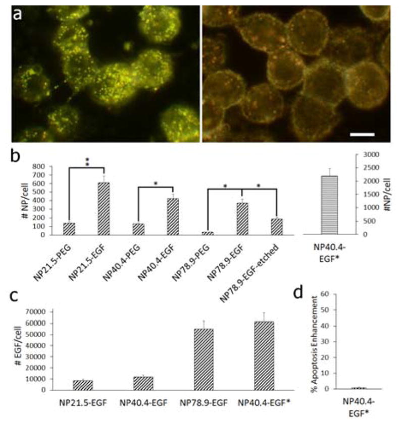
a) Darkfield image of MDA-MB-468 cells after incubation with NP78.9-EGF (left) and NP78.9-PEG (right) under otherwise identical conditions. Scale bar is 10 μm. b) Average number of particles uptake into MDA-MB-468 cells determined by ICP-MS for NP-PEG and NP-EGF with core diameters (left to right) of 21.5 nm, 40.4 nm, 78.9 nm. For the NP diameter of 78.9 nm we also included the data obtained after mild KI/I2 etching that preferentially removes surface bound NPs (“etched”). The effective EGF concentration for all conditions was 1 nM, except for NP40.4-EGF*, which contained an effective EGF concentration of 1.2 μM. *p < 0.05; **p < 0.01. c) Average number of EGF molecules delivered per cell for (left to right): NP21.5-EGF, NP40.4-EGF, NP78.9-EGF, NP40.4-EGF*. d) Apoptosis enhancement obtained for NP40.4-EGF*. Data in (b) – (d) were collected from at least two independent experiments.
