

Introduction
In 1934 Professor James Ferguson-Smith, a dermatologist, first described1 self-healing multiple keratoacanthoma syndrome in a 23-year old miner in Scotland. This patient had suffered multiple lesions on his face, ears, arms, thighs, and legs since the age of 16. Each lesion started as a red macula; became papular, enlarged, and ulcerated, and ultimately healed, leaving a deeply pitted scar. This condition became known as Ferguson-Smith syndrome or multiple self-healing squamous epitheliomas (MSSE). Up to today, less than 100 cases of MSSE have been reported worldwide. Most of these patients were studied by Malcolm A. Ferguson-Smith (geneticist, son of the dermatologist) in 1971. Malcolm Ferguson-Smith showed that this was an autosomal dominant syndrome.2
In 1993 MSSE was mapped to a region on chromosome 9q22, but the gene is yet unidentified.3 Molecular analyses found that the MSSE gene is a tumor suppressor gene.4 A recent study found a digenic or multilocus etiology for MSSE.5
The age of onset of MSSE ranges from 8 to 62 years. Most patients have MMSE by 34 years of age in women and 41 years in men. The number of tumors each patient has varies. Some patients only have 1 or 2 tumors during their lifetime, whereas others have more than 100. Individual lesions normally last for months. Tumors most commonly affect the nose, ears, and mouth regions, where they leave a deep pit as a scar. On limbs, they normally leave a flat scar. The trunk is rarely affected, and it is more common for lesions to appear in exposed areas of skin, suggesting ultraviolet light might be an important factor.6 Histologically, MSSE tumors are indistinguishable from well-differentiated squamous carcinomas, with epithelial permeation deep into the dermis, which is later infiltrated with lymphocytes. Each epithelial column shows central keratinization with the formation of cornified nests, which, as the tumor involutes, coalesce into a horny plug. The healing ulcer shows marked fibrosis of the dermis with loss of elastic fibers and absorption of small areas of keratinization.6
Case report
A 62-year-old woman presented with multiple tumors developing in a surgical margin on her forearm. Previous histology findings showed highly differentiated squamous cell carcinomas that were completely excised. This was the third time tumors recurred in the surgical margin. (Fig 1).
Fig 1.

Multiple tumors reoccurring in a previous surgical margin on the forearm.
The patient's history revealed a total of 62 cutaneous surgeries performed during the last 25 years, with histology reports showing both highly differentiated squamous cell carcinomas and keratoacanthomas. Ferguson-Smith syndrome was suspected and subsequently confirmed after blood sampling, DNA extraction, and Sanger sequencing of exon 1-9 and exon/intron boundaries of the transforming growth factor-β receptor 1 (TGFBR1) gene (NM_004612.2), which found a heterozygote variant, c.343+1G>A, affecting the splice donor of intron 1 and predicting loss of the functionally important extracellular ligand-binding domain. Because surgery seemed to aggravate our patient's condition, we decided to refrain from performing further surgical procedures and instead treat her conservatively. She was put on a low dose of acitretin (10 mg/d) because she was unable to tolerate a higher dose. All tumors regressed completely (Fig 2). Approximately 12 months after treatment was started, acute jaundice developed. She was admitted to our emergency department, and a computed tomography scan found a pancreatic tumor (Fig 3). She later underwent Whipple surgery. The removed tumor was histologically consistent with a pancreatic adenocarcinoma. She had no family history of pancreatic cancer. She was a nonobese smoker with low alcohol consumption and no history of pancreatitis or diabetes. Pyrosequencing of the TGFBR1 gene using DNA from the patient's blood and pancreatic tumor tissue showed an equal A-to-G ratio between the 2 tissues, indicating no loss of heterozygosity (LOH), which is the most frequent mechanism for somatic inactivation of tumor suppressor wild-type alleles in tumor cells.
Fig 2.

Complete regression of all tumors after conservative treatment.
Fig 3.
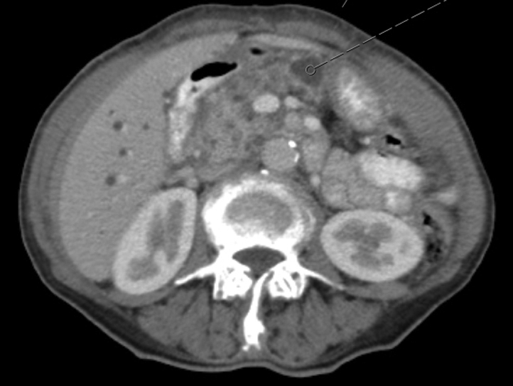
Axial image from abdominal computed tomography scan shows tumor (grey dotted arrow) within the pancreatic head.
Discussion
Ferguson-Smith's first patient died in 1948 of suppurative meningitis. An autopsy found lymphatic infiltration of anal and aural keratoacanthomas. The anal keratoacanthoma infiltrated the sphincter and the muscle layers of the anal canal.7
One case of a deep local invasion arising from a single keratoacanthoma has been described in a patient8 who presented with a keratoacanthoma involving his left ear and scalp that had completely destroyed the ear. Magnetic resonance imaging found the keratoacanthoma extended past the temporal bone into the cranial cavity. Postmortem autopsy found no distant metastases. No cases have ever been found to metastasize or cause death from carcinomatosis.
To the best of our knowledge, no previous literature has reported the occurrence of an internal malignancy in a Ferguson-Smith patient. The novelty of the occurrence led us to report this case, even as we understand no deductions or conclusions can be made regarding possible associations. Although the absence of LOH for TGFRB1 in the current patient's internal cancer supports no association with Ferguson-Smith syndrome, the current genetic investigation does not rule out other less common somatic second-hit mechanisms, such as promoter hypermethylation, small insertions or deletions, or a whole gene deletion. This case was discussed with Professor Malcolm Ferguson-Smith (personal communication, February 7, 2017) who encouraged publication, as it might help uncover examples of the same or a different association.
Acknowledgments
We thank professor Åke Borg for LOH analysis of tumor tissue DNA.
Footnotes
Funding sources: None.
Conflicts of interest: None disclosed.
References
- 1.Smith J.F. A case of multiple primary squamous-celled carcinomata of the skin in a young man, with spontaneous healing*. Br J Dermatol. 1934;46:267–272. [Google Scholar]
- 2.Ferguson-Smith M.A., Wallace D.C., James Z.H. Multiple self-healing squamous epithelioma. Birth Defects Orig Artic Ser. 1971;7:157–163. [PubMed] [Google Scholar]
- 3.Goudie D.R., Yuille M.A., Leversha M.A. Multiple self-healing squamous epitheliomata (ESS1) mapped to chromosome 9q22-q31 in families with common ancestry. Nat Genet. 1993;3:165–169. doi: 10.1038/ng0293-165. [DOI] [PubMed] [Google Scholar]
- 4.Bose S., Morgan L.J., Booth D.R. The elusive multiple self-healing squamous epithelioma (MSSE) gene: further mapping, analysis of candidates, and loss of heterozygosity. Oncogene. 2006;25:806–812. doi: 10.1038/sj.onc.1209092. [DOI] [PubMed] [Google Scholar]
- 5.Kang H.C., Quigley D.A., Kim I.J. Multiple self-healing squamous epithelioma (MSSE): rare variants in an adjacent region of chromosome 9q22.3 to known TGFBR1 mutations suggest a digenic or multilocus etiology. J Invest Dermatol. 2013;133:1907–1910. doi: 10.1038/jid.2013.45. [DOI] [PMC free article] [PubMed] [Google Scholar]
- 6.Ferguson-Smith M.A., Goudie D.R. Digenic/multilocus aetiology of multiple self-healing squamous epithelioma (Ferguson-Smith disease): TGFBR1 and a second linked locus. Int J Biochem Cell Biol. 2014;53:520–525. doi: 10.1016/j.biocel.2014.04.007. [DOI] [PubMed] [Google Scholar]
- 7.Currie A.R., Smith J.F. Multiple primary spontaneous-healing squamous-cell carcinomata of the skin. J Pathol Bacteriol. 1952;64:827–839. doi: 10.1002/path.1700640415. [DOI] [PubMed] [Google Scholar]
- 8.Chakrabarty K.H., Perks A.G. Ferguson-Smith syndrome: the importance of long term follow-up. Br J Plast Surg. 1996;49:497–498. doi: 10.1016/s0007-1226(96)90041-7. [DOI] [PubMed] [Google Scholar]