Abstract
Purpose
To demonstrate the presence of macular pigment in the retina of premature infants, and to examine its changes with age.
Methods
The participants included 40 premature infants. Infants who had received laser photocoagulation for retinopathy of prematurity were excluded. Macular pigment optical density (MPOD) was measured by fundus reflectometry using RetCam3, a digital fundus camera. The reflection imaging was performed for ROP screening. The imaging time points were from a post menstrual age (PMA) of 29 weeks 0 days to 46 weeks 5 days.
Results
The MPOD levels could be obtained from 39 premature infants. The levels at the first measurement ranged from 0 to 0.18 (mean 0.076, SD 0.044). The earliest time, when a nonvanishing MPOD level was obtained, was at a PMA of 33 weeks and 2 days, and that level was 0.05. The initial examination MPOD levels showed a moderate correlation with age (R2 = 0.32, P < 0.00017). The mean MPOD levels measured each week during the follow-up period showed a very strong correlation with age (R2 = 0.91, P < 0.0001). A regression line of MPOD = 0.0069 × age − 0.1783 was derived, where age is counted in PMA days.
Conclusions
The MPOD levels of premature infants were for the first time measured in living eyes. Macular pigment increased linearly with age.
Translational Relevance
Macular pigment increased with the development of macular morphology. This result suggested the importance of nutritional management of infants and mothers during perinatal period.
Keywords: macular pigment, imaging, premature infant
Introduction
The human macula has macular pigment consisting of the following three carotenoids: lutein ([3R, 3′R, 6′R]-lutein), zeaxanthin ([3R, 3′R]-zeaxanthin), and meso-zeaxanthin ([3R, 3′S; meso]-zeaxanthin).1,2 The macular pigment has its maximum absorption wavelength at 460 nm, and consequently acts as a filter for absorbing blue light in the retina. This function eliminates damage of retinal photoreceptors by singlet oxygen and radicals,3,4 and helps preserve good contrast sensitivity and decreased night glare.5
Bone et al.6 detected lutein and zeaxanthin in the retinas of 17- to 22-week-old fetuses by analyzing fetal autopsy tissue specimens with high-performance liquid chromatography (HPLC). Bernstein et al.7 measured macular pigment optical density (MPOD) in children ranging from preterm to 7 years using blue-light reflectometry. They reported that macular pigment increases with age postpartum that it is correlated with serum zeaxanthin concentration and skin carotenoid density, and that the morphologic and functional maturation of the retina in children is correlated with the level of macular pigment. In their studies, 11 premature infant eyes were imaged; however, no macular pigment was found in any of the images.
In the present study, we revisited the issue of macular pigment in the prematurely born infant investigating living subjects with a high sensitivity blue-light reflectometry device. Also, we used a large number of subjects for statistical significance of the findings.
Methods
Subjects
Forty-five premature infants who were consecutively admitted to the Seirei Hamamatsu General Hospital Neonatal Intensive Care Unit (NICU) between March 2016 and August 2017 were enrolled in this cross-sectional study. We excluded one whose parents did not give consent and four who underwent laser photocoagulation for retinopathy of prematurity (ROP), for a final total of 40 subjects (17 males and 23 females). No cases had a history of receiving anti-vascular endothelial growth factor therapy. The institutional review board of Seirei Hamamatsu General Hospital (No. 2206) approved this study. The parents/guardians of all the subjects signed a parental consent form, and the study complied with the tenets in the Declaration of Helsinki.
MPOD Measurement Methods
A digital fundus camera, RetCam3 (Clarity Medical System Inc., Pleasanton, CA), was used to measure MPOD. The camera features an RGB (red, green, blue) sensor chip, which under blue reflection imaging has no sensitivity in either the green or red channel, and therefore leads to reflection images with optimum image contrast. The measurements were performed per protocol as previously reported.8 The pupils were dilated using Caputo drops9 (a solution containing a mixture of 1% cyclopentolate, 0.5% tropicamide, and 5% phenylephrine at the ratio of 1:1:2). A lens having a field of view of 80° was placed on the cornea, and imaging of the macula was performed using blue light excitation (center wavelength of 484 nm). Blue-light reflection imaging could be accomplished within 1 minute for each eye; total imaging time including color photography was kept to below 5 minutes per eye in line with safety limits specified in the RetCam3 user's manual.
Anesthetic eye drops (0.4% Oxybuprocaine Hydrochloride) were used for the imaging; the subjects were not sedated. All imaging was carried out by the same investigator (HS) at Seirei Hamamatsu General Hospital. All images were analyzed and the MPOD values calculated by the same investigator (MS).
MPOD Measurement Timing
The subjects were examined in accordance with management guidelines for ROP.10,11 Infants delivered before the postmenstrual age (PMA) of 26 weeks were first examined at PMA of 29 weeks. Infants delivered after the PMA of 26 weeks were first examined approximately 2- to 3-weeks postpartum. From then on, examinations were repeated at 3- to 5-week intervals, depending on the maturation of the retinal blood vessels and degree of ROP, up to a maximum of 46 weeks and 5 days (327 days). In the examinations, color imaging of the fundus of both eyes and blue-light imaging to measure MPOD were carried out using the RetCam3. Imaging was cancelled when the systemic condition of the subject infant was poor.
Statistical Analysis
The correlations between PMA and MPOD value, between PMA and weight, between MPOD value and weight, and between the MPOD value of the left and right eyes were tested using Pearson's correlation coefficient test. PMA and mean MPOD value at the various PMAs, as well as PMA and mean weight at the various PMAs, were examined using simple linear regression analyses. All statistical analyses were carried out using IBM SPSS Statistics ver. 25 software (IBM Corp., Armonk, NY).
Results
Supplementary Table S1 presents the demographic data of the subjects. The age at birth ranged from a PMA of 24 weeks and 6 days (174 days) to 38 weeks and 4 days (270 days; mean 228 days, SD 20 days), and birth weight ranged from 632 to 2280 g (mean 1570 g, SD 365 g). Nine infants had systemic diseases. The highest stage and zone of ROP during the measurement period were shown in Supplementary Table S1. The absence of changes in ROP, such as line and ridge in the macular region, was confirmed by the examination with binocular ophthalmoscopy and images taken with RetCam3 in all cases.
Timing and Frequency of MPOD Measurements
The first imaging of the 40 premature infants was performed between a PMA of 29 weeks and 0 days (203 days) and a PMA of 41 weeks and 5 days (292 days). The final imaging was performed between a PMA of 35 weeks and 3 days (248 days) and 46 weeks and 2 days (324 days). During these periods, imaging was performed a total of 141 times. Imaging was performed once for four infants, twice for 13 infants, three times for seven infants, four times for eight infants, five times for three infants, six times for one infant, eight times for two infants, nine times for one infant, and 12 times for one infant. Among the 141 times of imaging, good images from which MPOD was measurable were obtained from both eyes 54 times, from only one eye 49 times, and from neither eye 38 times.
The Initial MPOD Values
An image from which MPOD was measurable was obtained at least once in 39 of 40 premature infants. For the one remaining infant, imaging was performed five times, from a PMA of 34 weeks and 4 days to a PMA of 44 weeks and 4 days; however, the obtainable images were degraded as a result of body and ocular movements.
The time at which the first measurable MPOD was obtained for the other 39 infants was a PMA of 33 weeks for four infants, 34 weeks for four, 35 weeks for nine, 36 weeks for seven, 37 weeks for three, 38 weeks for six, 40 weeks for two, 41 weeks for one, 42 weeks for one, and 43 weeks for one infant. The median time of first measurable MPOD was a PMA of 36 weeks (254 days). The MPOD values ranged from 0 to 0.18 (mean 0.076, SD 0.044). Three infants showed 0. The earliest age at which an MPOD value greater than 0 was obtained was a PMA of 33 weeks and 2 days (233 days), and the MPOD value was 0.05 (Supplementary Table S1, infant No. 18). Earlier measurements were attempted a total of seven times in four infants; however, images with a measurable MPOD could not be obtained. The imaging times of three premature infants for whom the MPOD value was 0 ranged from a PMA of 33 weeks and 4 days to a PMA of 35 weeks and 4 days. However, repetition of imaging in these three infants at PMAs of 36 weeks and 3 days or more yielded MPOD values greater than 0.
The relationship between the first MPOD value for each infant and the PMA is shown in Figure 1. When an MPOD value was obtained for both eyes of an infant, the higher value was used. A moderate but statistically significant correlation was found between the age and MPOD value (R2 [correlation of determination] = 0.32, P = 0.00017).
Figure 1.
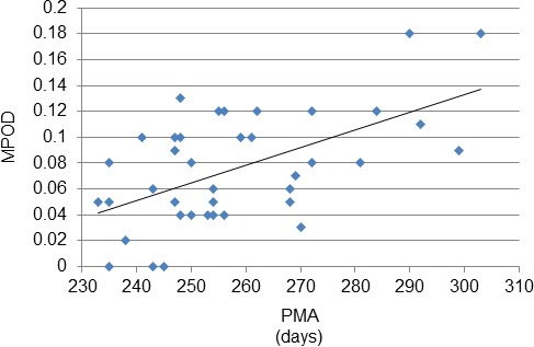
Correlation between PMA and MPOD (N = 39, R2 = 0.32, P = 0.00017, Pearson's correlation coefficient test).
The weights of the 39 premature infants at the time when the first MPOD value was measurable ranged from 1281 to 2372 g (mean 1865 g, SD 312 g). The minimum weight at which an MPOD value greater than 0 was obtained was 1281 g (Supplementary Table S1, infant No. 26, PMA of 38 weeks and 2 days). The relationship between weight and MPOD value is shown in Figure 2; a moderate but significant correlation was found (R2 = 0.27, P = 0.0009). A moderate but significant correlation was also found between age and weight (R2 = 0.20, P = 0.057; Fig. 3).
Figure 2.
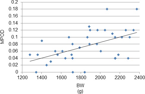
Correlation between body weight (BW) and MPOD (N = 37, R2 = 0.27, P = 0.0009, Pearson's correlation coefficient test).
Figure 3.

Correlation between PMA and BW (N = 37, R2 = 0.20, P = 0.0057, Pearson's correlation coefficient test).
Comparison of MPOD Values for the Left and Right Eyes
MPOD values could be obtained for both eyes on the same day from 32 premature infants. Figure 4 shows the MPOD values for the left and right eyes when they were first obtainable from both eyes. There was a strong and highly significant correlation between the values for the two eyes (R2 = 0.73, P < 0.0001).
Figure 4.
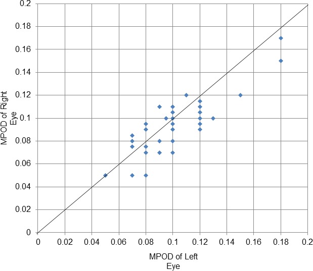
Correlation of MPOD between right and left eyes (N = 32, R2 = 0.73, P < 0.0001, Pearson's correlation coefficient test).
MPOD values were obtained four times for both eyes for three infants. Changes in MPOD values for the right and left eyes of these three infants are shown in Figure 5. The trend of increase in MPOD values for both eyes was similar.
Figure 5.
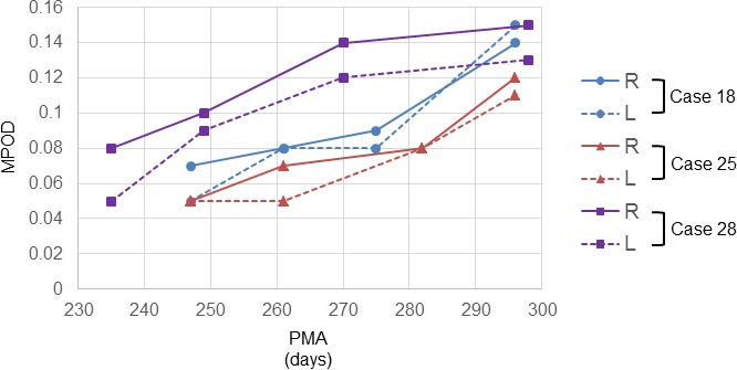
Changes of MPOD with PMA in right and left eyes of three cases.
Changes in MPOD Values With Age
MPOD values could be obtained at least twice from 31 premature infants. The frequency of measurement was twice for 12 infants, three times for 11, four times for three, five times for four, and six times for one infant. Changes in MPOD values from these 31 infants are presented in Figure 6. When MPOD values were obtained for both eyes, the higher value was used. In one premature infant (infant No. 33 in Supplementary Table S1, arrow in Fig. 6), the MPOD value was measured twice in 3 weeks, but showed the same value both times. In the remaining 30 infants, the MPOD value increased with age.
Figure 6.
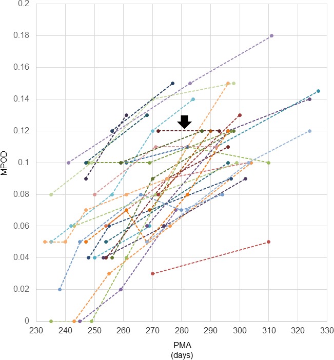
Changes of MPOD with PMA in 31 cases that were obtained MPOD values more than two times. The MPOD value increased with age except for one subjects (arrow).
When the age at which MPOD was measured in the 31 infants was classified by number of weeks, the result was a PMA of 33 weeks for four infants, 34 weeks for six, 35 weeks for 13, 36 weeks for 10, 37 weeks for eight, 38 weeks for 16, 39 weeks for seven, 40 weeks for nine, 41 weeks for six, 42 weeks for 13, 43 weeks for five, 44 weeks for three, and 45 weeks for zero, and 46 weeks for three infants. The mean MPOD values by age in weeks are shown by the blue line in Figure 7. There was a very strong correlation between age in weeks and mean MPOD value (R2 = 0.91, P < 0.0001). The regression formula that best estimates MPOD value as a function of age in weeks was: y = 0.0069x − 0.1783 (y: MPOD value, x: number of weeks). The SD of the value estimated using this regression equation was 0.0094.
Figure 7.
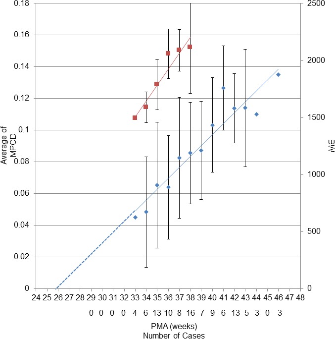
Changes in the average of MPOD (blue line) and BW (red line) with PMA. Bars represent SD that was calculated in case subjects' number was more than five. Blue dashed line represents the extension of the regression line. The PMA for MPOD of 0 is 26 weeks (MPOD, R2 = 0.91, P < 0.0001; BW, R2 = 0.92, P = 0.0025, Pearson's correlation coefficient test).
Weight was measured at all the measurement time points up to a PMA of 38 weeks. Mean weight by age in weeks is shown by the red line in Figure 7. There was a strongly significant correlation between weight and age in weeks (R2 = 0.92, P = 0.0025). The regression formula obtained to estimate weight from the age in weeks was: y = 138.79x − 3071 (y: weight, x: number of weeks). The SD of the value estimated using this regression equation was 86.08 g.
Discussion
We measured the MPOD of premature infants using fundus reflectometry. The measurement could be performed in 39 of 40 infants. The earliest time point at which an MPOD value greater than 0 was detectable was a PMA of 33 weeks and 2 days (233-days old), and at that age the MPOD value was 0.05. The mean of the first measurable value of MPOD was 0.076 ± 0.044, and the MPOD level increased with the age at which the measurement could be performed (Fig. 1). The mean MPOD value at every week correlated strongly with PMA in week (R2 = 0.91, P < 0.0001, Fig. 7). Thus, MPOD increased as a function of growth. There was a strong correlation between the left and right eyes over the range of MPOD values (R2 = 0.73, P < 0.0001, Fig. 4), and the growth patterns for both eyes were similar.
There are four main methods to measure MPOD in a living eye: a psychophysical method using a heterochromatic flicker photometry, fundus autofluorescence spectroscopy, resonance Raman spectroscopy, and fundus reflectometry. Of these, fundus reflectometry is the only applicable method for measurements of premature infants and neonates.7,8 In this study, MPOD could be measured in 39 of 40 infants. However, for images taken from PMAs of 29 to 32 weeks, sufficient contrast to measure MPOD could not be obtained. The imaging problems were caused by the tunica vasculosa lentis and/or by opacification of the vitreous body, which are suggested as limitations of this measurement method. Moreover, sedation was not performed during the imaging, and so there were instances in which qualified image was not obtained within 1 minute due to body or eye movements.
A previous report12 showed that the plasma concentration of carotenoids in mothers correlated with that in neonates. This fact suggests that carotenoids likely migrate from maternal blood to fetal blood via the placenta, and accumulate in the fetal retina. After delivery, infants are supplied with carotenoids through breast milk, which then accumulate in the retina. Considering these things, it is quite natural that premature infants have macular pigment. In fact, the presence of carotenoids has been confirmed in the fetal retina in a study that examined eyes by HPLC during autopsy.6 However, to the best of our knowledge, there is no report in which living eyes were used to demonstrate that macular pigment is present in premature infants. Bernstein et al.7 used fundus reflectometry to measure MPOD in a population ranging from premature infants of PMA 29 weeks to children aged 6 years, but did not find macular pigment in their premature infants. This study is, therefore, the first to demonstrate the presence of macular pigment in the living premature infant retina. Several facts may account for the inability to detect MPOD in previous reports. First, the time of measurement for the eight infants in the previous report was between PMA of 29 and 33 weeks, which are comparatively early ages. In the present study, MPOD measurements were performed before a PMA of 33 weeks in nine infants. Among them, images in which MPOD was measurable could be obtained for four eyes, and of these, the MPOD value was 0 for one eye, and greater than 0 (0.05–0.08) for the other three eyes. Another possible reason is the status of carotenoid supply to the infant. Of the eight infants in the previous report, three received carotenoids from breast milk or artificial milk, one did not receive any, and the situation was unknown for four. The 40 infants in this study were given breast milk immediately after birth, and so were already receiving a supply of carotenoids at the time MPOD was measured. In this study, however, the level of carotenoid supply is unknown because the amount of carotenoids in breast milk and the concentration in the maternal plasma were not measured. On the other hand, Figure 7 shows that MPOD increased in a parallel manner with weight gain. In other words, an increase in MPOD accompanies systemic growth, indicating that the nutritional supply from breast milk is distributed systemically, including to the retina. The previous study included images taken by RetCam2, in contrast we used RetCam3 in all cases. Because the image quality by RetCam3 was better than RetCam2 (Fig. 8), the difference in the device used might be another reason.
Figure 8.
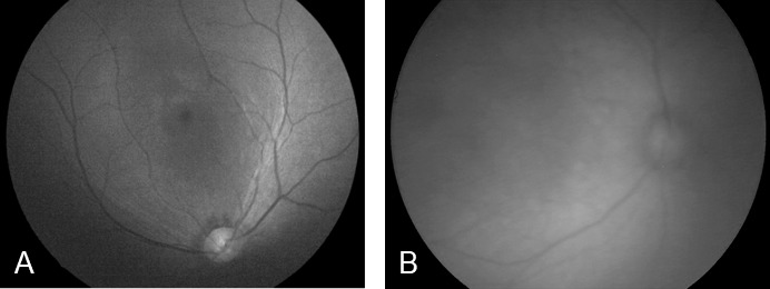
Blue-light reflectance images. (A) Image taken by RetCam3. (B) Image taken by RetCam2 in the previous study.7
The structure of the fetal retina has been studied histologically, and recent developments in optical coherence tomography (OCT) have also enabled detailed reports on the morphology of the premature infant retina.13–15 According to these reports, the thick inner retina composed from the ganglion cell layer (GCL), inner plexiform layer (IPL), and inner nuclear layer (INL), and the outer nuclear layer (ONL) composed of a single layer of cone cells can be seen in the center of the retina of embryos with a gestational age of 20 to 22 weeks. At a gestational age of 25 weeks, a shallow pit begins to form, and at 30 to 32 weeks, displacement of GCL, IPL, and INL to the periphery begins to occur. At 33 to 36 weeks, the central pit deepens, and at 37 to 39 weeks, the GCL, IPL, and INL disappear and the foveal pit is established. The PMA of 33 weeks and 2 days at which an MPOD value greater than 0 was first obtained in this study corresponds to the time when the foveal pit is deepening. The increase in MPOD that accompanies subsequent ages is suggested to be correlated with maturation of the fovea. Assuming that the increase in MPOD is linear even prior to a PMA of 33 weeks, the MPOD linear regression line shown in Figure 7 will elongate to the left, and looking at the time point when MPOD = 0, the PMA is 26 weeks. At this point in time, only a shallow pit can be seen in the central fovea. In other words, this corresponds to the time when the fovea starts to form. This indicates that macular pigment begins to exist when formation of the fovea starts.
Infants delivered between the PMAs of 37 weeks and 0 days and 41 weeks and 6 days are defined full-term deliveries. In addition to the report of Bernstein et al.,7 Henriksen et al.12 measured the MPOD of 16 full-term babies using fundus reflectometry. They reported that the MPOD values of the full-term babies ranged from 0.04 to 0.16 (mean 0.087, SD 0.032). In addition, extrapolating from the regression line they constructed using PMA and actual MPOD values, the MPOD value at 40 weeks and 0 days was predicted to be 0.0835 to 0.0887. In the present study, the actual MPOD value of the nine infants in whom MPOD was measured at a PMA of 40 weeks (PMA of 40 weeks and 0–6 days) was 0.07 to 0.15, with a mean of 0.1033. Moreover, the value determined from the regression formula was 0.0977. The present results suggested that the level of macular pigment in premature infants was lower than that in full-term infants; however, macular pigment increased with growth, and the value reached at a PMA of 40 weeks was equivalent or more than that of full-term infants. MPOD values of adults measured by fundus reflectance spectroscopy were reported to range from 0.13 to 0.8.16 However, the device used for adults differed from that used in this study in that the excitation wavelength and method of calculation were different. Therefore, no direct comparison can be made. However, the MPOD values for premature infants in this study and those for full-term infants in previous studies were less than the values for adults. Bernstein et al.7 reported that MPOD increases with age in children. This suggests that in premature infants and neonates, macular pigment likely increases with structural and functional maturation of the retina until the level approaches that in adults. Lutein and zeaxanthin, which are components of macular pigment, are supplied from breast milk postpartum. Therefore, it is important for mothers to eat vegetables, and infants should continue to eat vegetables after being weaned from breast milk.
Macular pigment in the left and right eyes of the same individual are reported to show similar pigment density7 and spatial distribution of pigment.17,18 In this study as well, MPOD values in the left and right eyes, and their manner of increase, were similar, suggesting that the left and right eyes develop in a similar manner.
A previous study19 suggested that lutein administration prevented the occurrence of ROP or lowed the severity of ROP, but others20,21 have not found that lutein administration reduces the incidence of ROP. Therefore, there is no consensus yet regarding the effectiveness of lutein administration to premature infants at risk for ROP. Future direct correlation studies between MPOD and the onset of ROP would be helpful in this respect.
There are some limitations to this study. As mentioned previously, it was difficult to obtain images of sufficient quality to measure MPOD using fundus reflectometry in premature infants of PMA less than 33 weeks. Therefore, whether or not macular pigment exists prior to 33 weeks and 2 days is unknown. To obtain accurate knowledge on the changes in macular pigment with age, it would be ideal to perform the measurements at the same ages and at constant intervals in all subjects. However, in this study, MPOD was measured during examinations carried out to diagnose the status of ROP. Consequently, the measurement times for the infants were irregular. The measured MPOD values were divided by the week of PMA, and the mean values used to construct Figure 7. Although a linear fit from which MPOD value could be determined as a function of age was obtained, the data for each age came from a different subset of infants. Consequently, the change in MPOD value with age was not examined for the same individual infant. The previous reports using OCT showed that macular edema was observed in 15% to 60% of premature infants.22–24 We did not observe any abnormalities in the macula by binocular ophthalmoscopy and images taken with RetCam3, but the absence of macular edema was not confirmed by OCT. If macula edema exists, the emission and reflection lights are affected in some extent, and there is a possibility that MPOD is underestimated. Because we had no opportunity to use OCT in this study, this issue has to be investigated in the future.
In this study, we were able for the first time to obtain MPOD values from living eyes of premature infants using fundus refractometry. The presumption is that macular pigment begins to exist from the stage at which the foveal pit starts to form and increases linearly with age as the fovea matures structurally. In premature infants, MPOD levels postpartum are lower than those of full-term infants in the early stage. However, it is evident that the levels increase thereafter with growth of the premature infant, and by the age of 40 weeks they reach the same levels as those of full-term infants.
Supplementary Material
Acknowledgments
Presented at ARVO2017 as a poster (No. 0727), May 2017, Baltimore, Maryland.
Disclosure: H. Sasano, None; A. Obana, None; M. Sharifzadeh, None; P.S. Bernstein, None; S. Okazaki, None; Y. Gohto, None; T. Seto, None; W. Gellermann; None
References
- 1.Bone RA, Landrum JT, Hime GW, Cains A, Zamor J. Stereochemistry of the human macular carotenoids. Invest Ophthalmol Vis Sci. 1993;34:2033–2040. [PubMed] [Google Scholar]
- 2.Landrum JT, Bone RA. Lutein, zeaxanthin, and the macular pigment. Arch Biochem Biophys. 2001;385:28–40. doi: 10.1006/abbi.2000.2171. [DOI] [PubMed] [Google Scholar]
- 3.Krinsky NI, Landrum JT, Bone RA. Biologic mechanisms of the protective role of lutein and zeaxanthin in the eye. Annu Rev Nutr. 2003;23:171–201. doi: 10.1146/annurev.nutr.23.011702.073307. [DOI] [PubMed] [Google Scholar]
- 4.Krinsky NI, Johnson EJ. Carotenoid actions and their relation to health and disease. Mol Aspects Med. 2005;26:459–516. doi: 10.1016/j.mam.2005.10.001. [DOI] [PubMed] [Google Scholar]
- 5.Nolan JM, Power R, Stringham J, et al. Enrichment of macular pigment enhances contrast sensitivity in subjects free of retinal disease: Central Retinal Enrichment Supplementation Trials - Report 1. Invest Ophthalmol Vis Sci. 2016;57:3429–3439. doi: 10.1167/iovs.16-19520. [DOI] [PubMed] [Google Scholar]
- 6.Bone RA, Landrum JT, Fernandez L, Tarsis SL. Analysis of the macular pigment by HPLC: retinal distribution and age study. Invest Ophthalmol Vis Sci. 1988;29:843–849. [PubMed] [Google Scholar]
- 7.Bernstein PS, Sharifzadeh M, Liu A, et al. Blue-light reflectance imaging of macular pigment in infants and children. Invest Ophthal Vis Sci. 2013;54:4034–4040. doi: 10.1167/iovs.13-11891. [DOI] [PMC free article] [PubMed] [Google Scholar]
- 8.Sharifzadeh M, Bernstein PS, Gellermann W. Reflection-based imaging of macular pigment distributions in infants and children. J Biomed Opt. 2013;18:116001. doi: 10.1117/1.JBO.18.11.116001. [DOI] [PMC free article] [PubMed] [Google Scholar]
- 9.Caputo AR, Schnitzer RE, Lindquist TD, Sun S. Dilation in neonates: a protocol. Pediatrics. 1982;69:77–80. [PubMed] [Google Scholar]
- 10.Takeuchi A, Nagata M, Terauchi H, et al. Multicenter prospective study of retinopathy of prematurity–II. Optimum timing of the first examination [article in Japanese] J Jpn Ophthalmol Soc. 1994;98:679–683. [PubMed] [Google Scholar]
- 11.Fierson WM. Screening examination of premature infants for retinopathy of prematurity. Pediatrics. 2013;131:189–195. doi: 10.1542/peds.2012-2996. [DOI] [PubMed] [Google Scholar]
- 12.Henriksen BS, Chan G, Hoffman RO, et al. Interrelationships between maternal carotenoid status and newborn infant macular pigment optical density and carotenoid status. Invest Ophthal Vis Sci. 2013;54:5568–5578. doi: 10.1167/iovs.13-12331. [DOI] [PMC free article] [PubMed] [Google Scholar]
- 13.Vinekar A, Mangalesh S, Jayadev C, Maldonado RS, Bauer N, Toth CA. Retinal imaging of infants on spectral domain optical coherence tomography. Biomed Res Int. 2015;2015:782420. doi: 10.1155/2015/782420. [DOI] [PMC free article] [PubMed] [Google Scholar]
- 14.Vajzovic L, Hendrickson AE, O'Connell RV, et al. Maturation of the human fovea: correlation of spectral-domain optical coherence tomography findings with histology. Am J Ophthalmol. 2012;154:779–789. doi: 10.1016/j.ajo.2012.05.004. [DOI] [PMC free article] [PubMed] [Google Scholar]
- 15.Hendrickson A, Possin D, Vajzovic L, Toth CA. Histologic development of the human fovea from midgestation to maturity. Am J Ophthalmol. 2012;154:767–778. doi: 10.1016/j.ajo.2012.05.007. [DOI] [PMC free article] [PubMed] [Google Scholar]
- 16.Berendschot TT, van Norren D. Objective determination of the macular pigment optical density using fundus reflectance spectroscopy. Arch Biochem Biophys. 2004;430:149–155. doi: 10.1016/j.abb.2004.04.029. [DOI] [PubMed] [Google Scholar]
- 17.Berendschot TT, van Norren D. Macular pigment shows ringlike structures. Invest Ophthalmol Vis Sci. 2006;47:709–714. doi: 10.1167/iovs.05-0663. [DOI] [PubMed] [Google Scholar]
- 18.Delori FC, Goger DG, Keilhauer C, Salvetti P, Staurenghi G. Bimodal spatial distribution of macular pigment: evidence of a gender relationship. J Opt Soc Am A Opt Image Sci Vis. 2006;23:521–538. doi: 10.1364/josaa.23.000521. [DOI] [PubMed] [Google Scholar]
- 19.Manzoni P, Guardione R, Bonetti P, et al. Lutein and zeaxanthin supplementation in preterm very low-birth-weight neonates in neonatal intensive care units: a multicenter randomized controlled trial. Am J Perinatol. 2013;30:25–32. doi: 10.1055/s-0032-1321494. [DOI] [PubMed] [Google Scholar]
- 20.Dani C, Lori I, Favelli F, et al. Lutein and zeaxanthin supplementation in preterm infants to prevent retinopathy of prematurity: a randomized controlled study. J Matern Fetal Neonatal Med. 2012;25:523–527. doi: 10.3109/14767058.2011.629252. [DOI] [PubMed] [Google Scholar]
- 21.Romagnoli C, Giannantonio C, Cota F, et al. A prospective, randomized, double blind study comparing lutein to placebo for reducing occurrence and severity of retinopathy of prematurity. J Matern Fetal Neonatal Med. 2011;24(Suppl 1):147–150. doi: 10.3109/14767058.2011.607618. [DOI] [PubMed] [Google Scholar]
- 22.Maldonado RS, O'Connell RV, Sarin N, et al. Dynamics of human foveal development after premature birth. Ophthalmology. 2011;118:2315–2325. doi: 10.1016/j.ophtha.2011.05.028. [DOI] [PMC free article] [PubMed] [Google Scholar]
- 23.Lee AC, Maldonado RS, Sarin N, et al. Macular features from spectral-domain optical coherence tomography as an adjunct to indirect ophthalmoscopy in retinopathy of prematurity. Retina. 2011;31:1470–1482. doi: 10.1097/IAE.0b013e31821dfa6d. [DOI] [PMC free article] [PubMed] [Google Scholar]
- 24.Vinekar A, Avadhani K, Sivakumar M, et al. Understanding clinically undetected macular changes in early retinopathy of prematurity on spectral domain optical coherence tomography. Invest Ophthalmol Vis Sci. 2011;52:5183–5188. doi: 10.1167/iovs.10-7155. [DOI] [PubMed] [Google Scholar]
Associated Data
This section collects any data citations, data availability statements, or supplementary materials included in this article.


