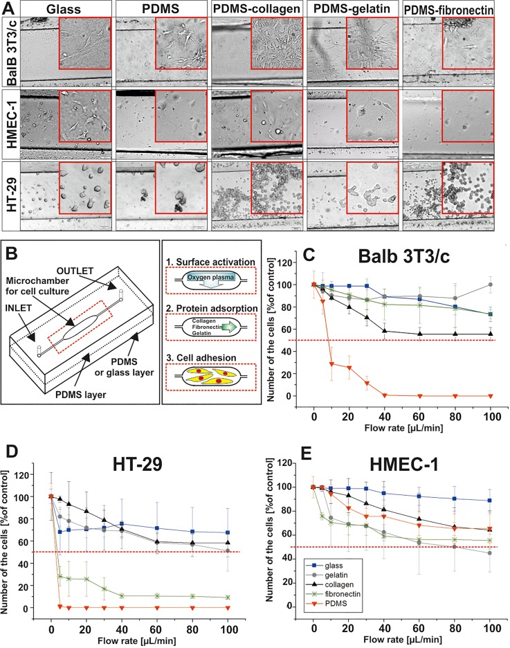
FIG. 6.
(a) Balb 3T3/c, HMEC-1, and HT-29 cell adhesion on the non-modified PDMS surface, glass, and PDMS surfaces modified with gelatin, collagen, and fibronectin. (b) Scheme of the experiments performed in the microsystems. (c) Balb 3T3/c, (d) HMEC-1, and (e) HT-29 cell detachment test performed in the microsystems 48 h after cell seeding. Flow rates in the range of 5–100 μl/min were tested. (n ≥ 3).

