Superficial siderosis of the central nervous system is a rare chronic progressive disease caused by continuous repeated hemorrhage in the subarachnoid space. The ability of the brain to biosynthesize ferritin in response to prolonged contact with hemoglobin iron is important in the pathogenesis of superficial siderosis. This frequently results in cerebellar ataxia and sensorineural deafness. Causes of superficial siderosis include head or back injury, brachial plexus or nerve root injury, dural defect, tumors, vascular disease such as anteriovenous malformation/fistula, and prior intradural surgery.1 Some reports have described superficial siderosis due to spinal dural defect with fluid collection in the spinal canal, and also the surgical closure of dural defects for treatment of superficial siderosis associated with abnormal communication between the subarachnoid space and epidural fluid collection. Thus, it is important to detect dural defects by imaging examination.
A 58-year-old man was admitted to our hospital for further examination of an unsteady gait. Screening brain MRI had revealed superficial siderosis (Fig. 1A). Audiometry revealed bilateral sensorineural hearing impairment that the patient was not aware of in daily life. Lumbar puncture indicated an opening pressure of 130 mm H2O. Cerebral spinal fluid (CSF) was clear and colorless, and CSF examination revealed elevated protein (72 mg/dl) but no other abnormalities.
Fig. 1.
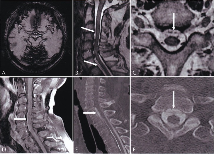
(A) Axial gradient echo image (TR / TE, 660.00 ms / 20.00 ms) shows superficial siderosis. (B) Sagittal T2-weighted spinal image (TR / TE, 3320.00 ms / 124.70 ms) shows epidural fluid collection from C3 to Th10. (C) Axial 3D T2-weighted images obtained with Cube (Cube T2, 1.4 mm, TR / TE, 1820 ms / 92.87 ms) of the spine demonstrates a dural defect (arrow) at the ventral dural sac at the Th1 level. (D) Sagittal T1-weighted images with fat suppression after gadolinium administration (T1-weighted images with fat suppression after gadolinium administration [T1WI-Gd], TR / TE, 540.00 ms / 7.96 ms) show dilation of the anterior spinal vein. (E–F) CT myelography confirms cerebral spinal fluid (CSF) leak around the dural defect.
The patient underwent brain and spinal MR imaging using a 3 Tesla unit (MRI Signa HDxt 3.0T; General Electric medical Systems, USA), which showed no tumor or vascular disease. Axial T2-weighted spinal imaging showed epidural fluid collection from C3 to Th10 (Fig. 1B), and axial Cube T2 of the spine revealed a dural defect at the ventral dural sac at the Th1 level (Fig. 1C). Sagittal T1-weighted images with fat suppression after gadolinium administration (T1WI-Gd) showed dilation of the anterior spinal vein (Fig. 1D). CT myelography was performed and confirmed a CSF leak around the dural defect (Fig. 1E and 1F). The patient was diagnosed with superficial siderosis due to spinal dural defect. He decided against surgery and was kept under observation.
In cases of superficial siderosis with dural defect, it is often difficult to detect the location of the dural defect. High-resolution constructive interference in steady-state (CISS) MR images were reported to be useful in detecting the dural defect preoperatively.2 We also successfully identified dural defect sites in a superficial siderosis case by Cube T2 and CT myelography. Cube is a relatively new 3D fast spin-echo pulse sequence with parallel imaging and extended echo train acquisition that can be reformatted into any plane to visualize even small and low-contrast lesions without a partial-volume effect. Imaging of the cervical spine using the 3D T2-weighted technique reportedly conferred superior delineation of anatomical structure compared to 2D T2-weighted sequences.3 Although CT myelography is needed to confirm the site of dural defect in preoperative or follow-up cases, Cube T2 is very useful in detecting the suspected site of dural defect compared with other 2D sequences prior to CT myelography.
Footnotes
Conflicts of Interest
The authors declare that they have no conflict of interest in this manuscript.
References
- 1.Kumar N. Neuroimaging in superficial siderosis: an in-depth look. AJNR Am J Neuroradiol 2010; 31:5–14. [DOI] [PMC free article] [PubMed] [Google Scholar]
- 2.Egawa S, Yoshii T, Sakaki K, et al. Dural closure for the treatment of superficial siderosis. J Neurosurg Spine 2013; 18:388–393. [DOI] [PubMed] [Google Scholar]
- 3.Meindl T, Wirth S, Weckbach S, Dietrich O, Reiser M, Schoenberg SO. Magnetic resonance imaging of the cervical spine: comparison of 2D T2-weighted turbo spin echo, 2D T2* weighted gradient-recalled echo and 3D T2-weighted variable flip-angle turbo spine echo sequences. Eur Radiol 2009; 19:713–721. [DOI] [PubMed] [Google Scholar]


