Abstract
Background
The effect of smoking on microbial dysbiosis and the potential consequence of such shift on markers of HIV disease is unknown. Here we assessed the relationship of microbial dysbiosis with smoking and markers of HIV disease.
Methods
Oral wash was collected from: (1) HIV-infected smokers (HIV-SM, n = 48), (2) HIV-infected non-smokers (HIV-NS, n = 24), or (3) HIV-uninfected smokers (UI-SM, n = 24). Microbial DNA was extracted and their bacterial and fungal microbiota (bacteriome and mycobiome, respectively) were characterized using Ion-Torrent sequencing platform. Sequencing data were compared using clustering, diversity, abundance and inter-kingdom correlations analyses.
Results
Bacteriome was more widely dispersed than mycobiome, there was no noticeable difference in clustering between groups. Richness of oral bacteriome in HIV-SM was significantly lower than that of UI-SM (P ≤ .03). Diversity of HIV-NS was significantly lower than that of HIV-SM or UI-SM at phylum level (P ≤ .02). Abundance of Phylum Firmicutes was significantly decreased in HIV-NS compared to HIV-SM and UI-SM (P = .007 and .027, respectively), while abundance of Proteobacteria was significantly increased in HIV-NS compared to HIV-SM and UI-SM (P = .0005 and .011, respectively). Fungal phyla did not differ significantly between the three cohorts. Cumulative smoking was positively correlated with Facklamia but negatively with Enhydrobacter, and current alcohol use was negatively correlated with Geniculata. Bacteria Facklamia exhibited weakly positive correlation with longer PI duration (r = 0.094, P = 0.012), and a negative correlation with nadir CD4 count (r = -0.345; P = 0.004), while Granulicatella was negatively correlated with nadir CD4 count (r = -0.329; P = 0.007). Fungus Stemphylium correlated negatively with nadir CD4 (r = -0.323; P = 0.008).
Conclusions
Dysbiosis of the oral microbiota is associated with clinical and immunologic variables in HIV-infected patients.
Introduction
HIV infection has been associated with dysbiosis of oral microbiome, with increased levels of pathogenic bacteria and fungi [1, 2], but the effect of smoking on microbiome shift and the consequence of such shift on HIV disease markers is unknown. Bacterial microbiome of healthy individuals has been characterized [3, 4], and our group was the first to characterize the oral mycobiome of healthy individuals [5]. Previous studies have suggested that nonculturable bacteria may be associated with oral diseases [6, 7], obesity [8], gastrointestinal cancers [9], and CVD [10, 11].
The effect of smoking on the composition and pro-inflammatory characteristics of the oral flora in the healthy population has been recently investigated [12–15], with studies suggesting that smoking favors colonization of pathogens like Fusobacterium and Porphyromonas in oral biofilms, and a concomitant pro-inflammatory response. In the general population, only two smoking cessation studies (n = 11 and 15) are available, showing a decrease in the prevalence of some pathogenic bacteria like Porphyromonas, and an increase in Veillonella, the latter being favorable for periodontal health as it inhibits colonization by pathogenic pathogens [12, 13]. No alteration in the microbiome was found following periodontal therapy in those who continued to smoke [12], suggesting the changes observed were the result of smoking cessation. These studies did not assess fungal species, nor the effect of the change in oral microbiome on markers of HIV infection. Here we present the first comprehensive study that assessed the effect of smoking on oral bacterial and fungal microbiome in HIV-infected subjects, and the relationship between oral microbiome and markers of HIV infection (higher viral load, nadir and current CD4 level) and cardiovascular risk.
Results
Sample demographics
Oral wash samples were collected from HIV-infected (n = 72) and uninfected individuals (n = 24). The HIV-positive group included 48 smokers and 24 non-smokers, while all 24 uninfected individuals were active smokers (Table 1).
Table 1. Study demographics*.
| Variable | HIV-infected Non-smoker (HIV-NS) |
HIV-infected Smoker (HIV-SM) |
Uninfected Smoker (UI-SM) |
P¶ |
|---|---|---|---|---|
| N | 24 | 48 | 24 | |
| Age (years, mean ± SD) | 44.99 ± 11.98 | 46.44 ± 10.56 | 42.74 ± 13.77 | .458 |
| BMI | 28.54 ± 4.03 | 25.96 ± 5.49 | 28.29 ± 4.92 | .010§ |
| CD4, Current | 808.83 ± 382.58 | 838.87 ± 344.85 | - | .564 |
| CD4, Nadir | 240.3 ± 206.76 | 189.46 ± 189.65 | - | .202 |
| Pack Years | - | 24.68 ± 17.1 | 21.58 ± 14.52 | .000† |
| ARV Duration (Months) | 135.62 ± 77.83 | 118.2 ± 75.26 | - | .444 |
| Gender (N, %) | .075‡ | |||
| Female | 3 (12.5%) | 8 (16.6%) | 9 (37.5%) | |
| Male | 21 (87.5%) | 40 (83.3%) | 15 (62.5%) | |
| Race (N, %) | .704‡ | |||
| African American | 15 (62.5%) | 35 (72.9%) | 17 (70.8%) | |
| Caucasian | 8 (33.3%) | 10 (20.8%) | 7 (29.2%) | |
| Hispanic | 1 (4.1%) | 2 (4.1%) | 0 (0%) | |
| Other | 0 (0%) | 1 (2.1%) | 0 (0%) | |
| Percent on PI (N, %) | .660‡ | |||
| No | 13 (54.2%) | 27 (56.2%) | - | |
| Yes | 11 (45.8%) | 21 (43.7%) | - |
*For numerical variables (Age, BMI, Current CD4, Nadir CD4, Pack years, and ARV duration) cell entries are sample mean ± standard deviation.
¶P-values for numerical variables are based on Kruskal-Wallis test across all groups. For pairwise comparisons, P-values are based on Wilcoxon test, adjusted for multiple comparisons (Holm)
§P = .014 for HIV-SM vs. HIV-NS
†P < .001 for HIV-SM vs. HIV-NS or UI-SM. P > .05 for all other pairwise comparisons. For categorical variables (Race, Gender and Percent on PI), percentages in cell entries are within group, with Race categories combined to African American versus non-African American.
‡P-values for categorical variables are based on two-sided Fisher’s exact test.
Clustering analyses
Principal coordinate analysis (PCoA) showed that while the bacteriome data were more widely dispersed than the mycobiome, there was no noticeable difference in clustering between the HIV-infected smokers, HIV-infected non-smokers, and uninfected smokers groups (Fig 1). Moreover, 99% variation was explained by three variables in bacteriome (Proteobacteria, Firmicutes and Bacteroidetes) and one variable in the mycobiome (Ascomycota), for all three groups (data not shown). We found that in the mycobiome, the uninfected smoker group exhibited significantly different clustering (based on distance to centroid) compared to both HIV-infected groups at Family and Genus levels (panels J,K in S1 Fig, P ≤ .018). At the species level of mycobiome, this difference was detected only between uninfected smokers and HIV-infected smokers (panel L in S1 Fig, P = .037).
Fig 1.
Principal coordinates analysis (PCoA) of (A-F) bacteriome and (G-L) mycobiome data at different taxa in the three groups. Confidence ellipses are shown for each group, at 0.95% confidence.
Diversity and richness analyses
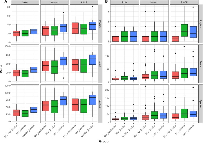
Shannon diversity index of bacteriome did not differ significantly between the smoker and non-smoker groups of HIV-infected patients (data not shown), while richness of oral bacteriome in HIV-infected smokers was significantly lower than that of uninfected smokers (compared using Chao1 and ACE measures, P ≤ .03, Fig 2A). Diversity of mycobiome in HIV-infected non-smokers was significantly lower than that of HIV-infected or uninfected smokers at phylum level (P ≤ .02), but not at genus level. There was no difference in richness of the mycobiome at any taxon level (Fig 2B).
Fig 2.
Boxplots showing richness estimates (observed, chao1 and ACE) of (A) bacteriome and (B) mycobiome at phylum, genus, and species levels.
Core bacteriome and mycobiome
The core bacteriome (comprising taxa detected with a frequency of at least 20%, at an abundance > 1%) comprised eight bacterial phyla, 65 genera, and 86 species common in all three study groups (Fig 3A–3C). Genus Pelomonas was unique to HIV-infected non-smoker and uninfected smoker groups (Fig 3B). Bacterial species Pseudomonas pseudoalcaligenes was detected only in HIV-infected non-smokers, while two Pelomonas species were present in only the uninfected smoker group (Pelomonas puraquae and Pelomonas sp., Fig 3C).
Fig 3.
Venn diagrams showing frequency distribution of (A-C) core bacterial and (D-F) core fungal taxa (detected at abundance > 1%) in the three study groups. Frequency distribution in the core microbiota are shown for (A,D) Phylum, (B,E) Genus and (C,F) Species levels. HIV-SM: HIV-infected smokers, HIV-nSM: HIV-infected non-smokers, nHIV-SM: uninfected smokers.
Analysis of core mycobiome revealed four fungal phyla were common to all three groups, and one (Chytridiomycota) was unique to the HIV-infected and uninfected smoker groups (Fig 3D). At the genus level, 11 fungal genera were common to all cores in the three groups, 7 were unique to uninfected smoker group, while 4 (Cladosporium, Nakaseomyces, Scleroderma, and Rhodotorula) were detected only in the HIV-infected smoker group (Fig 3E). At the species level, the mycobiome showed 17 species to be common to all three groups, two species (Candida glabrata and Scleroderma sp) were unique to HIV-infected smoker group, while three species (Stemphylium solani, Debaryomyces hansenii, and Olpidium brassicae) were detected only in the uninfected smoker group (Fig 3F).
Abundance profiles
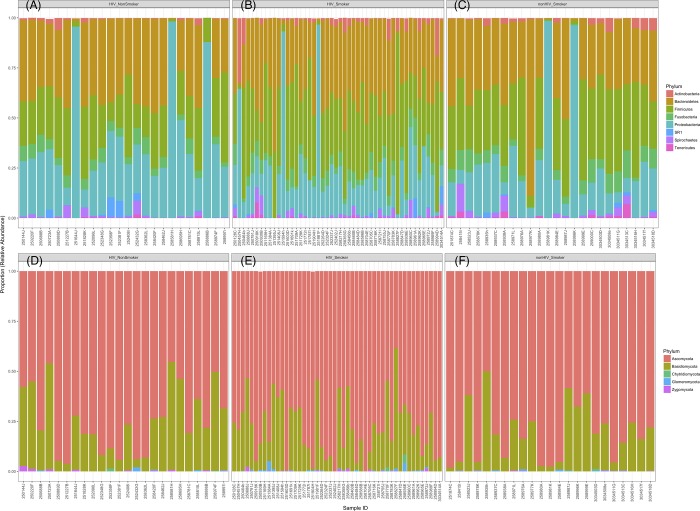
Most abundant phyla in HIV-infected smokers were Firmicutes > Bacteroidetes > Proteobacteria, while in HIV-infected non-smokers were Proteobacteria > Bacteroidetes > Firmicutes (Fig 4). The distribution of phyla in the non-HIV infected smokers were Bacteroidetes > Firmicutes > Proteobacteria. In the mycobiome, Ascomycota followed by Basidiomycota were the most abundant phyla for all three groups (Fig 4). Abundance of Firmicutes was significantly decreased in HIV-infected nonsmokers compared to HIV-infected smokers and uninfected smokers (P = .007 and .027, respectively, Fig 5A). In contrast, abundance of Proteobacteria was significantly increased in HIV-infected nonsmokers compared to HIV-infected smokers and uninfected smokers (P = .0005 and .011, respectively, Fig 5A). Fungal phyla did not differ significantly between the three cohort groups (Fig 5A). Comparisons of bacterial genera showed that levels of Granulicatella, Lactobacillus, Veillonella, and Enhydrobacter were significantly increased, while that of Neisseria were significantly decreased, in HIV-infected smokers compared to infected non-smokers (Table 2). Levels of four fungal genera were significantly different between the three cohorts in the study (Table 3), including an unidentified Myxotrichaceae, Stemphylium, Candida, and Chroogomphus. Interestingly, levels of Candida were increased in both HIV-infected and uninfected smoker groups (34.7% and 38.4%, respectively) compared to the HIV-infected non-smoker group (19.2%, P = 0.12 and 0.04, respectively). Our results also revealed a non-significant increase in levels of C. dubliniensis in the HIV-infected smokers (7.3%) compared to HIV-infected non-smokers (1.8%, P = 0.1; Table 4). In addition, C. rugosa was detected only in the uninfected smoker group (0.2%). We also found that ratio of the abundance of Fusobacteria:Proteobacteria (F:P) was significantly decreased in HIV-infected non-smokers compared to both smoker groups (P ≤ .006), while the abundance ratio of Bacteriodetes:Proteobacteria (B:P) was significantly decreased in HIV-infected non-smokers compared to HIV-infected smoker group (P = .028, Fig 5B).
Fig 4.
Stacked bar charts showing distribution of (A-C) bacterial and (D-F) fungal phyla across the tested samples in the three groups. Phyla present at an abundance of at least 1% relative to the total abundance in each sample were included in the analyses.
Fig 5. Abundance profile of bacterial and fungal phyla in study groups.
(A) Boxplots showing relative abundance of bacterial and fungal phyla, (B) abundance ratio of Fusobacteria:Proteobacteria (ratio F:P) and Bacteriodetes:Proteobacteria (ratio B:P).
Table 2. Bacterial genera with significantly different abundance between the three groups.
| Bacterial Genus | HIV-infected Non-Smoker (A) |
HIV-infected Smoker (B) |
Uninfected Smoker (C) |
P (A vs B) |
P (A vs C) |
P (B vs C) |
|---|---|---|---|---|---|---|
| Prevotella | 22.1% | 23.8% | 19.1% | 1.000 | 0.255 | 0.175 |
| Streptococcus | 16.2% | 20.6% | 19.6% | 0.430 | 0.551 | 1.000 |
| Haemophilus | 10.0% | 8.1% | 7.9% | 0.855 | 1.000 | 1.000 |
| Neisseria | 7.0% | 2.8% | 2.8% | 0.012 | 0.012 | 1.000 |
| Granulicatella | 1.5% | 2.6% | 2.4% | 0.033 | 0.186 | 1.000 |
| Lactobacillus | 0.4% | 2.7% | 2.7% | 0.009 | 0.019 | 1.000 |
| Parvimonas | 0.3% | 0.3% | 0.7% | 1.000 | 0.068 | 0.014 |
| Veillonella | 0.2% | 0.7% | 0.3% | 0.010 | 0.823 | 0.140 |
| Enhydrobacter | 0.1% | 0.1% | 5.0% | 0.003 | 1.000 | 0.050 |
| Pelomonas | 0.1% | 0.2% | 0.0% | 0.445 | 1.000 | 0.036 |
| Stenotrophomonas | 0.0% | 1.6% | 0.0% | 0.290 | 1.000 | 0.048 |
| Facklamia | 0.0% | 0.0% | 0.1% | 0.205 | 0.002 | 0.155 |
Table 3. Fungal genera with significantly different abundance between the three groups.
| Fungal Genus | HIV-infected Non-Smoker (A) |
HIV-infected Smoker (B) |
Uninfected Smoker (C) |
P (A vs B) |
P (A vs C) |
P (B vs C) |
|---|---|---|---|---|---|---|
| Unidentified Myxotrichaceae | 47.2% | 33.1% | 25.6% | 0.022 | 0.005 | 0.281 |
| Candida | 19.2% | 34.7% | 38.4% | 0.118 | 0.044 | 1.000 |
| Chroogomphus | 18.2% | 12.9% | 9.6% | 0.361 | 0.024 | 0.318 |
| Saccharomyces | 1.7% | 1.1% | 3.7% | 1.000 | 1.000 | 1.000 |
| Stemphylium | 0.4% | 0.0% | 0.1% | 1.000 | 0.782 | 0.040 |
Table 4. Abundance of Candida species in the tested samples.
| Candida species | HIV-infected Non-Smoker (A) |
HIV-infected Smoker (B) |
Uninfected Smoker (C) |
P-value (A vs. B) |
P-value (A vs. C) |
P-value (B vs. C) |
|---|---|---|---|---|---|---|
| Candida albicans | 6.8% | 11.8% | 14.5% | 1.000 | 0.310 | 0.896 |
| Candida dubliniensis | 1.8% | 7.3% | 5.1% | 0.105 | 0.346 | 1.000 |
| Candida ethanolica | 0.0% | 0.0% | 0.1% | NA | 0.655 | 0.314 |
| Candida orthopsilosis | 0.1% | 0.2% | 0.3% | 1.000 | 0.144 | 0.483 |
| Candida oxycetoniae | 0.0% | 0.1% | 0.0% | 1.000 | 1.000 | 1.000 |
| Candida parapsilosis | 0.2% | 0.1% | 0.4% | 1.000 | 0.557 | 0.375 |
| Candida rugosa | 0.0% | 0.0% | 0.2% | NA | 0.306 | 0.083 |
| Candida sp. | 8.5% | 11.2% | 16.1% | 1.000 | 0.170 | 0.259 |
| Candida sp 3 2 | 0.0% | 0.0% | 0.1% | NA | 0.655 | 0.314 |
| Candida sp HN 26 | 0.0% | 0.1% | 0.0% | 1.000 | 1.000 | 1.000 |
| Candida sp L2683B | 0.0% | 0.0% | 0.1% | NA | 0.655 | 0.314 |
| Candida tropicalis | 1.5% | 3.7% | 1.4% | 1.000 | 1.000 | 1.000 |
| Candida versatilis | 0.0% | 0.0% | 0.0% | 0.996 | NA | 1.000 |
| Candida lusitaniae | 0.0% | 0.1% | 0.0% | 0.157 | 0.110 | 1.000 |
Intra- and inter-kingdom correlations
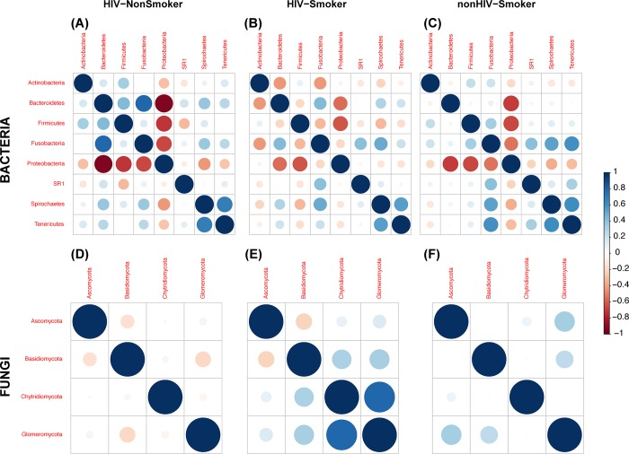
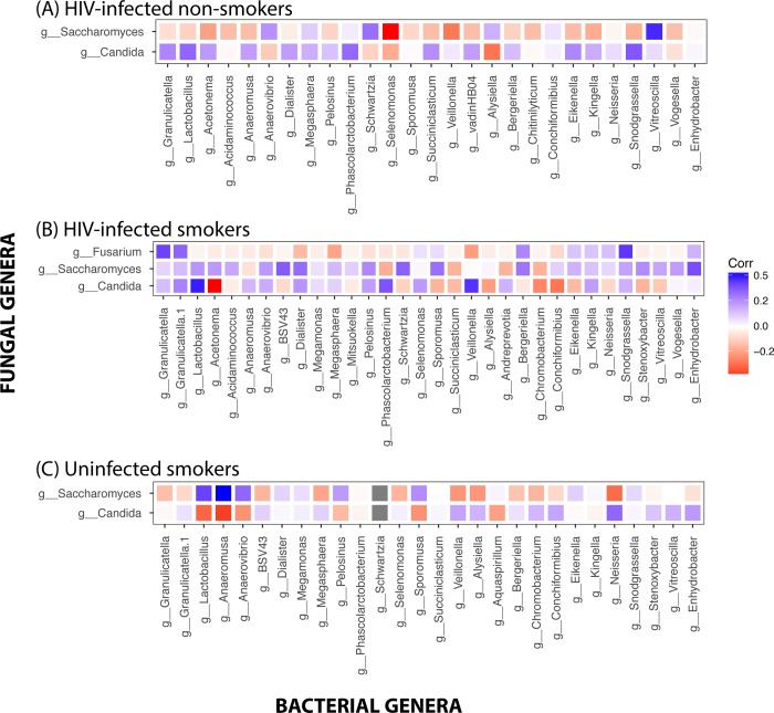
Next, we determined the significant intra- and inter-kingdom correlations in the bacteriome and mycobiome at phylum and genus levels. Correlations of Actinobacteria with other phyla differed between the three cohort groups, where negative correlations were detected with Bacteroidetes and Fusobacteria in HIV-infected smoker group, while positive correlations were noted in the other two groups (Fig 6A–6C). In the mycobiome, correlations of Ascomycota and Basidiomycota with the bacterial phyla exhibited distinct patterns in the three study groups (Fig 6D–6F). Analyses of inter-kingdom interactions revealed that fungal Phylum Glomeromycota exhibited significant positive correlation with bacterial Phylum Tenericutes (r = 0.88, P < .001) in HIV-infected non-smokers. We also explored the inter-kingdom interactions of fungal and bacterial genera that were significantly different between the groups. Abundance of Candida was correlated negatively with Neisseria in HIV-infected smokers, positively in infected non-smokers, and uninfected smokers (Fig 7). Interestingly, Candida was positively correlated with Lactobacillus in HIV-infected smokers and non-smokers, but negatively correlated in uninfected smokers. In contrast, Saccharomyces was negatively correlated with Neisseria in uninfected smokers, but positively correlated in both HIV-infected (smoker and non-smoker) groups. However, these inter-kingdom correlations of Candida and Saccharomyces did not reach statistical significance.
Fig 6.
Intra-kingdom correlations within the bacteriome and mycobiome for (A,D) HIV-infected non-smokers, (B,E) HIV-infected smokers, and (C,F) uninfected smokers. Spearman’s correlation for each comparison was determined for the three groups. Blue circles indicate positive correlations; red circles indicate negative correlation; diameter of circles represent the absolute value of correlation for each pair of the microbe-microbe matrix.
Fig 7.
Inter-kingdom correlations between the bacteriome and mycobiome for (A) HIV-infected non-smokers, (B) HIV-infected smokers, and (C) uninfected smokers. Spearman’s correlation for each comparison was determined for the three groups. Blue tiles indicate positive correlation; red tiles indicate negative correlations; tile sizes represent the absolute value of correlation for each pair of the microbe-microbe matrix.
Correlations between microbial genera and clinical variables
Next, we determined the correlations between microbial (bacterial and fungal) genera and clinical variables. Facklamia correlated positively with age (r = 0.209, P = 0.048) and cumulative smoking (in pack-years), (r = 0.303, P = 0.004), while Enhydrobacter exhibited negative correlation with pack-years (r = -0.241, P = 0.024). Geniculata was negatively correlated with current alcohol use (r = -.267, P = 0.046). No significant correlations were observed between any bacteria and BMI. Several bacteria correlated with HIV-immunologic parameters markers and HIV treatment factors; Facklamia also exhibited weakly positive correlation with longer PI duration (r = 0.094, P = 0.012), and a negative correlation with nadir CD4 count (r = -0.345; P = 0.004). Granulicatella also was negatively correlated with nadir CD4 count (r = -0.329; P = 0.007). No bacteria correlated with current CD4 counts, or ART or NRTI duration. Several fungi correlated with markers of HIV-clinical and immunologic parameters, with Stemphylium correlated negatively with nadir CD4 (r = -0.323; P = 0.008). No fungi correlated significantly with current alcohol use, cumulative smoking, age, BMI, current CD4 cell count, or ART or HIV duration.
Discussion
To our knowledge, our study is the first to comprehensively characterize differences in the oral bacteriome and mycobiome concomitantly in HIV-infected smokers and non-smokers. We found that while the within-group variation in microbiota was similar between the three groups, fungal diversity of the mycobiome was significantly increased in HIV-infected smokers compared to non-smokers, suggesting that smoking induces changes in the oral mycobiome. Previous investigations have also shown that oral bacteriome varies between smokers and non-smokers. One such study conducted by the Lung HIV Microbiome Project (LHMP), Morris et al [16] focused on healthy individuals, and compared the bacterial microbiome of oral washes and bronchoscopic alveolar lavages (BAL) (representing the upper and lower respiratory tract, respectively) in healthy nonsmokers and smokers (n = 64). These investigators reported that majority of the bacteria were common between the mouth and the lung and that levels of Porphyromonas, Neisseria, and Gemella in the oral communities were significantly decreased in smokers compared to nonsmokers, which is in agreement with our finding that abundance of Neisseria was significantly decreased in HIV-infected smokers compared to infected non-smokers. A subsequent study by the same group comparing the oral and lung bacteriomes of uninfected and HIV-infected individuals [17] reported that differences in microbial populations were detected only in the oral washes among the subject groups (Streptococcus, Actinomyces, Rothia, and Atopobium), but not in the lower respiratory tract (BAL). Our results are in agreement with these findings, since we also found that abundance of specific bacterial taxa (Granulicatella, Lactobacillus, Veillonella, Enhydrobacter, Streptococcaceae and Comamonadaceae) were significantly increased in HIV-infected smokers compared to infected non-smokers. In addition, abundance of the fungal genus Candida was increased in HIV-infected smokers. While previous studies characterized the oral bacteriome in healthy smokers or HIV-infected patients (compared to healthy non-smokers or uninfected healthy individuals, respectively) our results provide insight about dysbiosis of bacteriome and mycobiome concomitantly in HIV-infected smokers and non-smokers, as well as uninfected smokers. Inter-kingdom correlations between two known pathogenic genera (Candida and Neisseria) were negative in HIV-infected smokers but positive in infected non-smokers. While this correlation did not reach statistical significance, it raises the possibility that the two genera may interact and influence microbial dysbiosis in the oral cavity.
Cumulative smoking was positively correlated with Facklamia and negatively correlated with Enhydrobacter, but did not correlate with Fusobacterium and Porphyromonas, as was seen in studies of non-HIV infected populations. Additionally, both bacteria (Facklamia and Granulicatella) and fungi (Chroogomphus, Microbotryum, Stemphylium) correlated with worse HIV immunologic parameters, higher viral load and more advanced disease (i.e. lower nadir CD4). Consumption of alcohol did not correlate positively with any bacteria or fungi, which was unexpected negative finding that is worth investigating further. Our findings are notable for the fact that we identified fungi other than Candida (the most common fungal colonizer in HIV infection), to be correlated with HIV disease markers. The clinical relevance of these correlations remains to be explored.
One potential caveat of the current study is that the results are derived based on mouthwash samples. While oral wash collection is non-invasive and relatively simple to implement, it does not account for the high diversity in local bacterial niches in the oral cavity, including biofilms, gingival, plaque. Despite this limitation, our results provide a global snapshot of the oral microbiome and may reveal novel linkages between the microbiota and HIV infection and its covariates. Samples were collected at a single time point in the current study, therefore, it was not possible to determine whether changes in microbiome were related to progression of disease or response to therapy. In addition, since the intra-kingdom correlations are based on compositional data, these correlations need to be validated in larger cohort studies and targeted functional assays.
Smoking is associated with periodontal disease, which in turn is also related to HIV infection, and could be a confounder in some of our findings. However, none of the patients in the current study had any clinical symptoms of periodontal disease (redness, swelling or bleeding). However, a formal exam performed by a dentist to rule out mild periodontal disease was not part of the clinical protocol.
The clinical relevance of these findings is unknown, and the consequences of such changes, including on cardiovascular disease risk and systemic inflammation, need to be investigated in future studies.
Materials and methods
Study population
The study was approved by the IRB of University Hospital Cleveland Medical Center, and all participants signed a written informed consent before any evaluation was performed. Participants were recruited for this study and by design assigned to three different groups as follows: (1) HIV-infected smokers (HIV-SM), (2) HIV-infected non-smokers (HIV-NS), and (3) HIV-uninfected smokers (nonHIV-SM).
Inclusion criteria
(1) For HIV-infected smokers (n = 48): men and women 18 years of age or older were recruited. They were receiving stable Antiretroviral therapy (ART) for at least 12 weeks prior to study entry, with cumulative prior ART of at least 6 months, with plasma HIV-1 RNA level of ≤ 400 copies/ml, and were smoking on average ≥10 cigarettes/day for ≥6 months and had a history of ≥5 cumulative years of smoking.
(2) For HIV-infected non-smokers (n = 24): criteria were similar as above, except they were non-Smokers, defined as having never smoked or smoked for less than 3 months cumulative during lifetime and not within the last 12 months.
(3) For HIV uninfected smokers (n = 24): HIV-uninfected healthy men and women smokers, 18 years of age or older, with no regular use who currently smoke ≥10 cigarettes/day for ≥6 continuous months and history of ≥5 cumulative years of smoking.
Exclusion criteria
History of myocardial infarction, stroke, or coronary revascularization procedure, pregnancy/ breastfeeding, Any active or chronic uncontrolled inflammatory condition, Current alcohol or recreational drug use which interferes with the subject’s ability to comply with dosing schedule and protocol evaluations, se of the systemic cancer chemotherapy, or immumodulating agents, uncontrolled thyroid disease or diabetes, liver enzymes > 2.5 x upper limit of normal, Hemoglobin < 9.0 g/dL, or calculated creatinine clearance <50 mL/min. None of the patients had any clinical symptoms of periodontal disease (redness, swelling or bleeding) however we did not have formal exam performed by a dentist to rule out mild periodontal disease.
Collection of oral wash samples
Oral wash samples were collected from the above three groups as describe earlier[18]. Briefly 20–25 ml saline was emptied into separately labelled blue capped 50-mL Falcon™ centrifuge tubes (Fisher Scientifics Co.). Each subject swished and gargled the saline from the tube in mouth for 2 minutes and expectorated the rinse into the tube. The tubes were closed tightly and stored in -80 till further use. Before use, all the tubes with oral wash samples were completely thawed on ice. Tubes were then centrifuged at 4000 rpm for 15 minutes and 20–25 ml supernatant transferred into fresh 50-mL centrifuge tubes. The pellet left in each tube was used to extract DNA for microbiome studies.
DNA extraction
Fungal and bacterial genomic DNA was isolated and purified with the QiaAmp DNA Mini Kit (Qiagen) following the manufacturer’s instructions with minor modifications. Briefly, 3 additional bead-beating steps (Sigma-Aldrich beads, diameter = 500 μm) with the MP Fastprep-24 (speed setting of 6, 3 runs of 60s) were performed. The quality and purity of the isolated genomic DNA was confirmed spectrophotometrically using NanoDrop 2000 device (Fisher Scientific SAS, Illkirch, France). DNA concentration was quantified using the Qubit 2.0 instrument applying the Qubit dsDNA HS Assay (Life Technologies, USA). Extracted DNA samples were stored at -20°C.
Microbiome analyses
Analysis of the microbiome profile in the extracted DNA samples was conducted as described previously by our group [1, 18]. A brief summary of the method is provided below.
Amplicon library preparation
The Internal Transcribed Spacer 1 (ITS1) and 16S rDNA regions for fungi and bacteria, respectively, were amplified as described previously [1]. Briefly, ITS1 region was amplified using ITS1F (CTTGGTCATTTAGAGGAAGTAA) and ITS 2 (GCTGCGTTCTTCATCGATGC) primers, and the V4 region of the 16S rRNA gene was amplified using16S-515F: GTGCCAGCMGCCGCGGTAA and 16s-806R: GGACTACHVGGGTWTCTAAT primers. PCR reactions were carried out on 100 ng template DNA, in 50 μl (final volume) reaction mixture consisting of Dream Taq Green PCR Master Mix (ThermoScientific), 0.1g/L bovine serum albumin, 1% of dimethyl sulfoxide (DMSO), 6 mM MgCl2, and a final primer concentration of 400nM. PCR conditions were: initial denaturation at 94°C for 3 min, 35 cycles of denaturation for 30s each at 94°C, annealing at 50°C for 30 s, extension at 72°C for 1 min, final extension of 5 min at 72°C. PCR products were sheared for 20 min, using Ion Shear Plus Fragment Library Kit (Life Technologies, NY, USA). Amplicon library was generated with sheared PCR products using Ion plus Fragment Library kits (<350 bp) according to the manufacturer’s instructions, and the library was barcoded with Ion Xpress™ Barcode Adapter, and ligated with the A and P1 adaptors.
Sequencing, classification and analysis
The adapted barcoded libraries were equalized using the Ion Library Equalizer kit to a final concentration of 100 pM, pooled and diluted to 26 pM, and attached to the surface of Ion Sphere particles (ISPs) using an Ion PGM Template OT2 200bp kit v2 (Life Technologies, USA) according to the manufacturer's instructions, via emulsion PCR. Quality of ISPs templates was checked using Ion Sphere™ Quality Control Kit (Part no. 4468656) with the Qubit 2.0 device. Sequencing of the pooled libraries was carried out on the Ion Torrent Personal Genome Machine (PGM) system using the Ion Sequencing 200 kit v2 (all from Life Technologies) for 150 cycles (600 flows), with a 318 chip following the manufacturer's instructions. De-multiplexing and classification was performed using the Qiime 1.6 platform. The resulting sequence data were trimmed to remove adapters, barcodes and primers during the de-multiplexing process. In addition, the bioinformatics process filters were applied to the sequence data for the removal of low-quality reads below Q25 Phred score and denoised to exclude sequences with read length below 100 bp[19]. De novo operational taxonomic units (OTUs) were clustered using Uclust algorithm and defined by 97% sequence similarity[20]. Classification at the species level was referenced using the UNITE 5.8s database and taxa assigned using the nBlast method with a 90% confidence cut-off [21, 22].
Bioinformatics and statistical analyses
The statistical programming language R and related packages [23] were used for diversity and correlation analyses, and Kruskal-Wallis (non-parametric) analysis of variance using abundance data. Principal coordinates analysis (PCoA) was conducted with identified reads/OTUs using classical multidimensional scaling (functions dist/euclidean, cmdscale) to analyze distribution of dissimilarities and hierarchical clustering in dendrograms in the R package vegan. Statistical significance in differences between clustering groups were determined using adonis/permanova and betadisper functions followed by Tukey's HSD test for pairwise comparisons. Diversity was analyzed using Shannon diversity index (characterizes species diversity) and abundance-based richness estimates (observed, bias-corrected Chao and ACE) were calculated using vegan [24]. Taxa with a frequency of at least 20% (and relative abundance >1%) were considered to comprise the core biome in our study. Intra- and inter-kingdom correlations were determined using the corrplot package in R. For hypothesis tests, P < .05 was considered statistically significant, except for pairwise comparisons among groups, where a Bonferroni correction was applied, and P < .0167 was considered the significance threshold. All group-wise comparisons were conducted with SPSS (ver. 24). Sequencing data files are available at https://drive.google.com/drive/folders/1aPWhf6DDB6FGaCd8d1lCtXfLCj1g4LEs?usp=sharing.
Supporting information
Clustering differences, based on distance to centroid, in samples from the three groups at different taxa in bacteria (A-F) and fungi (G-L).
(TIF)
Acknowledgments
The authors wish to thank the study participants for their providing the samples in the study.
Data Availability
All relevant data are within the paper and its Supporting Information files.
Funding Statement
We gratefully acknowledge funding support from the National Institutes of Health grant R56HL126526 (to GM), R01DE024228 (to MAG and PKM), U01 AI105937-01, the Center for AIDS Research (CFAR, P30 AI036219), and Steris Foundation grant (to PKM). The funders had no role in study design, data collection and analysis, decision to publish, or preparation of the manuscript.
References
- 1.Mukherjee PK, Chandra J, Retuerto M, Sikaroodi M, Brown RE, Jurevic R, et al. Oral mycobiome analysis of HIV-infected patients: identification of Pichia as an antagonist of opportunistic fungi. PLoS Pathog. 2014;10(3):e1003996 Epub 2014/03/15. doi: 10.1371/journal.ppat.1003996 ; PubMed Central PMCID: PMC3953492. [DOI] [PMC free article] [PubMed] [Google Scholar]
- 2.Aas JA, Barbuto SM, Alpagot T, Olsen I, Dewhirst FE, Paster BJ. Subgingival plaque microbiota in HIV positive patients. J Clin Periodontol. 2007;34(3):189–95. Epub 2007/02/21. doi: CPE1034 [pii] doi: 10.1111/j.1600-051X.2006.01034.x . [DOI] [PubMed] [Google Scholar]
- 3.Aas JA, Paster BJ, Stokes LN, Olsen I, Dewhirst FE. Defining the normal bacterial flora of the oral cavity. J Clin Microbiol. 2005;43(11):5721–32. Epub 2005/11/08. doi: 43/11/5721 [pii] doi: 10.1128/JCM.43.11.5721-5732.2005 ; PubMed Central PMCID: PMC1287824. [DOI] [PMC free article] [PubMed] [Google Scholar]
- 4.Nasidze I, Li J, Quinque D, Tang K, Stoneking M. Global diversity in the human salivary microbiome. Genome Res. 2009;19(4):636–43. Epub 2009/03/03. doi: gr.084616.108 [pii] doi: 10.1101/gr.084616.108 ; PubMed Central PMCID: PMC2665782. [DOI] [PMC free article] [PubMed] [Google Scholar]
- 5.Ghannoum MA, Jurevic RJ, Mukherjee PK, Cui F, Sikaroodi M, Naqvi A, et al. Characterization of the oral fungal microbiome (mycobiome) in healthy individuals. PLoS Pathog. 2010;6(1):e1000713 Epub 2010/01/15. doi: 10.1371/journal.ppat.1000713 ; PubMed Central PMCID: PMC2795202. [DOI] [PMC free article] [PubMed] [Google Scholar]
- 6.Paster BJ, Olsen I, Aas JA, Dewhirst FE. The breadth of bacterial diversity in the human periodontal pocket and other oral sites. Periodontol 2000. 2006;42:80–7. Epub 2006/08/26. doi: PRD174 [pii] doi: 10.1111/j.1600-0757.2006.00174.x . [DOI] [PubMed] [Google Scholar]
- 7.Paster BJ, Boches SK, Galvin JL, Ericson RE, Lau CN, Levanos VA, et al. Bacterial diversity in human subgingival plaque. J Bacteriol. 2001;183(12):3770–83. Epub 2001/05/24. doi: 10.1128/JB.183.12.3770-3783.2001 ; PubMed Central PMCID: PMC95255. [DOI] [PMC free article] [PubMed] [Google Scholar]
- 8.Zeigler CC, Persson GR, Wondimu B, Marcus C, Sobko T, Modeer T. Microbiota in the oral subgingival biofilm is associated with obesity in adolescence. Obesity (Silver Spring). 2012;20(1):157–64. Epub 2011/10/15. doi: oby2011305 [pii] doi: 10.1038/oby.2011.305 . [DOI] [PubMed] [Google Scholar]
- 9.Ahn J, Chen CY, Hayes RB. Oral microbiome and oral and gastrointestinal cancer risk. Cancer Causes Control. 2012;23(3):399–404. Epub 2012/01/25. doi: 10.1007/s10552-011-9892-7 . [DOI] [PMC free article] [PubMed] [Google Scholar]
- 10.Ordovas JM, Mooser V. Metagenomics: the role of the microbiome in cardiovascular diseases. Curr Opin Lipidol. 2006;17(2):157–61. Epub 2006/03/15. doi: 10.1097/01.mol.0000217897.75068.ba 00041433-200604000-00009 [pii]. . [DOI] [PubMed] [Google Scholar]
- 11.Koren O, Spor A, Felin J, Fak F, Stombaugh J, Tremaroli V, et al. Human oral, gut, and plaque microbiota in patients with atherosclerosis. Proc Natl Acad Sci U S A. 2011;108 Suppl 1:4592–8. Epub 2010/10/13. doi: 1011383107 [pii] doi: 10.1073/pnas.1011383107 ; PubMed Central PMCID: PMC3063583. [DOI] [PMC free article] [PubMed] [Google Scholar]
- 12.Delima SL, McBride RK, Preshaw PM, Heasman PA, Kumar PS. Response of subgingival bacteria to smoking cessation. J Clin Microbiol. 2010;48(7):2344–9. Epub 2010/04/23. doi: JCM.01821-09 [pii] doi: 10.1128/JCM.01821-09 ; PubMed Central PMCID: PMC2897479. [DOI] [PMC free article] [PubMed] [Google Scholar]
- 13.Fullmer SC, Preshaw PM, Heasman PA, Kumar PS. Smoking cessation alters subgingival microbial recolonization. J Dent Res. 2009;88(6):524–8. Epub 2009/07/10. doi: 0022034509338676 [pii] doi: 10.1177/0022034509338676 . [DOI] [PubMed] [Google Scholar]
- 14.Shchipkova AY, Nagaraja HN, Kumar PS. Subgingival microbial profiles of smokers with periodontitis. J Dent Res. 2010;89(11):1247–53. Epub 2010/08/27. doi: 0022034510377203 [pii] doi: 10.1177/0022034510377203 ; PubMed Central PMCID: PMC3318026. [DOI] [PMC free article] [PubMed] [Google Scholar]
- 15.Kumar PS, Matthews CR, Joshi V, de Jager M, Aspiras M. Tobacco smoking affects bacterial acquisition and colonization in oral biofilms. Infect Immun. 2011;79(11):4730–8. Epub 2011/08/24. doi: IAI.05371-11 [pii] doi: 10.1128/IAI.05371-11 ; PubMed Central PMCID: PMC3257914. [DOI] [PMC free article] [PubMed] [Google Scholar]
- 16.Morris A, Beck JM, Schloss PD, Campbell TB, Crothers K, Curtis JL, et al. Comparison of the respiratory microbiome in healthy nonsmokers and smokers. Am J Respir Crit Care Med. 2013;187(10):1067–75. doi: 10.1164/rccm.201210-1913OC ; PubMed Central PMCID: PMCPMC3734620. [DOI] [PMC free article] [PubMed] [Google Scholar]
- 17.Beck JM, Schloss PD, Venkataraman A, Twigg H 3rd, Jablonski KA, Bushman, et al. Multicenter Comparison of Lung and Oral Microbiomes of HIV-infected and HIV-uninfected Individuals. Am J Respir Crit Care Med. 2015;192(11):1335–44. doi: 10.1164/rccm.201501-0128OC ; PubMed Central PMCID: PMCPMC4731698. [DOI] [PMC free article] [PubMed] [Google Scholar]
- 18.Chandra J, Retuerto M, Mukherjee PK, Ghannoum M. The Fungal Biome of the Oral Cavity. Methods Mol Biol. 2016;1356:107–35. doi: 10.1007/978-1-4939-3052-4_9 . [DOI] [PubMed] [Google Scholar]
- 19.Caporaso JG, Kuczynski J, Stombaugh J, Bittinger K, Bushman FD, Costello EK, et al. QIIME allows analysis of high-throughput community sequencing data. Nat Methods. 2010;7(5):335–6. Epub 2010/04/13. doi: 10.1038/nmeth.f.303 ; PubMed Central PMCID: PMC3156573. [DOI] [PMC free article] [PubMed] [Google Scholar]
- 20.Edgar RC. Search and clustering orders of magnitude faster than BLAST. Bioinformatics. 2010;26(19):2460–1. Epub 2010/08/17. doi: 10.1093/bioinformatics/btq461 . [DOI] [PubMed] [Google Scholar]
- 21.Koljalg U, Nilsson RH, Abarenkov K, Tedersoo L, Taylor AF, Bahram M, et al. Towards a unified paradigm for sequence-based identification of fungi. Mol Ecol. 2013;22(21):5271–7. Epub 2013/10/12. doi: 10.1111/mec.12481 . [DOI] [PubMed] [Google Scholar]
- 22.Altschul SF, Gish W, Miller W, Myers EW, Lipman DJ. Basic local alignment search tool. J Mol Biol. 1990;215(3):403–10. doi: 10.1016/S0022-2836(05)80360-2 . [DOI] [PubMed] [Google Scholar]
- 23.Morgan XC, Huttenhower C. Chapter 12: Human microbiome analysis. PLoS Comput Biol. 2012;8(12):e1002808 Epub 2013/01/10. doi: 10.1371/journal.pcbi.1002808 ; PubMed Central PMCID: PMC3531975. [DOI] [PMC free article] [PubMed] [Google Scholar]
- 24.Oksanen J, Blanchet FG, Kindt R, Legendre P, Minchin PR, O'Hara RB, et al. vegan: Community Ecology Package. R package version 2.0–10. 2013.
Associated Data
This section collects any data citations, data availability statements, or supplementary materials included in this article.
Supplementary Materials
Clustering differences, based on distance to centroid, in samples from the three groups at different taxa in bacteria (A-F) and fungi (G-L).
(TIF)
Data Availability Statement
All relevant data are within the paper and its Supporting Information files.


