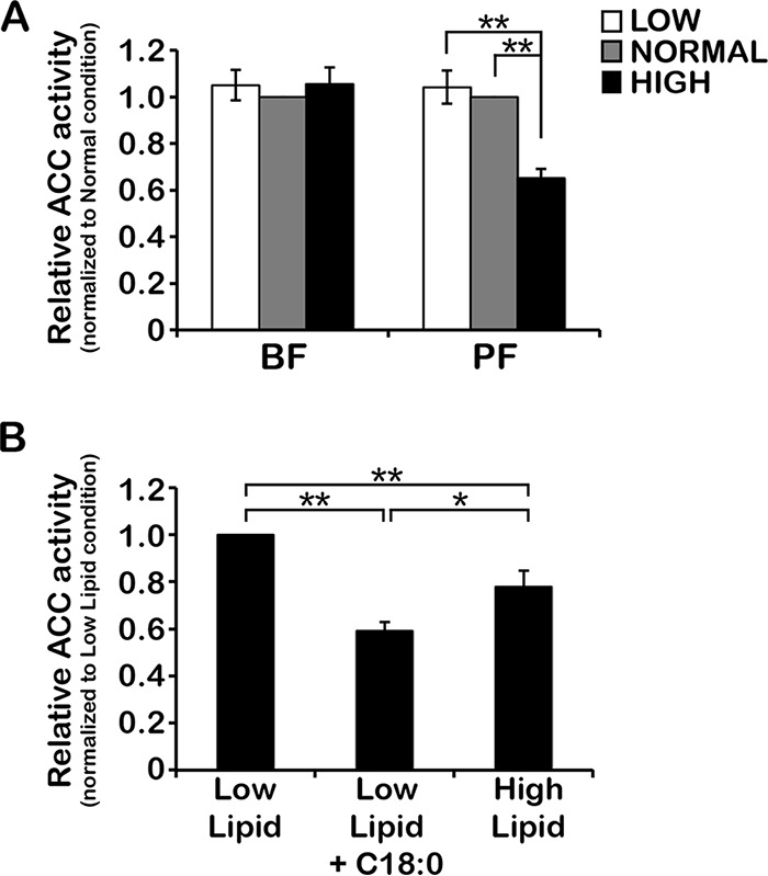
FIG 2 .

Environmental lipids affect TbACC activity in PF but not BF. (A) BF and PF WT cells were grown under low-, normal-, and high-lipid conditions to the mid-log phase (~3 days). Lysates prepared in the presence of phosphatase inhibitor cocktail (equal total levels of protein, 2 to 10 µg) were assayed for TbACC activity by measuring incorporation of [14C]NaHCO3 into the acid-resistant malonyl-CoA product in the presence of ATP and acetyl-CoA. Values were first normalized to the no-ATP negative control before averaging was performed. Average values were then expressed relative to that of normal-lipid media. Means ± SEM of results from three independent experiments performed in pentuplicate are shown. **, P ≤ 0.005 (two-tailed Student’s t test). (B) PF WT cells grown under low-lipid conditions to the mid-log phase (~2 days) were subdivided into three cultures and grown for an additional 24 h, with one culture maintained in low-lipid media (Low Lipid), one supplemented with a final concentration of 20% FBS (High Lipid), and one supplemented with 35 µM stearate fatty acid (Low Lipid + C18:0). Hypotonic lysates were prepared and assayed for TbACC activity as described above. Values were normalized to the no-ATP control before averaging was performed, and averaged values are expressed relative to the low-lipid condition. Means ± SEM of results from three independent experiments are shown. *, P ≤ 0.05; **, P ≤ 0.005 (two-tailed Student’s t test).
