

A 35-year-old housewife presented with multiple small translucent cysts, 2–5 mm in size, over the bilateral infraorbital regions [Figure 1]. A fewcysts had a distinct blue color. Clear fluid oozed out on puncturing these with a sterile needle. Dermoscopy (Dinolite AM 4113 ZT-R4) was done with the patient in a sitting position using both polarized and nonpolarized modes. The lesions appeared as clear cysts with a central crateriform indentation on nonpolarized mode [Figure 2a]. Switching to the polarized mode made the cysts inconspicuous with only a faint pallor being perceptible [Figure 2b]. Nonpolarized dermoscopy of the blue vesicles showed a hypopyon-like appearance, with accumulation of dark fluid in the lower half [Figure 2c]. The central indentation was well appreciable in these as well. In the polarized mode, the same area was seen as a bluish black flush with no other distinctive characteristics [Figure 2d]. There were thick tortuous vessels all over the region, compatible with the patient's history of having applied potent topical steroids for 2–3 months. However, no specific vascular pattern was associated with the lesions.
Figure 1.
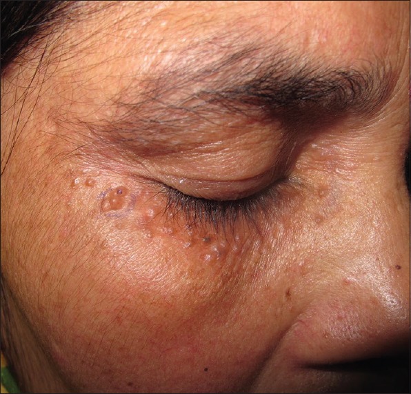
Clear to bluish-colored small cysts in the infraorbital region
Figure 2.
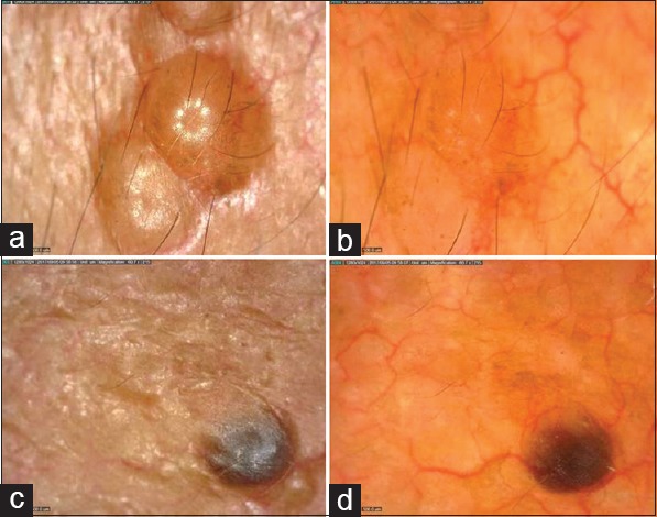
(a) Clear fluid-filled cysts with indentation in the centre are seen in nonpolarized mode and (b) only a nondescript pale area seen in polarized mode (×60). (c and d) The bluish papule on nonpolarized (c) and polarized (d) mode (×60). It appears as a partly-filled cyst with a hypopyon appearance showing clearing in the upper portion. Central indentation can still be appreciated (c)
Histopathology revealed a unilocular cyst lined by two layered flattened cuboidal epithelium suggestive of eccrine hidrocystomas (EC) [Figure 3a]. There was no evidence of decapitation secretion, thus ruling out apocrine hidrocystoma [Figure 3b]. The patient was started on 1% atropine ointment application twice a day. Marked lesion resolution was observed after 10 days of treatment.
Figure 3.
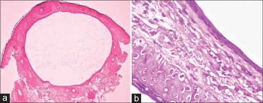
(a) Unilocular cyst in dermis lined by two layers of flattened epithelium. Lumen showed small amounts of pale eosinophilic secretions (H and E, ×40); (b) Cyst wall lined by two layers of flattened epithelium with no evidence of decapitation secretion or luminal papillary projections, thus ruling out an apocrine origin (H and E, ×400)
EC were first described by Robinson in women working in hot and humid environment.[1] They probably arise from the intradermal portion of the eccrine sweat duct, which gets blocked due to hyperhidrosis. Dermoscopic appearance of ECs has been described in three Turkish patients[2] and one Portugese patient[3] previously and may be helpful in differentiating ECs from other common lesions located in the same region such as syringomas and sebaceous hyperplasia. Duman et al. described ECs as homogeneous bluish-purplish central areas surrounded by pale halos.[2] Similarly, Correia et al. described them as well-demarcated bluish cystic spaces.[3] We did not observe surrounding pallor in any lesion. Further, the lesions were predominantly clear appearing rather than bluish in our patient. The prominent central indentation, giving the lesions a molluscoid appearance and a hypopyon-like appearance was not observed in the previous two reports. Among the differentials, multiple trichoepitheliomas show arborizing telangiectasias and an ivory-white background.[4] Eruptive vellus hair cysts demonstrate central yellowish rounded lesions surrounded by gray circles.[4] Syringomas show nonspecific features with pigment network with a reddish tinge and occasionally rosette structures.[4] Arborizing vessels is a prominent feature of basal cell carcinomas on the head and neck, while apocrine hidrocystomas have been described as showing bluish homogenous structures with whitish areas and arborizing vessels on top.[5]
To summarize, we describe the dermoscopic appearance of ECs as molluscoid appearing clear cysts, with a hypopyon-like appearance in darker lesions. The described features are better appreciated on a nonpolarized mode than polarized mode. The findings, however, need to be validated in larger patient groups.
Declaration of patient consent
The authors certify that they have obtained all appropriate patient consent forms. In the form the patient(s) has/have given his/her/their consent for his/her/their images and other clinical information to be reported in the journal. The patients understand that their names and initials will not be published and due efforts will be made to conceal their identity, but anonymity cannot be guaranteed.
Financial support and sponsorship
Nil.
Conflicts of interest
There are no conflicts of interest.
References
- 1.Robinson AR. Hidrocystoma. J Cutan Genitourin Dis. 1893;11:293–303. [Google Scholar]
- 2.Correia O, Duarte AF, Barros AM, Rocha N. Multiple eccrine hidrocystomas—from diagnosis to treatment: The role of dermatoscopy and botulinum toxin. Dermatology. 2009;219:77–9. doi: 10.1159/000218155. [DOI] [PubMed] [Google Scholar]
- 3.Duman N, Duman D, Sahin S. Pale halo surrounding a homogeneous bluish- purplish central area: Dermoscopic clue for eccrine hidrocystoma. Dermatol Pract Concept. 2015;5:43–5. doi: 10.5826/dpc.0504a11. [DOI] [PMC free article] [PubMed] [Google Scholar]
- 4.Zhong P, Tan C. Dermoscopic features of eruptive milium-like syringoma. Eur J Dermatol. 2015;25:203–4. doi: 10.1684/ejd.2014.2506. [DOI] [PubMed] [Google Scholar]
- 5.Zaballos P, Bañuls J, Medina C, Salsench E, Serrano P, Guionnet N. Dermoscopy of apocrine hidrocystomas: A morphological study. J Eur Acad Dermatol Venereol. 2014;28:378–81. doi: 10.1111/jdv.12044. [DOI] [PubMed] [Google Scholar]