Abstract
Damage to the cerebral microvasculature is a feature of cerebral malaria. Circulating endothelial progenitor cells are needed for microvascular repair. Based on this knowledge, we hypothesized that the failure to mobilize sufficient circulating endothelial progenitor cells to the cerebral microvasculature is a pathophysiologic feature of cerebral malaria. To test this hypothesis, we compared peripheral blood levels of CD34+/VEGFR2+ and CD34+/CD133+ cells and plasma levels of the chemokine stromal cell–derived growth factor 1 (SDF-1) in 214 children in Accra, Ghana. Children with cerebral malaria had lower levels of CD34+/VEGFR2+ and CD34+/CD133+ cells compared with those with uncomplicated malaria, asymptomatic parasitemia, or healthy controls. SDF-1 levels were higher in children with acute malaria compared with healthy controls. Together, these results uncover a potentially novel role for endothelial progenitor cell mobilization in the pathophysiology of cerebral malaria.
INTRODUCTION
Cerebral malaria is a serious complication of Plasmodium falciparum infection and contributes significantly to the morbidity and mortality of this pandemic. Primarily young children develop a diffuse potentially rapidly reversible encephalopathy associated with loss of consciousness, seizures, and few localizing neurologic signs.1 Despite anti-parasitic treatment, 15–30% die.1,2 In most survivors detectable neurologic sequelae resolve within 4–6 weeks.2,3
A pathophysiologic hallmark of P. falciparum infection is the microvascular sequestration of infected red blood cells (IRBCs) in the cerebral microvasculature. Sequestration is thought to specifically contribute to the pathophysiology of cerebral malaria. This same evidence, however, supports an equally important role for microvascular damage. Indeed, electron micrographs of cerebral malaria patients’ brains show that the intimate interactions between the IRBCs and endothelial cells1,3–5 are also accompanied by diffuse activation of the vascular endothelium and perturbations of the blood–brain barrier.6–10 Elevated plasma von Willebrand factor (vWF) levels in P. falciparum–infected patients and pathology indicates that endothelial cell damage occurs.1,3,4,11–13
Seminal work from Asahara and others14 showed that circulating endothelial progenitor cells (CEPCs) in the peripheral blood of humans are key mediators of microvascular homeostasis.15 It has been established that damaged endothelial cells are replaced not only by the replication of local endothelial cells but also by bone marrow–derived CEPCs that migrate to sites of damage and are incorporated into the microvasculature.14,16,17 They augment the local response, which may be insufficient to repair extensive or chronic injury by replication or migration.
More recent work has shown that recovery from endothelial damage is further defined by a balance between the magnitude of microvascular damage and the capacity for repair.18 Accordingly, CEPCs have been implicated in the pathogenesis of cardiovascular disease, tissue hypoxia, cancer, vasculitis, and sickle cell disease.18–26 Insufficient or dysfunctional CEPCs and levels of chemokines, which mediate CEPC mobilization and migration from the bone marrow, correlate with poor clinical outcomes.18,27–29
Based on this information, we hypothesized that low levels of CEPCs might be also associated with the development of cerebral malaria. Children able to maintain the balance between damage and repair would not develop cerebral malaria. If this equilibrium was disturbed, however, cerebral malaria might develop because of breaches in the integrity of the brain microvasculature.
To test this hypothesis, we measured and compared levels of CEPCs by flow cytometry in Ghanaian children with cerebral malaria (CM), uncomplicated malaria (UM), asymptomatic parasitemia (AP), severe malaria anemia (SA), and uninfected healthy controls (HC). To detect CEPCs, we used the traditional dual expression of CD34 and the vascular endothelial cell growth factor 2 (VEGFR2).14 However, because the CD34+/VEGFR2+ cell population may include mature endothelial cells shed from the vessel walls, we also measured levels of CD34+/CD133+ cells to enhance the detection of immature progenitor cells.15,17,30,31 To discern if the host was attempting to mobilize CEPCs to sites of microvascular damage, we measured levels of the stromal cell–derived growth factor 1 (SDF-1) chemokine by ELISA.
MATERIALS AND METHODS
Study population and enrollment
The University of Ghana Medical School (UGMS)/Korle-Bu Teaching Hospital (KBTH) in the capital city of Accra is the leading hospital in Ghana and has a referral unit for pediatric cases including malaria. Malaria transmission is perennial and hyperendemic. Children with acute malaria were recruited at the UGMS/KBTH during the peak transmission season (May–August) in 2005 and 2006. Patients with uncomplicated malaria were recruited from the Hospital’s Outpatient Clinic. Children with asymptomatic parasitemia and healthy community controls were recruited in community schools in 2005 from October to November and in 2006 in June and August. Enrollment of patients and collection and use of samples were done according to protocols approved by the Institutional Review Boards of the Weill Medical College of Cornell University and the Noguchi Memorial Institute for Medical Research, the Ethics and Protocol Review Committee of the University of Ghana Medical School, and local school officials and parent boards. Individual informed consent was obtained from the parent or guardian of each child before enrollment.
Children between the ages of 1 and 12 years of age were screened for inclusion by a team of study nurses and physicians. Children presenting with suspected malaria were evaluated with a complete history and physical examination. Children at local schools were assessed by history and a brief physical examination. Inclusion criteria for malaria were a history of fever or current fever (axillary > 37.5°C) and malaria parasitemia of 2,500/μL. Uncomplicated malaria patients were parasitemic and fully conscious without any WHO criteria for severe malaria.32 Children with cerebral malaria (CM) had to be unconscious, with a Blantyre coma scale score (BCS) of ≤ 3 and duration of coma > 60 minutes, and no record of recent severe head trauma or other cause of coma or neurologic diseases including meningitis/encephalitis. Children with CM could have other concomitant severe malaria syndromes as defined by the WHO.32 Children with asymptomatic parasitemia and normal controls were with and without malaria parasitemia, respectively. Exclusion criteria were sickle cell trait/disease, history of HIV infection, trauma, surgery, diabetes or cardiovascular disease, bacteremia, meningitis/encephalitis, or any other disease besides malaria. Blood cultures and lumbar punctures were obtained in all patients with acute malaria and suspected CM, respectively. Blood samples for study purposes were not collected from children with suspected severe malaria anemia (SA, hemoglobin < 5 g/dL) on the basis of severe clinical pallor. If CM patients with concomitant SA had available residual blood from diagnostic or other studies, they were eligible for inclusion in the study. SA patients from an independent study of malaria anemia in Year 1 of the study with available residual blood were also included.
Sample collection
Venous blood was collected on the initial clinical evaluation for the determination of white blood cell count (WBC), hemoglobin concentration (Hb), platelet count, blood sugar, parasite density, malaria smear, and sickle cell anemia test (sodium metabisulfite). An aliquot was transported to the NMIMR and used for flow cytometry and plasma obtained by centrifugation was stored at −80°C for ELISA analysis.
Flow cytometry
CEPCs were analyzed as previously described.33 Aliquots of 50 μL whole peripheral blood per reaction were incubated for 15 minutes in the dark with mouse anti-human phycoerythrin (PE)- or fluorescein isothiocyanate (FITC)-conjugated antibody pairs: CD34-FITC (MiltenyiBiotec, Auburn, CA ) and VEGFR2-PE (R&D Systems, Minneapolis, MN) or CD34-FITC (MiltenyiBiotec) and CD133-PE (MiltenyiBiotec). Aliquots of cells incubated without antibodies or either with mouse anti-human CD15-PE and CD15-FITC (BD Biosciences, San Diego, CA) were used as controls. After incubation, RBCs were lysed with FACS lysis solution (BD Biosciences), washed with FACS flow (BD Biosciences, San Diego, CA), and immediately analyzed. Samples were assayed on a FACScan (BD Biosciences), and each analysis included 20,000 events.
Chemokine ELISA
Plasma concentrations of SDF-1 were assayed in duplicate using Quantikine ELISA assays (R&D Systems). Subject plasma (100 μL/reaction) was tested in parallel with SDF-1 standards and a human plasma SDF-1 control (R&D Systems). Absorbance was measured at 450 nm. A plot of the SDF-1 concentrations of the standards versus their absorbance readings was used to determine the SDF-1 concentrations in the subject’s samples.
Statistical analysis
The primary objective of the analysis was to compare the mean level of each parameter (% EPC, WBC count, parasite density, and hemoglobin (Hb) among the five groups: CM, UM, AP, SA, and HC. Because age was considered a potential confounder, because of the higher prevalence of CM in young children, all analyses made an adjustment for age by using age as a covariate. Because of concerns that disease severity and or anemia could be confounders, all analyses also included adjustments using parasite density and hemoglobin as covariates. To adjust for any systematic shift in measurements from Year 1 to Year 2 of the study, the study year was used as a blocking factor. Accordingly, the analysis was conducted as a two-way analysis of variance (ANOVA) where the factors were clinical group and study year and the covariates were age, parasitic density, and hemoglobin.
In all the analyses, the parallelism assumption was not rejected, and accordingly, analysis of covariance (ANCOVA) was deemed appropriate. On finding any significant group differences (P < 0.05), Tukey-adjusted pairwise comparisons were carried out to determine which groups differed from one another. It was determined that a log-transformation of the data conformed to the standard ANOVA assumptions. Accordingly, all analyses were conducted using the log-transformed data, but the summaries and graphs were based on geometric means and their associated 95% confidence limits expressed in the original, untransformed units of measurement.
Pre-study sample size/power calculations for the 2-year study were made based on studies of CEPCs in adult men, because no sizable amount of data was available regarding CEPCs in children. Based on the adult data, we designed the study to achieve 80% power to detect an effect size of 0.61, which led to a requisite sample size of 34 subjects in each clinical group.
RESULTS
Baseline subject characteristics
Baseline patient characteristics of the 214 children included in the study are given in Table 1. Fifty-one percent were male. The children ranged in age from 1 to 12 years, with a mean age of 6.1 ± 2.8 years. Two patients with bacteremia (Salmonella typhi and Shigella flexneri) and 4 UM patients with suspected intravascular hemolysis (IVH) caused by the passage of “cola colored” urine were removed from the analysis.
Table 1.
Patient baseline characteristics
| Cerebral malaria | Uncomplicated malaria | Asymptomatic parasitemia | Severe malaria anemia | Healthy controls | |
|---|---|---|---|---|---|
| N | 42 | 51 | 44 | 14 | 63 |
| Age (years) | 5.2 ± 2.9 (min 1, max 12) | 5.7 ± 3.0 (min 1.25, max 12) | 7.3 ± 2.6 (min 2, max 12) | 3.25 ± 2.0 (min 1, max 9) | 6.8 ± 2.2 (min 3, max 12) |
| Male sex [N (%)] | 25 (60%) | 28 (55%) | 23 (52%) | 11 (79%) | 22 (35%) |
| WBC (×109/L) | 12.5 ± 5.5 | 8.9 ± 4.5 | 6.5 ± 1.7 | 10.2 ± 5.1 | 7.0 ± 1.7 |
| Parasite density (/μL) | 196,073 ± 398,886 | 106,061 ± 130,166 | 1001 ± 1021 | 183,534 ± 215,561 | 0 |
| Hemoglobin (g/dL) | 8.1 ± 2.0 | 9.7 ± 1.8 | 11.9 ± 1.0 | 4.2 ± 0.4 | 11.6 ± 1.0 |
Baseline characteristics were as expected
The parasite densities differed significantly across the five study groups (CM, UM, AP, SA, and HC; P < 0.0001); they were significantly increased in the CM, UM, and SA groups compared with the AP or HC children, respectively, and the AP children had increased levels compared with the HC children (P < 0.0001). There was no significant difference in parasite densities between CM and UM or between CM and SA children.WBC counts differed significantly across the five groups (P < 0.0001); they were significantly increased in patients with CM compared with children with UM (P < 0.0001), AP (P < 0.0001), or HC (P < 0.0001). Hemoglobin levels (Hb) differed significantly across the five groups (P < 0.0001). Hb levels were significantly lower in children with CM than in children with UM, AP, or HC (all P < 0.0001) and in children with UM than in children with AP or HC (P < 0.0001). Levels of Hb were significantly lower in the SA children compared with either CM, UM, AP, or HC (P < 0.0001). Levels of Hb were not different between AP and HC children.
Of the 42 CM patients enrolled in the study, the BCSs were as follows: 0 (N = 2), 1 (N = 4), 2 (N = 21), and 3 (N = 15). Eight of the CM patients had concomitant evidence of other severe malaria syndromes. Three of these patients had IVH and one had respiratory distress. Four also had SA; one of these also had IVH and another had respiratory distress. Six CM patients died; of these, three also had SA, IVH, or respiratory distress.
Two of the 14 children who had SA without cerebral malaria also had intravascular hemolysis.
Flow cytometry of circulating endothelial progenitor cells
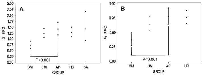
ANCOVA showed a significant difference for %CEPC levels detected using the CD34/VEGFR2 antibody combination (P < 0.0001) among the five subject groups tested (CM, UM, AP, HC, and SA) as well as using the CD34/CD133 antibody combination (P < 0.002) for the four subject groups tested (CM, UM, AP, and HC). Further pairwise comparisons showed that the mean %CEPC levels detected using the traditional antibody combination of CD34/VEGFR2 were significantly lower in children with CM compared with those with UM (P < 0.0004), AP (P < 0.001), HC children (P < 0.004), or those with SA (P < 0.006; Figure 1A). Using the more progenitor cell-specific antibody combination CD34/CD133, the %CEPC levels were also significantly lower in CM children compared pairwise with those with UM (P < 0.003), AP (P < 0.001), or HC (P < 0.006; Figure 1B). Pairwise comparisons of the mean %CEPC levels between other groups were not statistically significant (e.g., UM versus AP or AP versus HC).
Figure 1.
Percentage of circulating endothelial progenitor cells in each study group. A, Flow cytometry of CD34+/VEGFR2+ cells. CM: N = 31, geometric mean = 0.72; UM: N = 46, geometric mean = 1.24; AP: N = 39, geometric mean = 1.42; HC: N = 57, geometric mean = 1.28; SA: N = 14, geometric mean = 1.42 (P < 0.0001) B, Flow cytometry of CD34+/CD133+ cells. CM: N = 21, geometric mean = 0.37; UM: N = 31, geometric mean = 0.64; AP: N = 38, geometric mean = 0.77; HC: N = 57 geometric mean = 0.76 (P < 0.002). Geometric means and 95% confidence limits are presented. All means are adjusted for age, hemoglobin, parasite density, and study year. Because of the volume of blood available from young children, not all antibody combinations could be tested on each patient sample and therefore the number of patients (N) varies.
Chemokine levels
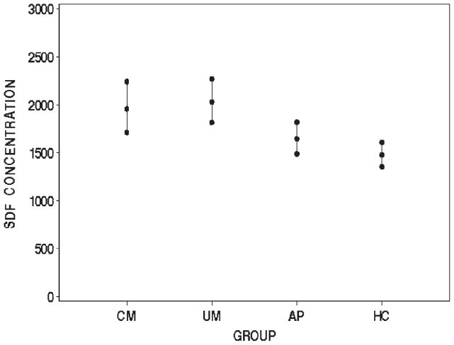
ANCOVA showed a significant difference in SDF-1 levels among the four subject groups (P < 0.001). Further analysis showed that levels of SDF-1 were significantly elevated in patients with CM compared with healthy controls (P < 0.02; Figure 2). SDF-1 levels were also elevated in patients with UM compared with healthy controls (P < 0.0004). SDF-1 levels were elevated (but not statistically significant) in patients with UM compared with those with AP (P = 0.06). There were no significant differences in the SDF-1 levels of patients with CM and UM or between those with CM and AP or AP and HC.
Figure 2.
SDF-1 plasma levels in each study group. CM: N = 33, geometric mean = 1,959; UM: N = 33, geometric mean = 2,031; AP: N = 44, geometric mean = 1,645; HC: N = 60, geometric mean = 1,476 (P < 0.001). SDF-1 concentrations given are in pg/mL. All means are adjusted for age, hemoglobin, parasitic density, and study year.
DISCUSSION
We hypothesized that low levels of CEPCs might be associated with the development of cerebral malaria. Children in a P. falciparum–endemic region in Ghana with cerebral malaria have significantly lower levels of CEPCs, as defined by the dual expression of CD34 and VEGFR2, than children with uncomplicated malaria, asymptomatic parasitemia, severe malaria anemia, or uninfected controls, and they also have lower levels of CD34+/CD133+ cells than children with uncomplicated malaria, asymptomatic parasitemia, or uninfected controls. In addition, plasma levels of SDF-1 in children with acute malaria (UM and CM) were significantly higher than in healthy controls.
These results permit the placement of cerebral malaria pathogenesis within the context of the current paradigms of microvascular homeostasis, thereby providing a new way of looking at the pathophysiology of the disease. Although microvascular damage is common to the major proposed mechanisms of cerebral malaria: hypoxia, a cytokine-induced sepsis-like syndrome and sequestration; how the host response to microvascular damage might contribute to the development of and recovery from this serious complication of P. falciparum infection has not previously been considered.
Our analysis suggests that the findings are syndrome specific for cerebral malaria and not merely an epiphenomenon. We assessed the possibility that the low levels of CEPCs observed were merely caused by severe disease by using parasitic density as a covariate in our statistical analysis. In addition, we performed flow cytometry analysis on a group of children with another severe malaria syndrome: severe malaria anemia. Similar to UM, AP, and healthy controls, children with severe malaria anemia have significantly higher levels of CEPCs (CD34+/VEGFR2+) compared with those with cerebral malaria. Therefore, the low levels of CEPCs in children with cerebral malaria do not seem to be caused by severe disease.
It was also possible that general bone marrow suppression, which is believed to contribute to anemia in malaria, might effect levels of the bone marrow–derived CEPCs.34 The FACS analysis of patients with severe malaria anemia, however, does not support this thesis. To further control for the potential impact of bone marrow suppression and anemia on our findings, hemoglobin was used as a covariate in our statistical analysis.
Our hypothesis is further supported by the finding that SDF-1 levels are high in patients with acute malaria (CM and UM) compared with healthy controls. SDF-1 serves a critical role in the mobilization of EPCs from the bone marrow and their retention at sites of microvascular damage.35–37 The results therefore reinforce the contention that the P. falciparum host is attempting to repair microvascular damage.
The results also provide plausible novel explanations for some clinical epidemiologic observations of the disease. For example, in Ghana, as in much of sub-Saharan Africa, beginning in infancy, children experience multiple consecutive P. falciparum infections. However, children between the ages of 2 and 5 years are more prone to develop cerebral malaria.38,39 It is interesting, therefore, to consider whether this finding might be explained by the exhaustion of endothelial progenitor cells in the BM or the impairment of the BM’s ability to produce EPCs after repeated infections but before the development of anti-parasite immunity. In addition, with age and repeated challenges children might develop an enhanced capacity for microvascular repair, which contributes to their immunity to cerebral malaria. It might further be postulated that the observed diminution in age-related immunity on leaving an endemic area could be associated with the waning of this enhanced capacity caused by the absence of persistent microvascular damage.
The results also suggest that chemotherapeutics and novel therapies being developed for other diseases associated with alterations of microvascular hemostasis might be used in the treatment of cerebral malaria.40–42 The etiology of the observed low levels of CEPCs, however, needs to be determined. EPC reserves in the bone marrow may become depleted, but the disease milieu may also lead to functional impairments and the cells’ inability to mature or migrate from the bone marrow.25,27 Therapies that merely augment the number of CEPCs might not be effective, whereas those designed to enhance CEPC function could be of benefit.
In addition, the time course of the host response to P. falciparum–induced microvascular damage needs to be determined. Because our study was designed to specifically determine whether low CEPCs were associated with cerebral malaria, measurements were determined only at a single time point. However, P. falciparum infection results in a spectrum of disease severity. The patient with AP might progress to UM, and the UM patient may or may not progress to CM or develop SA. Based on the findings, we would anticipate that patients who progress on the continuum to develop CM have decreasing levels of CEPCs and that those that recover have increasing levels. Prospective measurements of CEPC levels before the development of and after the recovery from CM are necessary to verify that this is the case. This information will permit a determination of when therapeutics would be most effective.
Such studies may also permit the establishment of CEPC levels at which patients are at risk for the development of cerebral malaria. In patients with cardiovascular disease, low levels of CEPCs correlate with disease progression and are predictive of cardiovascular events.18,28,29 CEPC levels could similarly be predictive of who will develop and recover from cerebral malaria.
It is recognized that the development of CM may be caused by a constellation of factors of which low CEPC levels are merely one contributor. Indeed, low CEPC levels may not reflect microvascular damage as postulated, but be from another cause. Further study is needed. However, despite these limitations, our study provides a new conceptual lens through which to view malaria pathogenesis that may also hold potential diagnostic and therapeutic relevance.
Acknowledgments
The authors thank Tatiana Peshansky for bioinformatics support, Marshall Glesby and William Rogers for useful discussions, and Warren Johnson, Dan Fitzgerald, Kyu Rhee, Jorgen Kurtzhals, and Heidi Stuhlmann for critical review of the manuscript.
Financial support: The study was funded by the National Institutes of Health NS054243 with support from the Weill Medical College General Clinical Research Center (MO1 RR00047).
References
- 1.Turner G. Cerebral malaria. Brain Pathol. 1997;7:569–582. doi: 10.1111/j.1750-3639.1997.tb01075.x. [DOI] [PMC free article] [PubMed] [Google Scholar]
- 2.Newton CR, Taylor TE, Whitten RO. Pathophysiology of fatal falciparum malaria in African children. Am J Trop Med Hyg. 1998;58:673–683. doi: 10.4269/ajtmh.1998.58.673. [DOI] [PubMed] [Google Scholar]
- 3.Pongponratn E, Turner GD, Day NP, Phu NH, Simpson JA, Stepniewska K, Mai NT, Viriyavejakul P, Looareesuwan S, Hien TT, Ferguson DJ, White NJ. An ultrastructural study of the brain in fatal Plasmodium falciparum malaria. Am J Trop Med Hyg. 2003;69:345–359. [PubMed] [Google Scholar]
- 4.MacPherson GG, Warrell MJ, White NJ, Looareesuwan S, Warrell DA. Human cerebral malaria. A quantitative ultrastructural analysis of parasitized erythrocyte sequestration. Am J Pathol. 1985;119:385–401. [PMC free article] [PubMed] [Google Scholar]
- 5.Taylor TE, Fu WJ, Carr RA, Whitten RO, Mueller JS, Fosiko NG, Lewallen S, Liomba NG, Molyneux ME. Differentiating the pathologies of cerebral malaria by postmortem parasite counts. Nat Med. 2004;10:143–145. doi: 10.1038/nm986. [DOI] [PubMed] [Google Scholar]
- 6.Adams S, Brown H, Turner G. Breaking down the blood-brain barrier: signaling a path to cerebral malaria? Trends Parasitol. 2002;18:360–366. doi: 10.1016/s1471-4922(02)02353-x. [DOI] [PubMed] [Google Scholar]
- 7.Brown H, Hien TT, Day N, Mai NT, Chuong LV, Chau TT, Loc PP, Phu NH, Bethell D, Farrar J, Gatter K, White N, Turner G. Evidence of blood-brain barrier dysfunction in human cerebral malaria. Neuropathol Appl Neurobiol. 1999;25:331–340. doi: 10.1046/j.1365-2990.1999.00188.x. [DOI] [PubMed] [Google Scholar]
- 8.Combes V, Taylor TE, Juhan-Vague I, Mege JL, Mwenechanya J, Tembo M, Grau GE, Molyneux ME. Circulating endothelial microparticles in Malawian children with severe falciparum malaria complicated with coma. JAMA. 2004;291:2542–2544. doi: 10.1001/jama.291.21.2542-b. [DOI] [PubMed] [Google Scholar]
- 9.Muanza K, Traore B, Gay F, Krudsood S, Danis M, Looareesuwan S. Circulating receptors implicated in the cytoadherence occurring in severe Plasmodium falciparum malaria in Thailand. Ann Trop Med Parasitol. 1999;93:449–455. doi: 10.1080/00034989958186. [DOI] [PubMed] [Google Scholar]
- 10.Turner GD, Ly VC, Nguyen TH, Tran TH, Nguyen HP, Bethell D, Wyllie S, Louwrier K, Fox SB, Gatter KC, Day NP, White NJ, Berendt AR. Systemic endothelial activation occurs in both mild and severe malaria. Correlating dermal microvascular endothelial cell phenotype and soluble cell adhesion molecules with disease severity. Am J Pathol. 1998;152:1477–1487. [PMC free article] [PubMed] [Google Scholar]
- 11.Horstmann RD, Dietrich M. Haemostatic alterations in malaria correlate to parasitaemia. Blut. 1985;51:329–335. doi: 10.1007/BF00320043. [DOI] [PubMed] [Google Scholar]
- 12.Mohanty D, Ghosh K, Nandwani SK, Shetty S, Phillips C, Rizvi S, Parmar BD. Fibrinolysis, inhibitors of blood coagulation, and monocyte derived coagulant activity in acute malaria. Am J Hematol. 1997;54:23–29. doi: 10.1002/(sici)1096-8652(199701)54:1<23::aid-ajh4>3.0.co;2-6. [DOI] [PubMed] [Google Scholar]
- 13.de Mast Q, Groot E, Lenting PJ, de Groot PG, McCall M, Sauerwein RW, Fijnheer R, van der Ven A. Thrombocytopenia and release of activated von Willebrand Factor during early Plasmodium falciparum malaria. J Infect Dis. 2007;196:622–628. doi: 10.1086/519844. [DOI] [PubMed] [Google Scholar]
- 14.Asahara T, Murohara T, Sullivan A, Silver M, van der Zee R, Li T, Witzenbichler B, Schatteman G, Isner JM. Isolation of putative progenitor endothelial cells for angiogenesis. Science. 1997;275:964–967. doi: 10.1126/science.275.5302.964. [DOI] [PubMed] [Google Scholar]
- 15.Urbich C, Dimmeler S. Endothelial progenitor cells: characterization and role in vascular biology. Circ Res. 2004;95:343–353. doi: 10.1161/01.RES.0000137877.89448.78. [DOI] [PubMed] [Google Scholar]
- 16.Lin Y, Weisdorf DJ, Solovey A, Hebbel RP. Origins of circulating endothelial cells and endothelial outgrowth from blood. J Clin Invest. 2000;105:71–77. doi: 10.1172/JCI8071. [DOI] [PMC free article] [PubMed] [Google Scholar]
- 17.Rafii S. Circulating endothelial precursors: mystery, reality, and promise. J Clin Invest. 2000;105:17–19. doi: 10.1172/JCI8774. [DOI] [PMC free article] [PubMed] [Google Scholar]
- 18.Hill JM, Zalos G, Halcox JP, Schenke WH, Waclawiw MA, Quyyumi AA, Finkel T. Circulating endothelial progenitor cells, vascular function, and cardiovascular risk. N Engl J Med. 2003;348:593–600. doi: 10.1056/NEJMoa022287. [DOI] [PubMed] [Google Scholar]
- 19.Hristov M, Erl W, Weber PC. Endothelial progenitor cells: mobilization, differentiation, and homing. Arterioscler Thromb Vasc Biol. 2003;23:1185–1189. doi: 10.1161/01.ATV.0000073832.49290.B5. [DOI] [PubMed] [Google Scholar]
- 20.Mutunga M, Fulton B, Bullock R, Batchelor A, Gascoigne A, Gillespie JI, Baudouin SV. Circulating endothelial cells in patients with septic shock. Am J Respir Crit Care Med. 2001;163:195–200. doi: 10.1164/ajrccm.163.1.9912036. [DOI] [PubMed] [Google Scholar]
- 21.Rosenzweig A. Endothelial progenitor cells. N Engl J Med. 2003;348:581–582. doi: 10.1056/NEJMp020175. [DOI] [PubMed] [Google Scholar]
- 22.Solovey A, Lin Y, Browne P, Choong S, Wayner E, Hebbel RP. Circulating activated endothelial cells in sickle cell anemia. N Engl J Med. 1997;337:1584–1590. doi: 10.1056/NEJM199711273372203. [DOI] [PubMed] [Google Scholar]
- 23.Sowemimo-Coker SO, Meiselman HJ, Francis RB., Jr Increased circulating endothelial cells in sickle cell crisis. Am J Hematol. 1989;31:263–265. doi: 10.1002/ajh.2830310409. [DOI] [PubMed] [Google Scholar]
- 24.Takahashi T, Kalka C, Masuda H, Chen D, Silver M, Kearney M, Magner M, Isner JM, Asahara T. Ischemia- and cytokine-induced mobilization of bone marrow-derived endothelial progenitor cells for neovascularization. Nat Med. 1999;5:434–438. doi: 10.1038/7434. [DOI] [PubMed] [Google Scholar]
- 25.Vasa M, Fichtlscherer S, Aicher A, Adler K, Urbich C, Martin H, Zeiher AM, Dimmeler S. Number and migratory activity of circulating endothelial progenitor cells inversely correlate with risk factors for coronary artery disease. Circ Res. 2001;89:E1–E7. doi: 10.1161/hh1301.093953. [DOI] [PubMed] [Google Scholar]
- 26.Woywodt A, Streiber F, de Groot K, Regelsberger H, Haller H, Haubitz M. Circulating endothelial cells as markers for ANCA-associated small-vessel vasculitis. Lancet. 2003;361:206–210. doi: 10.1016/S0140-6736(03)12269-6. [DOI] [PubMed] [Google Scholar]
- 27.Heeschen C, Lehmann R, Honold J, Assmus B, Aicher A, Walter DH, Martin H, Zeiher AM, Dimmeler S. Profoundly reduced neovascularization capacity of bone marrow mononu-clear cells derived from patients with chronic ischemic heart disease. Circulation. 2004;109:1615–1622. doi: 10.1161/01.CIR.0000124476.32871.E3. [DOI] [PubMed] [Google Scholar]
- 28.Schmidt-Lucke C, Rossig L, Fichtlscherer S, Vasa M, Britten M, Kamper U, Dimmeler S, Zeiher AM. Reduced number of circulating endothelial progenitor cells predicts future cardiovascular events: proof of concept for the clinical importance of endogenous vascular repair. Circulation. 2005;111:2981–2987. doi: 10.1161/CIRCULATIONAHA.104.504340. [DOI] [PubMed] [Google Scholar]
- 29.Werner N, Kosiol S, Schiegl T, Ahlers P, Walenta K, Link A, Bohm M, Nickenig G. Circulating endothelial progenitor cells and cardiovascular outcomes. N Engl J Med. 2005;353:999–1007. doi: 10.1056/NEJMoa043814. [DOI] [PubMed] [Google Scholar]
- 30.Peichev M, Naiyer AJ, Pereira D, Zhu Z, Lane WJ, Williams M, Oz MC, Hicklin DJ, Witte L, Moore MA, Rafii S. Expression of VEGFR-2 and AC133 by circulating human CD34(+) cells identifies a population of functional endothelial precursors. Blood. 2000;95:952–958. [PubMed] [Google Scholar]
- 31.Salven P, Mustjoki S, Alitalo R, Alitalo K, Rafii S. VEGFR-3 and CD133 identify a population of CD34+ lymphatic/vascular endothelial precursor cells. Blood. 2003;101:168–172. doi: 10.1182/blood-2002-03-0755. [DOI] [PubMed] [Google Scholar]
- 32.WHO. Severe and complicated malaria. World Health Organization, Division of Control of Tropical Diseases. Trans R Soc Trop Med Hyg. 1990;84(Suppl 2):1–65. [PubMed] [Google Scholar]
- 33.Vasa M, Fichtlscherer S, Adler K, Aicher A, Martin H, Zeiher AM, Dimmeler S. Increase in circulating endothelial progenitor cells by statin therapy in patients with stable coronary artery disease. Circulation. 2001;103:2885–2890. doi: 10.1161/hc2401.092816. [DOI] [PubMed] [Google Scholar]
- 34.Kurtzhals JA, Rodrigues O, Addae M, Commey JO, Nkrumah FK, Hviid L. Reversible suppression of bone marrow response to erythropoietin in Plasmodium falciparum malaria. Br J Haematol. 1997;97:169–174. doi: 10.1046/j.1365-2141.1997.82654.x. [DOI] [PubMed] [Google Scholar]
- 35.Yamaguchi J, Kusano KF, Masuo O, Kawamoto A, Silver M, Murasawa S, Bosch-Marce M, Masuda H, Losordo DW, Isner JM, Asahara T. Stromal cell-derived factor-1 effects on ex vivo expanded endothelial progenitor cell recruitment for ischemic neovascularization. Circulation. 2003;107:1322–1328. doi: 10.1161/01.cir.0000055313.77510.22. [DOI] [PubMed] [Google Scholar]
- 36.Shao H, Tan Y, Eton D, Yang Z, Uberti MG, Li S, Schulick A, Yu H. Statin and stromal cell-derived factor-1 additively promote angiogenesis by enhancement of progenitor cells incorporation into new vessels. Stem Cells. 2008;26:1376–1384. doi: 10.1634/stemcells.2007-0785. [DOI] [PubMed] [Google Scholar]
- 37.Kopp HG, Ramos CA, Rafii S. Contribution of endothelial progenitors and proangiogenic hematopoietic cells to vascularization of tumor and ischemic tissue. Curr Opin Hematol. 2006;13:175–181. doi: 10.1097/01.moh.0000219664.26528.da. [DOI] [PMC free article] [PubMed] [Google Scholar]
- 38.Marsh K, Snow RW. Host-parasite interaction and morbidity in malaria endemic areas. Philos Trans R Soc Lond B Biol Sci. 1997;352:1385–1394. doi: 10.1098/rstb.1997.0124. [DOI] [PMC free article] [PubMed] [Google Scholar]
- 39.Greenwood B, Marsh K, Snow R. Why do some African children develop severe malaria? Parasitol Today. 1991;7:277–281. doi: 10.1016/0169-4758(91)90096-7. [DOI] [PubMed] [Google Scholar]
- 40.Khakoo AY, Finkel T. Endothelial progenitor cells. Annu Rev Med. 2005;56:79–101. doi: 10.1146/annurev.med.56.090203.104149. [DOI] [PubMed] [Google Scholar]
- 41.Werner N, Nickenig G. Influence of cardiovascular risk factors on endothelial progenitor cells: limitations for therapy? Arterioscler Thromb Vasc Biol. 2006;26:257–266. doi: 10.1161/01.ATV.0000198239.41189.5d. [DOI] [PubMed] [Google Scholar]
- 42.Roncalli JG, Tongers J, Renault MA, Losordo DW. Endothelial progenitor cells in regenerative medicine and cancer: a decade of research. Trends Biotechnol. 2008;26:276–283. doi: 10.1016/j.tibtech.2008.01.005. [DOI] [PubMed] [Google Scholar]


