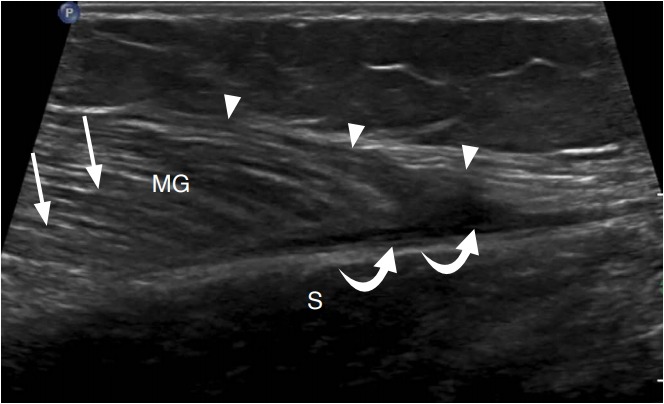
Fig. 10. A 30-year-old woman with medial head gastrocnemius tear (tennis leg).

Ultrasonography of the calf long axis to the distal medial head of the gastrocnemius (MG) demonstrates an irregular and hypoechoic distal myotendinous junction (arrowheads) with a small hypoechoic hematoma (curved arrows) between the MG and soleus (S). Compare to the normal muscle appearance (arrows).
