

Abstract
Rheumatoid pneumoconiosis, also known as Caplan's syndrome is defined as the association between silicosis and rheumatoid arthritis (RA). It is a rare and usually diagnosed in an advanced stage of RA course. It affects generally patients with long exposure to silica. In this article, we report a case of Caplan's syndrome.
Keywords: Caplan syndrome, Rheumatoid arthritis, Silicosis, Chest CT scan, Rituximab
Introduction
Occupational exposure to silica has been implicated in the development of autoimmune inflammatory diseases [1]. Its association with rheumatoid arthritis (RA) is well recognized [1], and is known as “Caplan syndrome” or also “rheumatoid pneumoconiosis.” It is a rare syndrome which can also occur in workers exposed to silica as well as in patients with silicosis or asbestosis [2]. It was identified in 1953 by Caplan [3] and consists of multiple well-defined rounded nodules on chest x-ray, distributed throughout the lungs predominantly at the periphery [4]. The prevalence of this syndrome among patients with pneumoconiosis is low [4]. The prevalence of 0.4% was found by Caplan [3]; more recently, in a comparative study, Honma and Vallyathan showed an incidence of 0.75% in Japan and 1.5% in the United States [5]. Here we reported a case of Caplan syndrome in a patient with a diagnosis of silicosis.
Case presentation
A 58-year-old man who did not smoke, had worked for 21 years as a truck driver with exposure to silica, until the age of 52, when he developed dyspnea with polyarthritis involving wrists and ankles. He was referred to the Pneumology department in Farhat Hached Hospital in Sousse in Tunisia. He had dyspnea in effort stage II based on New York Heart Association classification. The pulmonary auscultation revealed crepitus in the basal segments. The exploration of respiratory function showed a mild restrictive ventilatory deficit without alveolocapillary diffusion abnormalities. Chest x-rays revealed multiple micronodules distributed throughout the lungs but predominantly in the lower segments (Fig. 1). The bronchoalveolar lavage showed a predominance of macrophagic cells with a predominance of 30% of siderophages. The CT scan of the chest showed a pulmonary fibrosis with septal and intralobular cross-links in the 2 lung bases predominantly on the left side and at the upper lingual lobe (Fig. 2). The diagnosis of silicosis was made in 2011. Then, in 2016, the patient was referred to the rheumatology department for exploration of chronic symmetrical polyarthritis. On physical examination, the patient presented with synovitis in the shoulders, wrists, hands (Fig. 3), and the right knee. The x-ray showed bilateral erosions in both hands (Fig. 4) and feet (Fig. 5). Rheumatoid factor by the Latex and Rose-Waaler reaction and anticyclic citrullinated peptide antibodies measured by ELISA were highly positive (500 UI and 170 UI/L, respectively). The patient was diagnosed with RA based on the American College of Rheumatology/European League against Rheumatism classification criteria of RA [6]. The disease activity score (DAS28) [7] was up for 7.52. The diagnosis of Caplan syndrome was made. The patient received corticosteroids and rituximab (Mabthera) with an improvement in polyarthritis with a 12-month follow-up.
Fig. 1.
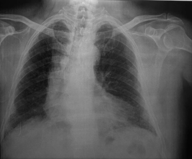
Chest x-ray showing multiple micronodules distributed throughout the lungs but predominantly in the lower segments.
Fig. 2.
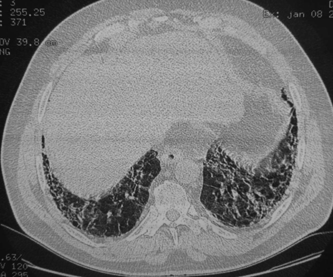
Computed tomography scan of the chest showing a pulmonary fibrosis with septal and intralobular cross-links in the 2 lung bases predominantly on the left side and at the upper lingual lobe.
Fig. 3.
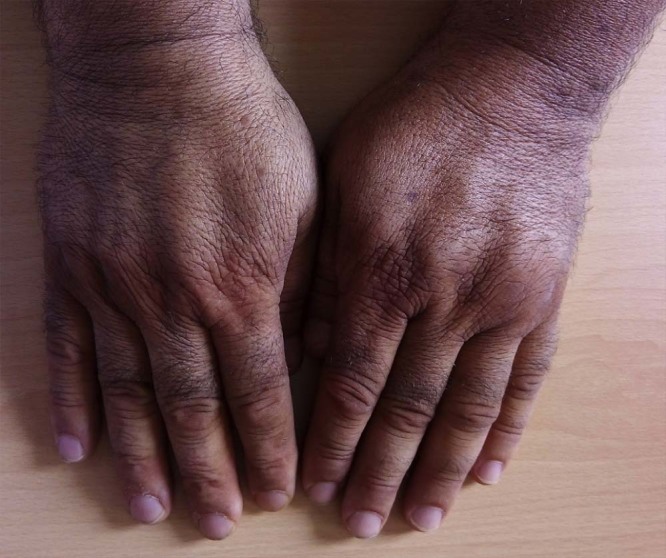
Picture of the patient's hands showing synovitis in both wrists and small joints of the hands.
Fig. 4.
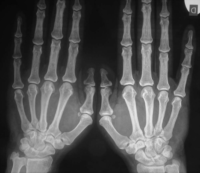
Radiography of hands showing erosions in the left radial extremity, in the right carpe, in the first left interphalangeal joint, and in the second and third right metacarpophalangeal joints.
Fig. 5.
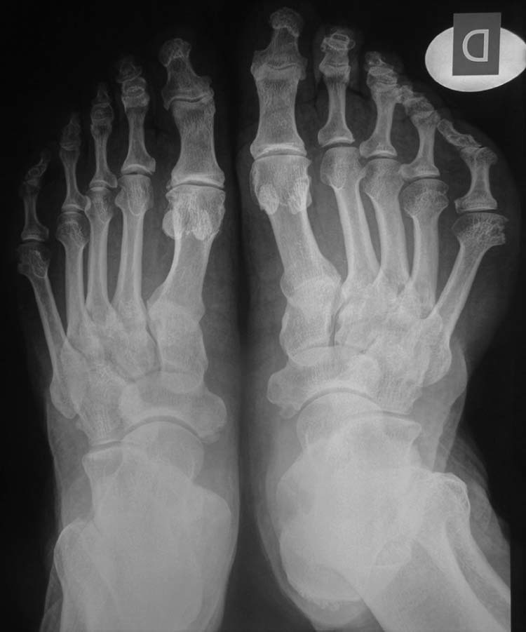
Radiography of feet showing concentric joint space narrowing in the first metatarsophalangeal joints with erosions in both the fifth metatarsophalangeal joint and the fourth right metatarsophalangeal joint.
Discussion
Caplan syndrome is first described by Caplan in 1953 [3], observed on coal workers of South Wales. The syndrome consists in the association between silicosis and RA [2]. The radiological features consist in multiple well-defined round opacities, measured between 0.5 and 5.0 cm in diameter, distributed throughout both lung fields but predominantly at the periphery [8]. The association between long exposure to silica, pneumoconiosis, and RA is proved [9]. In the study of Calvert et al. [10], the link between crystalline silica exposure and RA is well demonstrated. Both RA and rheumatoid factor is more frequent in patients with Caplan syndrome compared with exposed miners with or without simple pneumoconiosis [11].
The pathologic classic type of “Caplan nodules” is that they contain a central necrotic area that is surrounded by alternate necrotic tissue and layers of black coal dust, with a peripheral zone of cellular infiltration containing polymorphonuclear granulocytes, occasional giant cells, and macrophages [12]. These macrophages may contain dust particles [12]. The exact pathogenesis of RA is still unclear. The hypothesis is that silica particles are ingested by alveolar macrophages leading to inflammation and activation of fibroblasts. The macrophages destroyed the silica, which is again digested by new macrophages [13]. Activated macrophages by silica via the pathogen-associated molecular pattern will cause the production of several cytokines, including interleukin-1 and tumor necrosis factor-alpha. Following the activation of the innate immune system, dendritic cells will present antigens to CD4 T lymphocytes [13]. This repeated process causes chronic immune activity and fibrosis, and so facilitates the formation of autoantigens [12]. Pneumoconiosis results in the increase of autoantibodies, immune complexes, and excess production of immunoglobulins, including the rheumatoid factor [12]. Also positive anticyclic citrullinated peptides were frequently found in Caplan syndrome, and there is a link between exposure to silica and antibodies against citrullinated proteins-positive RA [14]. Radiological abnormalities and exposure history to silica allowed us to retain the diagnosis of Caplan syndrome in the clinical case we present by meeting the criteria established for the diagnosis of Caplan syndrome [13]. The occupational anamnesis in RA cases associated with radiological opacities allows differentiation of Caplan syndrome from pulmonary alterations due to RA [13]. This case also illustrates the potential of free silica and other mineral dusts to trigger autoimmune diseases. Concerning treatment, there is no conventional protocol. The treatment should be discussed case by case and performed according to the rheumatological guidelines, irrespective of Caplan syndrome [2]. Generally, as well as other pneumoconioses, silicosis does not respond to medical treatment [4].
Conclusion
Although rare, rheumatoid pneumoconiosis, also known as Caplan syndrome, can occur in workers exposed to silica, as well as in patients with silicosis. Aspects of diagnosis, classification, and occurrence of this syndrome are discussed, emphasizing the importance of the occupational anamnesis of patients with RA and lung opacities on chest x-rays.
Footnotes
Competing Interests: The authors have declared that no competing interests exist.
References
- 1.Costallat L.T.L., De Capitani E.M., Zambon L. Silicose pulmonaire et lupus érythémateux disséminé chez l'homme. À propos de deux observations. Rev Rhum. 2002;69(1):76–79. [Google Scholar]
- 2.Alaya R., Zouche I., Kaffel D., Riahi H., Hamdi W., Kchir M.M. 2017. Caplan's syndrome in an elderly-onset rheumatoid arthritis patient: About a new case. The Egyptian Rheumatologist. [Google Scholar]
- 3.Caplan A. Certain unusual radiological appearances in the chest of coal-miners suffering from rheumatoid arthritis. Thorax. 1953;8(1):29–37. doi: 10.1136/thx.8.1.29. [DOI] [PMC free article] [PubMed] [Google Scholar]
- 4.Schreiber J., Koschel D., Kekow J., Waldburg N., Goette A., Merget R. Rheumatoid pneumoconiosis (Caplan's syndrome) Eur J Intern Med. 2010;21(3):168–172. doi: 10.1016/j.ejim.2010.02.004. [DOI] [PubMed] [Google Scholar]
- 5.Honma K., Vallyathan V. Rheumatoid Pneumoconiosis: a comparative study of autopsy cases between Japan and North America. Ann Occup Hyg. 2002;46:65–67. [Google Scholar]
- 6.Aletaha D., Neogi T., Silman A.J., Funovits J., Felson D.T., Bingham C.O., 3rd 2010 Rheumatoid arthritis classification criteria: an American College of Rheumatology/European League Against Rheumatism collaborative initiative. Arthritis Rheum. 2010;62(9):2569–2581. doi: 10.1002/art.27584. [DOI] [PubMed] [Google Scholar]
- 7.Prevoo M.L., van ‘t Hof M.A., Kuper H.H., van Leeuwen M.A., van de Putte L.B., van Riel P.L. Modified disease activity scores that include twenty-eight-joint counts. Development and validation in a prospective longitudinal study of patients with rheumatoid arthritis. Arthritis Rheum. 1995;38(1):44–48. doi: 10.1002/art.1780380107. [DOI] [PubMed] [Google Scholar]
- 8.Tellesson W.G. Rheumatoid pneumoconiosis (Caplan's syndrome) in an asbestos worker. Thorax. 1961;16:372–377. doi: 10.1136/thx.16.4.372. [DOI] [PMC free article] [PubMed] [Google Scholar]
- 9.Otsuki T., Miura Y., Nishimura Y., Hyodoh F., Takata A., Kusaka M. Alterations of Fas and Fas-related molecules in patients with silicosis. Exp Biol Med (Maywood) 2006;231(5):522–533. doi: 10.1177/153537020623100506. [DOI] [PubMed] [Google Scholar]
- 10.Calvert G.M., Rice F.L., Boiano J.M., Sheehy J.W., Sanderson W.T. Occupational silica exposure and risk of various diseases: an analysis using death certificates from 27 states of the United States. Occup Environ Med. 2003;60(2):122–129. doi: 10.1136/oem.60.2.122. [DOI] [PMC free article] [PubMed] [Google Scholar]
- 11.Miall W.E., Caplan A., Cochrane A.L., Kilpatrick G.S., Oldham P.D. An epidemiological study of rheumatoid arthritis associated with characteristic chest x-ray appearances in coal-workers. Br Med J. 1953;2(4848):1231–1236. doi: 10.1136/bmj.2.4848.1231. [DOI] [PMC free article] [PubMed] [Google Scholar]
- 12.Weili G., Zhang N. A case report of rheumatoid pneumoconiosis (Caplan syndrome) Chest. 2016;149(4):A469. [Google Scholar]
- 13.De Capitani E.M., Schweller M., Silva C.M., Metze K., Cerqueira E.M., Bertolo M.B. Rheumatoid pneumoconiosis (Caplan's syndrome) with a classical presentation. J Bras Pneumol. 2009;35(9):942–946. doi: 10.1590/s1806-37132009000900017. [DOI] [PubMed] [Google Scholar]
- 14.Yahya A., Bengtsson C., Larsson P., Too C.L., Mustafa A.N., Abdullah N.A. Silica exposure is associated with an increased risk of developing ACPA-positive rheumatoid arthritis in an Asian population: evidence from the Malaysian MyEIRA case-control study. Mod Rheumatol. 2014;24(2):271–274. doi: 10.3109/14397595.2013.854076. [DOI] [PubMed] [Google Scholar]