Abstract
Abdominal compartment syndrome (ACS) is an uncommon complication of dengue haemorrhagic fever (DHF), described so far only in association with fluid refractory shock and high-volume resuscitation. We describe an unusual case of ACS in a patient of DHF where raised intra-abdominal pressure was due to spontaneous rectus sheath haematoma causing external compression. Early recognition of the haematoma, constant vigilance and timely decision for surgical intervention could salvage the patient with complete recovery of organ function.
Keywords: infectious diseases, tropical medicine (infectious disease), adult intensive care
Background
Presentation of dengue fever may mimic acute abdomen or not so uncommonly, patients with dengue may present with true surgical complications like acute cholecystitis, acute appendicitis or acute pancreatitis.1 Diagnosing a true surgical complication and determining the right timing for surgical intervention in such patients is of paramount importance for optimal outcome. We report a case of an elderly woman who presented with dengue haemorrhagic fever (DHF) and spontaneous rectus sheath haematoma (RSH) and developed abdominal compartment syndrome (ACS) where a desired outcome could be attained because of prompt recognition of the problem and timely surgical intervention. The major highlights of our case are, first, unusual nature of its presentation—ACS developing in a patient of DHF because of external compression by spontaneous RSH, never described earlier in the medical literature. Second, the present case emphasises the importance of continuous vigilance and clinical judgement to decide when to switch from conservative management to surgical exploration in cases of true surgical emergencies in patients with DHF.
Case presentation
A 74-year-old woman with only significant history of hypertension was admitted to our hospital with pain lower abdomen for past 3 days. Ten days before present admission, she had high-grade fever associated with bodyache and joint pain. Though the fever subsided within 2–3 days, she continued to remain symptomatic with extreme prostration and recurrent bouts of vomiting. There was no history of trauma. Apart from her usual prescription of amlodipine for hypertension, she had received paracetamol and pantoprazole as prescribed by her family physician. On examination, she was drowsy, Glasgow Coma Score scale of 10 (E3 V3 M4) with cold extremities, thready pulse 110/min regular, systolic blood pressure of 70 mm Hg and respiratory rate 12/min. She was pale with generalised petechiae especially on peripheries. She was moving all four limbs and nuchal rigidity was absent. Cardiac and lung auscultation were unremarkable. A non-tender firm swelling around 30 cm × 15 cm could be palpated on the right side of her abdomen without any local rise in temperature.
Investigations
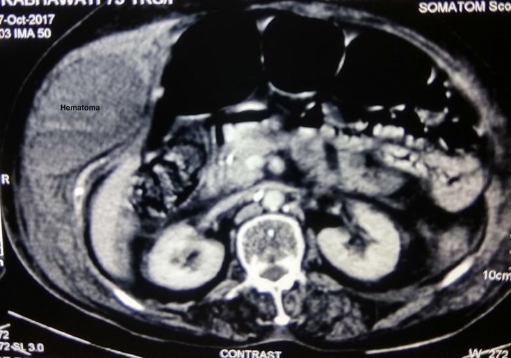
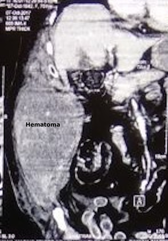
Initial laboratory investigations showed haemoglobin (Hb) 36 g/L, leucocyte count 12×109/L (65% neutrophils, 25% lymphocytes and 10% monocytes), platelet count 110×109/L, elevated aspartate/alanine aminotransferases (7.03/4.07 µkat/L) and lactate dehydrogenase (6.96 µkat/L), normal serum bilirubin, low albumin (22 g/L), deranged kidney function test (blood urea nitrogen 9.28 mmol/L and serum creatinine 123.53 µmol/L) and elevated prothrombin time (international normalised ratio 1.86). Arterial blood gas analysis revealed severe metabolic acidosis: pH 7.03, HCO35.3 mmol/L, PaCO22.71 kPa, normal oxygenation status, high lactate 10.42 mmol/L and normal glucose 6.55 mmol/L. Chest X-ray showed blunting of bilateral costophrenic angles. ECG and echocardiographic findings were unremarkable. A contrast-enhanced CT scan of the abdomen showed a large hyperdense cystic lesion involving abdominal wall on the right side likely to be a haematoma (figures 1 and 2). In view of an ongoing dengue epidemic, dengue serology was asked for that was positive for both dengue IgM and IgG, consistent with a recent primary dengue infection. Rapid card test for malaria antigen was negative. Two sets of blood cultures sent on admission were reported to be sterile after 5 days of incubation.
Figure 1.
Contrast-enhanced CT abdomen (axial plane) showing right-sided rectus sheath haematoma.
Figure 2.
Rectus sheath haematoma seen on the sagittal plane (CECT abdomen). CECT, contrast-enhanced CT.
Treatment
Provisional diagnosis of dengue shock syndrome with RSH was made and resuscitation was initiated with intravenous fluid, packed red cells, fresh frozen plasma and low-dose norepinephrine. She was intubated and mechanically ventilated in view of worsening shock and for airway protection. General surgery team was consulted for the abdominal wall haematoma and a conservative strategy was opted for the same after careful consideration of risk for further bleeding after release of the tamponade effect in a patient with thrombocytopaenia with DHF.
Outcome and follow-up
On day 2, she was haemodynamically stable, renal function parameters were normalised with normal urine output and low but stable Hb and platelet count (79 g/L and 66×109/L, respectively). By evening of third hospital day, her general condition worsened again with a fall in blood pressure requiring vasopressor support and a drop in her urine output. Abdominal wall haematoma by now had increased in size with a tense abdomen, sluggish bowel sound and a rise in intra-abdominal pressure measured to be 26 mm Hg. Hb level plummeted to 60 g/L with deterioration of kidney function test (blood urea nitrogen 10.71 mmol/L and serum creatinine 136.49 µmol/L). With a clear evidence of ACS, draining of haematoma was now considered to outweigh the risk of surgery. She was transfused 2 units of packed RBC and a single donor platelet and was taken up for the evacuation of haematoma. During operation, right-sided RSH was noted extending from right hypochondrium to right inguinal region; approximately 2000 mL of blood clots were drained and packing of the cavity was done. No active bleeding vessel could be seen. Pack was removed 72 hours after the clot evacuation without any re-bleeding. In subsequent days, she showed significant improvement in her general condition and was discharged finally from hospital on day 13, in a stable condition with normal haematological, renal and hepatic parameters.
Discussion
RSH is the accumulation of blood in sheath of rectus abdominis muscle, secondary to either shearing of the superior or inferior epigastric arteries or their branches anastomosing between the muscle and the posterior layer of the rectus sheath or due to direct injury to the underlying muscle itself.2 RSH is more common in the elderly due to the weakening of the abdominal wall muscles with age and female gender possibly reflecting the anatomic differences in the rectus sheath between the two sexes.2 Studies have identified various risk factors for developing RSH including anticoagulation, abdominal wall trauma (blunt trauma/surgery), muscle strain (like cough or repeated vomiting/retching or strenuous exercise), medical comorbidities (including chronic kidney disease or steroid/immunosuppression) and pregnancy.2 3 Surprisingly spontaneous RSH is rare in DHF, despite profound vasculopathy, thrombopathy and coagulopathy and are limited only to a countable number of case reports.4–8 First case was reported from Sri Lanka in 2001 in a patient who was also on aspirin.4 We postulate that RSH in our elderly female patient was precipitated by the shear stress on muscles due to repeated vomiting and retching and was further exaggerated by dengue-related coagulation abnormalities. No actively bleeding vessel could be identified during surgical exploration.
ACS is a life-threatening condition caused by rise in intra-abdominal pressure >20 mm Hg with evidence of organ dysfunction/failure.9 Rise in intra-abdominal pressure is usually associated with increase in volume of the peritoneal/retroperitoneal contents in the setting of trauma and high-volume resuscitation or not so commonly due to external compression of the abdominal wall. RSH as a cause of external compression and ACS was first documented by O’Mara et al, followed by few more anecdotal reports.10–13 ACS is a rare but known complication of DHF with shock, but in all the cases reported earlier, ACS in DHF was associated with fluid refractory shock and collection of peritoneal fluid.14 To the best of our knowledge, this is probably the first case report on ACS due to spontaneous RSH in a patient of DHF.
Treatment of spontaneous RSH is generally conservative including resuscitation, correction of coagulopathy, analgesia and treatment of the underlying condition. Interventional radiological intervention and embolisation is preferred in cases of continued bleeding with surgery being considered as the last option.11 13 But patients with RSH who develop ACS have poor outcome if managed conservatively, making surgical intervention mandatory.13
Learning points.
Abdominal compartment syndrome (ACS) should be considered as a possible cause of organ dysfunction in patients with dengue haemorrhagic fever (DHF).
Rectus sheath haematoma (RSH) though rare but may be a potentially life-threatening cause of ACS in DHF.
High index of suspicion and prompt imaging can establish diagnosis.
Careful monitoring of patients including monitoring of intra-abdominal pressure is a must for deciding timing of surgical intervention in patients of RSH who are otherwise managed conservatively, the priority in ACS.
Footnotes
Contributors: SG: conception and writing of the article. RS: contributed in compiling data and reviewing the manuscript. SG and AC: reviewing the manuscript. All authors contributed in the management of the patient.
Funding: The authors have not declared a specific grant for this research from any funding agency in the public, commercial or not-for-profit sectors.
Competing interests: None declared.
Patient consent: Obtained.
Provenance and peer review: Not commissioned; externally peer reviewed.
References
- 1.Jayasundara B, Perera L, de Silva A. Dengue fever may mislead the surgeons when it presents as an acute abdomen. Asian Pac J Trop Med 2017;10:15–19. 10.1016/j.apjtm.2016.12.010 [DOI] [PubMed] [Google Scholar]
- 2.Sheth HS, Kumar R, DiNella J, et al. . Evaluation of Risk Factors for Rectus Sheath Hematoma. Clin Appl Thromb Hemost 2016;22:292–6. 10.1177/1076029614553024 [DOI] [PubMed] [Google Scholar]
- 3.Cherry WB, Mueller PS. Rectus sheath hematoma: review of 126 cases at a single institution. Medicine 2006;85:105–10. 10.1097/01.md.0000216818.13067.5a [DOI] [PubMed] [Google Scholar]
- 4.Ganeshwaran Y, Seneviratne SM, Jayamaha R, et al. . Dengue fever associated with a haematoma of the rectus abdominis muscle. Ceylon Med J 2001;46:105–6. 10.4038/cmj.v46i3.8139 [DOI] [PubMed] [Google Scholar]
- 5.Waseem T, Latif H, Shabbir B. An unusual cause of acute abdominal pain in dengue fever. Am J Emerg Med 2014;32:819.e3–819.e4. 10.1016/j.ajem.2014.01.011 [DOI] [PubMed] [Google Scholar]
- 6.Sharma A, Bhatia S, Singh RP, et al. . Dengue Fever with rectus sheath hematoma: a case report. J Family Med Prim Care 2014;3:159–60. 10.4103/2249-4863.137665 [DOI] [PMC free article] [PubMed] [Google Scholar]
- 7.Bhat KJ, Shovkat R, Samoon HJ. Abdominal haematomas and dengue fever: Two different cases of spontaneous psoas muscle haematoma and bilateral rectus sheath haematoma complicating dengue haemorrhagic fever. J Vector Borne Dis 2015;52:339–41. [PubMed] [Google Scholar]
- 8.Nelwan EJ, Angelina F, Adiwinata R, et al. . Spontaneous rectus sheath hematomas in dengue hemorrhagic fever: A case report. IDCases 2017;10:35–7. 10.1016/j.idcr.2017.08.004 [DOI] [PMC free article] [PubMed] [Google Scholar]
- 9.Malbrain ML, Cheatham ML, Kirkpatrick A, et al. . Results from the International Conference of Experts on Intra-abdominal Hypertension and Abdominal Compartment Syndrome. I. Definitions. Intensive Care Med 2006;32:1722–32. 10.1007/s00134-006-0349-5 [DOI] [PubMed] [Google Scholar]
- 10.O’Mara MS, Semins H, Hathaway D, et al. . Abdominal compartment syndrome as a consequence of rectus sheath hematoma. Am Surg 2003;69:975–7. [PubMed] [Google Scholar]
- 11.Jafferbhoy SF, Rustum Q, Shiwani MH. Abdominal compartment syndrome--a fatal complication from a rectus sheath haematoma. BMJ Case Rep 2012;2012:bcr1220115332 10.1136/bcr.12.2011.5332 [DOI] [PMC free article] [PubMed] [Google Scholar]
- 12.Minaya Bravo AM, Medina Quintana RE, Mendoza Moreno F, et al. . Giant Rectus Sheath Hematoma Causing Abdominal Compartment Syndrome: Review and Management. J Surg 2017. JSUR-148. [Google Scholar]
- 13.Khan A, Strain J, Leung PS, et al. . Abdominal compartment syndrome due to spontaneous rectus sheath hematoma with extension into the retroperitoneal space. J Surg Case Rep 2017;2017:1–3. 10.1093/jscr/rjx226 [DOI] [PMC free article] [PubMed] [Google Scholar]
- 14.Kamath SR, Ranjit S. Clinical features, complications and atypical manifestations of children with severe forms of dengue hemorrhagic fever in South India. Indian J Pediatr 2006;73:889–95. 10.1007/BF02859281 [DOI] [PMC free article] [PubMed] [Google Scholar]


