Abstract
Objective
This study aims at identifying master regulators of transcriptional networks in autism spectrum disorders (ASDs).
Results
With two sets of independent RNA-Seq data generated on cerebellum from patients with ASDs and control subjects (N = 39 and 45 for set 1, N = 24 and 38 for set 2, respectively), we carried out a network deconvolution of transcriptomic data, followed by virtual protein activity analysis. We identified PPP1R3F (Protein Phosphatase 1 Regulatory Subunit 3F) as a candidate master regulator affecting a large body of downstream genes that are associated with the disease phenotype. Pathway analysis on the identified targets of PPP1R3F in both datasets indicated alteration of endocytosis pathway. Despite a limited sample size, our study represents one of the first applications of network deconvolution approach to brain transcriptomic data to generate hypotheses that may be further validated by large-scale studies.
Electronic supplementary material
The online version of this article (10.1186/s13104-018-3594-0) contains supplementary material, which is available to authorized users.
Keywords: Autism spectrum disorders, RNA-Seq, Next generation sequencing, Network deconvolution, Gene expression
Introduction
Autism spectrum disorders (ASD) comprise a set of highly inheritable neurodevelopmental conditions characterized by impairments in social communication, repetitive behaviors and restricted interests [1, 2]. ASDs are estimated to affect 1 in 68 children in the United States, and boys are 4.5 times more likely than girls to develop ASDs [3]. Several studies showed that the heritability of autistic phenotypes is estimated to be around 90% [4, 5]. The number of genes potentially implicated in ASDs is rapidly growing, mainly from large-scale genetic studies such as next generation sequencing (NGS) [6–12] and genome-wide association studies (GWAS) [13–16]. Although these studies have substantially advanced our understanding of the etiology of ASDs, the underlying molecular mechanisms remain elusive [17]. Transcriptome analysis is gaining momentum as a complementary approach to genetic association studies [17], enabling us to understand the molecular pathophysiology of ASDs.
A number of studies have evaluated whole-genome gene expression that may contribute to the onset of ASD. In a large-scale RNA-Seq effort, matched brain regions from subjects affected with ASDs and controls were utilized to identify neuronal genes strongly dysregulated in cortical regions [17]. Utilizing microarray technology, Voineagu et al. [18] demonstrated consistent differences in transcriptome organization between autistic/normal human brain tissues using gene co-expression network analysis. However, the potential molecular drivers of co-expressed modules have not been identified [18]. Despite applications of co-expression network approaches in the inference of regulatory machinery in ASD [19], state-of-the-art approaches such as network deconvolution methods are barely adopted in this area. Network deconvolution methods have been successfully used to study prostate differentiation [20] and cancers [21]. They can overcome limitations of the existing methods such as connecting genes with indirect interactions leaving their mutual causal effects aside as well as suffering from the exponentially increasing computational cost, etc. [22]. These methods can illuminate the underlying transcription circuitry of diseases and illustrate potential regulation drivers. For example, with transcriptional network deconvolution approach, we have recently provided novel insights on post-traumatic stress disorder (PTSD) [23] by identifying several genes as drivers of innate immune function. In the current study, we used ARACNe (algorithm for reconstruction of accurate cellular networks) [24] to deconvolve cellular networks. In this approach, gene–gene co-regulatory patterns are first identified using mutual information (MI), and the constructed networks are further pruned by removing indirect connections where two genes are co-regulated through one or more intermediaries. Using two of the largest transcriptomic datasets of postmortem brain tissues from ASD individuals and control subjects by Parikshak et al. [19] and Gupta et al. [17], we reconstructed the transcriptional networks followed by virtual protein activity analysis, to identify “master regulators” (MRs) that may differentially regulate the expression levels of multiple downstream genes in the cerebellum region of ASD individuals and controls.
Main text
Methods
Network construction and analysis tools are explained in the Additional file 1. Upon constructing the transcriptional networks, we used an algorithm called VIPER (virtual inference of protein-activity by enriched regulon analysis [21]). VIPER aims at inferring the protein activity of a MR by a systematic analysis of the expression patterns of its targets (regulons). VIPER directly integrates target mode of regulation indicating whether targets are repressed or activated given the statistical confidence in regulator–target interactions and target overlap between different regulators in order to obtain the enrichment of a protein regulon in differentially expressed genes [23]. Compared to the existing approaches such as T-profiler [25], gene set enrichment analysis (GSEA) [26], and Fisher’s exact test [27], VIPER supports seamless integration of genes with different likelihoods of representing activated, repressed or undetermined targets.
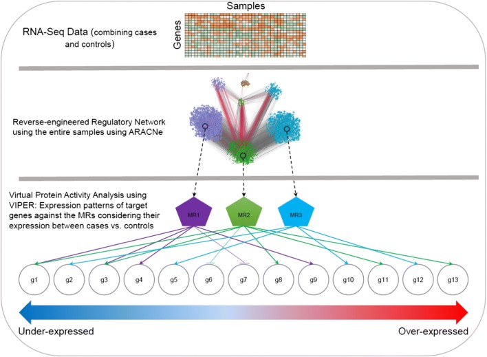
Both datasets contain multiple regions including cerebellum, which is relevant for ASDs since specific cerebellar zones can affect neocortical substrates for social interaction and cognitive functions such as language and executive functions [28–30]. Abnormalities of the cerebellum, which is believed to be involved in cognitive functions, can in part underlie autistic symptoms [31]. Several other brain regions, such as gyral surface of the anterior cingulate cortex and ventromedial prefrontal cortex [32], posterior superior temporal sulcus (pSTS) [33], amygdala, orbital frontal cortex, and fusiform gyrus [34] are also known to be ASD-relevant. We reasoned that in the same brain region, there should be highly active proteins whose expression regulates a large set of target genes and such patterns should be replicated in an independent dataset. Our preliminary finding indicates PPP1R3F (Protein Phosphatase 1 Regulatory Subunit 3F) as a potential master regulator (MR). The framework of the in silico experiments is illustrated in Fig. 1. Influence of dysregulation of this gene on ASD pathogenesis was then examined.
Fig. 1.
The overall process of network construction and virtual protein activity analysis to identify a master regulator
Results and discussion
We first used the data from Parikshak et al. [19] to construct the regulatory networks. This data is part of a large RNA-Seq repository on post-mortem human brain tissue (39 cases vs. 45 controls) from cerebellum, frontal cortex, temporal cortex, prefrontal cortex, and visual cortex. During the process of network deconvolution (see Methods in Additional file 1), pairwise MI between all of the available transcripts were obtained. Next, the constructed network was trimmed to remove genetic intermediaries, resulting in potential direct connections between MRs and their targets (we used the recommended P value threshold of 10−8, as a measure of confidence of regulatory relationships between two genes [24]). This analysis yielded a repertoire of 672,973 interactions, 23,935 regulators, and 24,847 targets in the constructed network using the dataset from Parikshak et al. [19]. We similarly analyzed the second dataset from Gupta et al. [17], a RNA-Seq data of post-mortem brain tissues with more samples of cerebellum region than other brain regions. Using the same network construction settings on this dataset [17] containing 24 cases and 38 controls, we deconvolved a network of 297,870 interactions containing 12,040 regulators and 12,529 targets. Both constructed networks are provided in Additional files 2 and 3.
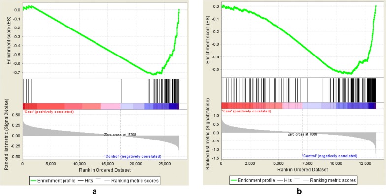
After applying VIPER, we compared the list of significant MRs at FDR ≤ 0.05. We identified PPP1R3F as the only MR shared between the two datasets. Given the small sample size of the data, it is possible that our analysis was underpowered and may have missed other relevant MRs in ASDs. Figure 2 illustrated how downregulation of this MR influences the expression of its regulons in the constructed networks of both data sets. PPP1R3F was significantly downregulated in Parikshak et al. data (FDR from one-sided t test: 0.029) as well as Gupta et al. data (FDR = 3.58 × 10⁻⁴).
Fig. 2.
Gene set enrichment analysis (GSEA) of PPP1R3F targets in the constructed networks using the data by a Parikshak et al. [16] and b Gupta et al. [14]. Black bars in the both figures depict the rank of the PPP1R3F targets in terms of correlation with the phenotype among the entire list of genes in the both datasets
PPP1R3F is one of the type-1 protein phosphatase (PP1) regulatory subunits. Protein phosphorylation is a key mechanism by which cells regulate signaling transduction pathways, and PPP1 family enzymes are associated with dephosphorylation of several proteins such as TGF-ß cascade [35]. PPP1R3F has been found to be important to neuronal activities [36]. A systematic resequencing of X-chromosome synaptic genes in a group of individuals with ASD (122 males and 20 females) has identified a rare non-synonymous variant in PPP1R3F that can predispose to developing ASDs [36]. This potentially damaging variant, c.733T > C, was observed in a boy with a diagnosis of asperger syndrome and was transmitted from a mother who suffered from learning disabilities and seizures [36].
Further, we examined the overlaps between PPP1R3F regulons and known candidate genes implicated in ASD and its related disorders (Table 1). The most significant overlap was found with SFARI gene list [37] (P = 8 × 10⁻⁴), followed by overlap with an intellectual disability database gene list (P = 0.072) [38]. The overlaps with other ASDs candidate gene lists also showed trends towards to being significant. These results suggest the potential relevance of the predicted PPP1R3F network to ASDs.
Table 1.
The overlap between the identified PPP1R3F regulons from both datasets (n = 177 genes) and several candidate gene lists of ASDs and ID (intellectual disability)
| Source of gene list | # Genes in the gene list | Overlap | P value | Fold enrichment | References |
|---|---|---|---|---|---|
| SFARI gene list (v 2.0) | 881 | 17 | 0.0008 | 2.4 | [37] |
| Intellectual disability database, University of Colorado Denver | 1095 | 11 | 0.268 | 1.2 | [41] |
| Intellectual disability database, University of Chicago | 1969 | 22 | 0.072 | 1.4 | [38] |
| Intellectual disabilities (IDS v. 1.0) | 897 | 11 | 0.097 | 1.5 | [42] |
| ASD de novo mutation list (v. 1.5)a | 1248 | 11 | 0.124 | 1.1 | [43] |
P values are calculated by two-sided Fisher’s exact test
aWe have removed de novo mutations in intergenic and intronic regions
Since PPP1R3F is a sex-linked gene, we accounted for differences between its expression in male and female samples with ASDs. In the Parikshak et al. data set (from Ref. [19]) there were 32 males and 7 females with ASDs while there were 39 male controls compared to 6 female controls. The gender information is not available on the Gupta et al. dataset [17]. We found no difference of PPP1R3F expression between male and female samples with ASDs in the Parikshak et al. dataset [19] (FDR = 0.644; two-sided t test), although this may be due to the small sample size. Nevertheless, to account for possible sex effects on the structure of the constructed network, we re-constructed the regulatory network using only male samples in the Parikshak et al. dataset [19] (i.e., 32 cases and 39 controls). Following the virtual protein activity analysis, we observed that PPP1R3F remained as a significant MR (VIPER enrichment P value = 0.0186). We note that constructing a network by using only female samples is significantly underpowered and leads to an unreliable network with a large number of false positive connections. These suggest that PPP1R3F likely acts independently from potential sex-based gene expression differences, and our observation of PPP1R3F as a MR was unlikely to be a sex-related artifact. Additionally, we conducted the same analyses on the gene expression data from prefrontal cortex, and did not find PPP1R3F as a significant MR (activity FDR = 0.1364). We should note that the number of samples from other brain regions were too small to be used for network analysis. Our finding thus suggests a potential role of PPP1R3F in developing ASDs by modulating a large body of genes in the cerebellum region.
We next conducted pathway enrichment analysis on the PPP1R3F regulons from both networks. We found that the gene targets are enriched for endocytosis pathway in both the Parikshak et al. dataset [19] (FDR = 5 × 10⁻³, fold enrichment = 8.26) and the Gupta et al. dataset [17] (FDR = 8 × 10⁻⁴, fold enrichment = 8.42). “Endocytosis” is the only significantly enriched pathway on both data sets. Combining both sets of gene targets (n = 177) (Supplementary Fig. 1 in Additional files 4 and 5) yielded a more significant enrichment of the endocytosis pathway (FDR = 4.85 × 10⁻⁴, fold enrichment = 8.97).
Since ASDs are commonly recognized as brain disorders, we further examined whether the identified MR is mainly expressed in the brain. We looked up PPP1R3F in GTEx consortium portal [39], and found that compared to other tissues, PPP1R3F is predominantly expressed in various brain regions such as frontal cortex and cerebellum (Supplementary Fig. 2 in Additional file 4). We also checked BrainSpan Atlas of the Developing Human Brain (http://brainspan.org) where we found that PPP1R3F is not expressed until 37 weeks post-conception. While remaining unexpressed in some brain regions, it is modestly expressed in 4 month postnatal stage in some brain regions including cerebellum. We further probed the expression of each of the 177 targets of PPP1R3F in GTEx and identified the tissues in which they are highly expressed. We found that 89 out of the 177 target genes of PPP1R3F are highly expressed in various brain regions compared to other tissues (P= 5.51 × 10⁻⁵, Fisher’s exact test; number of protein coding genes in GTEx = 20,900, number of protein coding genes highly expressed in the brain in GTEx = 7528). The enrichment of the expressed PPP1R3F target genes for those highly expressed in the brain supports the pathophysiological relevance of PPP1R3F to ASDs.
Conclusions
In this study, we performed exploratory analysis on two small-scale RNA-Seq data sets, and used a network deconvolution algorithm to construct regulatory networks. Applying virtual protein activity analysis on both networks, we identified PPP1R3F as a MR of 177 targets genes. Gene set enrichment analysis on the PPP1R3F regulons suggested that PPP1R3F may exert its functional effects through regulating endocytosis, a pathway that has been previously implicated in neuropsychiatric disorders [40].
Limitations
We acknowledge that our study is limited by the small sample size (due to the scarcity of brain tissues), and the results thus need further replications. Nonetheless, our study generates a testable hypothesis that may be validated by large-scale studies in the future. Additionally, further experimental validation of the regulatory effects of PPP1R3F on its downstream targets as predicted by our network analysis may provide novel insights on possible pathophysiological role of PPP1R3F as a MR of ASD gene network.
Additional files
Additional file 1. Detailed explanation of the methods being used in this study.
Additional file 2. The constructed networks from the Parikshak et al. dataset [19].
Additional file 3. The constructed networks from the Gupta et al. dataset [17].
Additional file 4. Supplementary figures.
Additional file 5. The list of the combined set of target genes of PPP1R3F.
Authors’ contributions
ADT designed the experiments, conducted the analysis and computations, and wrote the manuscript. JD advised on data analysis and edited the manuscript. KW designed the experiments, supervised data analysis and edited the manuscript. All authors read and approved the final manuscript.
Acknowledgements
The authors would like to thank the PsychENCODE consortium for providing the data. Data were generated as part of the PsychENCODE Consortium, supported by: U01MH103339, U01MH103365, U01MH103392, U01MH103340, U01MH103346, R01MH105472, R01MH094714, R01MH105898, R21MH102791, R21MH105881, R21MH103877, and P50MH106934 awarded to: Schahram Akbarian (Icahn School of Medicine at Mount Sinai), Gregory Crawford (Duke), Stella Dracheva (Icahn School of Medicine at Mount Sinai), Peggy Farnham (USC), Mark Gerstein (Yale), Daniel Geschwind (UCLA), Thomas M. Hyde (LIBD), Andrew Jaffe (LIBD), James A. Knowles (USC), Chunyu Liu (UIC), Dalila Pinto (Icahn School of Medicine at Mount Sinai), Nenad Sestan (Yale), Pamela Sklar (Icahn School of Medicine at Mount Sinai), Matthew State (UCSF), Patrick Sullivan (UNC), Flora Vaccarino (Yale), Sherman Weissman (Yale), Kevin White (UChicago) and Peter Zandi (JHU). We would also like to thank the providers of the Gupta et al. dataset (The Arking Lab at the McKusick-Nathans Institute of Genetic Medicine of Johns Hopkins University) to generate the brain gene expression data and make the data freely available.
Competing interests
The authors declare that they have no competing interests.
Availability of data and materials
All data used in this study are available from references [17, 19].
Consent for publication
Not applicable.
Ethics approval and consent to participate
Not applicable.
Funding
This study is supported by NIH grant MH108728 to K.W.
Publisher’s Note
Springer Nature remains neutral with regard to jurisdictional claims in published maps and institutional affiliations.
Abbreviations
- ASDs
autism spectrum disorders
- NGS
next generation sequencing
- GWAS
genome-wide association studies
- PTSD
post-traumatic stress disorder
- ARACNe
algorithm for reconstruction of accurate cellular networks
- MI
mutual information
- MR
master regulator
- VIPER
virtual inference of protein-activity by enhanced regulon analysis
- GSEA
gene set enrichment analysis
Footnotes
Electronic supplementary material
The online version of this article (10.1186/s13104-018-3594-0) contains supplementary material, which is available to authorized users.
Contributor Information
Abolfazl Doostparast Torshizi, doostparaa@email.chop.edu.
Jubao Duan, Email: jduan@uchicago.edu.
Kai Wang, wangk@email.chop.edu.
References
- 1.de la Torre-Ubieta L, Won HJ, Stein JL, Geschwind DH. Advancing the understanding of autism disease mechanisms through genetics. Nat Med. 2016;22:345–361. doi: 10.1038/nm.4071. [DOI] [PMC free article] [PubMed] [Google Scholar]
- 2.Li J, Wang L, Guo H, Shi L, Zhang K, Tang M, Hu S, Dong S, Liu Y, Wang T, et al. Targeted sequencing and functional analysis reveal brain-size-related genes and their networks in autism spectrum disorders. Mol Psychiatry. 2017;22:1282. doi: 10.1038/mp.2017.140. [DOI] [PubMed] [Google Scholar]
- 3.Christensen DL, Bilder DA, Zahorodny W, Pettygrove S, Durkin MS, Fitzgerald RT, Rice C, Kurzius-Spencer M, Baio J, Yeargin-Allsopp M. Prevalence and characteristics of autism spectrum disorder among 4-year-old children in the autism and developmental disabilities monitoring network. J Dev Behav Pediatry. 2016;37:1–8. doi: 10.1097/DBP.0000000000000235. [DOI] [PubMed] [Google Scholar]
- 4.Freitag CM. The genetics of autistic disorders and its clinical relevance: a review of the literature. Mol Psychiatry. 2007;12:2–22. doi: 10.1038/sj.mp.4001896. [DOI] [PubMed] [Google Scholar]
- 5.Sandin S, Lichtenstein P, Kuja-Halkola R, Larsson H, Hultman CM, Reichenberg A. The familial risk of autism. JAMA. 2014;311:1770–1777. doi: 10.1001/jama.2014.4144. [DOI] [PMC free article] [PubMed] [Google Scholar]
- 6.Iossifov I, Ronemus M, Levy D, Wang Z, Hakker I, Rosenbaum J, Yamrom B, Lee YH, Narzisi G, Leotta A, et al. De novo gene disruptions in children on the autistic spectrum. Neuron. 2012;74:285–299. doi: 10.1016/j.neuron.2012.04.009. [DOI] [PMC free article] [PubMed] [Google Scholar]
- 7.O’Roak BJ, Vives L, Girirajan S, Karakoc E, Krumm N, Coe BP, Levy R, Ko A, Lee C, Smith JD, et al. Sporadic autism exomes reveal a highly interconnected protein network of de novo mutations. Nature. 2012;485:246–250. doi: 10.1038/nature10989. [DOI] [PMC free article] [PubMed] [Google Scholar]
- 8.Yu TW, Chahrour MH, Coulter ME, Jiralerspong S, Okamura-Ikeda K, Ataman B, Schmitz-Abe K, Harmin DA, Adli M, Malik AN, et al. Using whole-exome sequencing to identify inherited causes of autism. Neuron. 2013;77:259–273. doi: 10.1016/j.neuron.2012.11.002. [DOI] [PMC free article] [PubMed] [Google Scholar]
- 9.De Rubeis S, He X, Goldberg AP, Poultney CS, Samocha K, Cicek AE, Kou Y, Liu L, Fromer M, Walker S, et al. Synaptic, transcriptional and chromatin genes disrupted in autism. Nature. 2014;515:209–215. doi: 10.1038/nature13772. [DOI] [PMC free article] [PubMed] [Google Scholar]
- 10.Iossifov I, O’Roak BJ, Sanders SJ, Ronemus M, Krumm N, Levy D, Stessman HA, Witherspoon KT, Vives L, Patterson KE, et al. The contribution of de novo coding mutations to autism spectrum disorder. Nature. 2014;515:216–221. doi: 10.1038/nature13908. [DOI] [PMC free article] [PubMed] [Google Scholar]
- 11.Yean RKC, Merico D, Bookman M, Howe JL, Thiruvahindrapuram B, Patel RV, Whitney J, Deflaux N, Bingham J, Wang Z, et al. Whole genome sequencing resource identifies 18 new candidate genes for autism spectrum disorder. Nat Neurosci. 2017;20:602–611. doi: 10.1038/nn.4524. [DOI] [PMC free article] [PubMed] [Google Scholar]
- 12.Yuen RK, Thiruvahindrapuram B, Merico D, Walker S, Tammimies K, Hoang N, Chrysler C, Nalpathamkalam T, Pellecchia G, Liu Y, et al. Whole-genome sequencing of quartet families with autism spectrum disorder. Nat Med. 2015;21:185–191. doi: 10.1038/nm.3792. [DOI] [PubMed] [Google Scholar]
- 13.Weiss LA, Arking DE, Gene Discovery Project of Johns H, the Autism C. Daly MJ, Chakravarti A. A genome-wide linkage and association scan reveals novel loci for autism. Nature. 2009;461:802–808. doi: 10.1038/nature08490. [DOI] [PMC free article] [PubMed] [Google Scholar]
- 14.Wang K, Zhang H, Ma D, Bucan M, Glessner JT, Abrahams BS, Salyakina D, Imielinski M, Bradfield JP, Sleiman PM, et al. Common genetic variants on 5p14.1 associate with autism spectrum disorders. Nature. 2009;459:528–533. doi: 10.1038/nature07999. [DOI] [PMC free article] [PubMed] [Google Scholar]
- 15.Autism Spectrum Disorders Working Group of The Psychiatric Genomics C: meta-analysis of GWAS of over 16,000 individuals with autism spectrum disorder highlights a novel locus at 10q24.32 and a significant overlap with schizophrenia. Mol Autism. 2017;8:21. doi: 10.1186/s13229-017-0137-9. [DOI] [PMC free article] [PubMed] [Google Scholar]
- 16.Anney R, Klei L, Pinto D, Regan R, Conroy J, Magalhaes TR, Correia C, Abrahams BS, Sykes N, Pagnamenta AT, et al. A genome-wide scan for common alleles affecting risk for autism. Hum Mol Genet. 2010;19:4072–4082. doi: 10.1093/hmg/ddq307. [DOI] [PMC free article] [PubMed] [Google Scholar]
- 17.Gupta S, Ellis SE, Ashar FN, Moes A, Bader JS, Zhan J, West AB, Arking DE. Transcriptome analysis reveals dysregulation of innate immune response genes and neuronal activity-dependent genes in autism. Nat Commun. 2014;5:5748. doi: 10.1038/ncomms6748. [DOI] [PMC free article] [PubMed] [Google Scholar]
- 18.Voineagu I, Wang X, Johnston P, Lowe JK, Tian Y, Horvath S, Mill J, Cantor RM, Blencowe BJ, Geschwind DH. Transcriptomic analysis of autistic brain reveals convergent molecular pathology. Nature. 2011;474:380–384. doi: 10.1038/nature10110. [DOI] [PMC free article] [PubMed] [Google Scholar]
- 19.Parikshak NN, Swarup V, Belgard TG, Irimia M, Ramaswami G, Gandal MJ, Hartl C, Leppa V, Ubieta LT, Huang J, et al. Genome-wide changes in lncRNA, splicing, and regional gene expression patterns in autism. Nature. 2016;540:423–427. doi: 10.1038/nature20612. [DOI] [PMC free article] [PubMed] [Google Scholar]
- 20.Dutta A, Le Magnen C, Mitrofanova A, Ouyang X, Califano A, Abate-Shen C. Identification of an NKX3.1-G9a-UTY transcriptional regulatory network that controls prostate differentiation. Science. 2016;352:1576–1580. doi: 10.1126/science.aad9512. [DOI] [PMC free article] [PubMed] [Google Scholar]
- 21.Alvarez MJ, Shen Y, Giorgi FM, Lachmann A, Ding BB, Ye BH, Califano A. Functional characterization of somatic mutations in cancer using network-based inference of protein activity. Nat Genet. 2016;48:838–847. doi: 10.1038/ng.3593. [DOI] [PMC free article] [PubMed] [Google Scholar]
- 22.Doostparast Torshizi A, Armoskus C, Zhang S, Zhang H, Evgrafov OV, Knowles JA, Duan J, Wang K. Deconvolution of transcriptional networks identified TCF4 as a master regulator in schizophrenia. BioRxiv. 2017 doi: 10.1126/sciadv.aau4139. [DOI] [PMC free article] [PubMed] [Google Scholar]
- 23.Doostparast Torshizi A, Wang K. Deconvolution of transcriptional networks in post-traumatic stress disorder uncovers master regulators driving innate immune system function. Sci Rep. 2017;7:14486. doi: 10.1038/s41598-017-15221-y. [DOI] [PMC free article] [PubMed] [Google Scholar]
- 24.Margolin AA, Wang K, Lim WK, Kustagi M, Nemenman I, Califano A. Reverse engineering cellular networks. Nat Protoc. 2006;1:662–671. doi: 10.1038/nprot.2006.106. [DOI] [PubMed] [Google Scholar]
- 25.Boorsma A, Foat BC, Vis D, Klis F, Bussemaker HJ. T-profiler: scoring the activity of predefined groups of genes using gene expression data. Nucleic Acids Res. 2005;33:W592–W595. doi: 10.1093/nar/gki484. [DOI] [PMC free article] [PubMed] [Google Scholar]
- 26.Subramanian A, Tamayo P, Mootha VK, Mukherjee S, Ebert BL, Gillette MA, Paulovich A, Pomeroy SL, Golub TR, Lander ES, Mesirov JP. Gene set enrichment analysis: a knowledge-based approach for interpreting genome-wide expression profiles. Proc Natl Acad Sci USA. 2005;102:15545–15550. doi: 10.1073/pnas.0506580102. [DOI] [PMC free article] [PubMed] [Google Scholar]
- 27.Abatangelo L, Maglietta R, Distaso A, D’Addabbo A, Creanza TM, Mukherjee S, Ancona N. Comparative study of gene set enrichment methods. BMC Bioinf. 2009;10:275. doi: 10.1186/1471-2105-10-275. [DOI] [PMC free article] [PubMed] [Google Scholar]
- 28.Wang SS, Kloth AD, Badura A. The cerebellum, sensitive periods, and autism. Neuron. 2014;83:518–532. doi: 10.1016/j.neuron.2014.07.016. [DOI] [PMC free article] [PubMed] [Google Scholar]
- 29.Becker EB, Stoodley CJ. Autism spectrum disorder and the cerebellum. Int Rev Neurobiol. 2013;113:1–34. doi: 10.1016/B978-0-12-418700-9.00001-0. [DOI] [PubMed] [Google Scholar]
- 30.Hampson DR, Blatt GJ. Autism spectrum disorders and neuropathology of the cerebellum. Front Neurosci. 2015;9:420. doi: 10.3389/fnins.2015.00420. [DOI] [PMC free article] [PubMed] [Google Scholar]
- 31.Rogers TD, McKimm E, Dickson PE, Goldowitz D, Blaha CD, Mittleman G. Is autism a disease of the cerebellum? An integration of clinical and pre-clinical research. Front Syst Neurosci. 2013;7:15. doi: 10.3389/fnsys.2013.00015. [DOI] [PMC free article] [PubMed] [Google Scholar]
- 32.Balsters JH, Apps MA, Bolis D, Lehner R, Gallagher L, Wenderoth N. Disrupted prediction errors index social deficits in autism spectrum disorder. Brain. 2017;140:235–246. doi: 10.1093/brain/aww287. [DOI] [PMC free article] [PubMed] [Google Scholar]
- 33.Alaerts K, Woolley DG, Steyaert J, Di Martino A, Swinnen SP, Wenderoth N. Underconnectivity of the superior temporal sulcus predicts emotion recognition deficits in autism. Soc Cogn Affect Neurosci. 2014;9:1589–1600. doi: 10.1093/scan/nst156. [DOI] [PMC free article] [PubMed] [Google Scholar]
- 34.Kaiser MD, Pelphrey KA. Disrupted action perception in autism: behavioral evidence, neuroendophenotypes, and diagnostic utility. Dev Cogn Neurosci. 2012;2:25–35. doi: 10.1016/j.dcn.2011.05.005. [DOI] [PMC free article] [PubMed] [Google Scholar]
- 35.Korrodi-Gregorio L, Silva JV, Santos-Sousa L, Freitas MJ, Felgueiras J, Fardilha M. TGF-beta cascade regulation by PPP1 and its interactors -impact on prostate cancer development and therapy. J Cell Mol Med. 2014;18:555–567. doi: 10.1111/jcmm.12266. [DOI] [PMC free article] [PubMed] [Google Scholar]
- 36.Piton A, Gauthier J, Hamdan FF, Lafreniere RG, Yang Y, Henrion E, Laurent S, Noreau A, Thibodeau P, Karemera L, et al. Systematic resequencing of X-chromosome synaptic genes in autism spectrum disorder and schizophrenia. Mol Psychiatry. 2011;16:867–880. doi: 10.1038/mp.2010.54. [DOI] [PMC free article] [PubMed] [Google Scholar]
- 37.Abrahams BS, Arking DE, Campbell DB, Mefford HC, Morrow EM, Weiss LA, Menashe I, Wadkins T, Banerjee-Basu S, Packer A. SFARI gene 2.0: a community-driven knowledgebase for the autism spectrum disorders (ASDs) Mol Autism. 2013;4:36. doi: 10.1186/2040-2392-4-36. [DOI] [PMC free article] [PubMed] [Google Scholar]
- 38.http://dnatesting.uchicago.edu/tests/intellectual-disability-exome-panel. Accessed 5 Jan 2018.
- 39.Consortium GT The genotype-tissue expression (GTEx) project. Nat Genet. 2013;45:580–585. doi: 10.1038/ng.2653. [DOI] [PMC free article] [PubMed] [Google Scholar]
- 40.Loebrich S. The role of F-actin in modulating Clathrin-mediated endocytosis: lessons from neurons in health and neuropsychiatric disorder. Commun Integr Biol. 2014;7:e28740. doi: 10.4161/cib.28740. [DOI] [PMC free article] [PubMed] [Google Scholar]
- 41.http://gfuncpathdb.ucdenver.edu/iddrc/iddrc/home.php. Accessed 4 Jan 2018.
- 42.Chiurazzi P, Pirozzi F. Advances in understanding - genetic basis of intellectual disability. F1000Res. 2016;5(F1000 Faculty Rev):599. doi: 10.12688/f1000research.7134.1. [DOI] [PMC free article] [PubMed] [Google Scholar]
- 43.Turner TN, Yi Q, Krumm N, Huddleston J, Hoekzema K, Stessman HAF, Doebley AL, Bernier RA, Nickerson DA, Eichler EE, Hoekzema K. denovo-db: a compendium of human de novo variants. Nucleic Acids Res. 2017;45:D804–D811. doi: 10.1093/nar/gkw865. [DOI] [PMC free article] [PubMed] [Google Scholar]
Associated Data
This section collects any data citations, data availability statements, or supplementary materials included in this article.
Supplementary Materials
Additional file 1. Detailed explanation of the methods being used in this study.
Additional file 2. The constructed networks from the Parikshak et al. dataset [19].
Additional file 3. The constructed networks from the Gupta et al. dataset [17].
Additional file 4. Supplementary figures.
Additional file 5. The list of the combined set of target genes of PPP1R3F.
Data Availability Statement
All data used in this study are available from references [17, 19].


