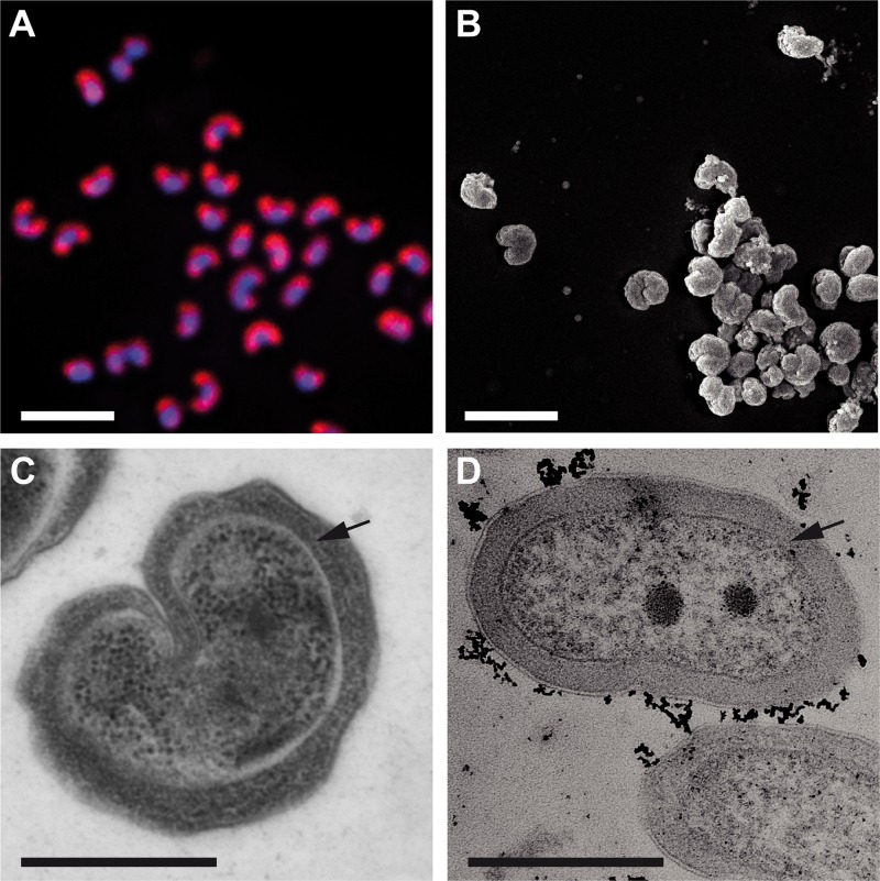
FIG 2 .
Morphology of “Ca. Nitrotoga fabula.” (A) Pure culture of “Ca. Nitrotoga fabula” visualized by FISH with the “Ca. Nitrotoga”-specific probe Ntoga122 (red) and by DAPI staining (blue). Bar, 2 µm. (B) Scanning electron micrograph imaged after chemical fixation (bar, 2 µm). (C and D) Transmission electron micrographs (after cryopreservation [C] and after chemical fixation [D]; bars, 0.5 µm). Black arrows indicate the periplasmic space.

