To the editor
Chemokine receptors (CKRs) are critical for the sensitization, elicitation, and resolution of allergic contact dermatitis (ACD). Endogenous chemokines of the CKR CXCR3, which is expressed primarily on T cells, are upregulated in ACD. T cells primarily regulate the pathophysiology of ACD by producing cytokines that induce inflammation and edema, directly killing cells, and activating other immune cell populations. Blocking T cell infiltration in the skin is a viable therapeutic strategy for treating ACD, and improved understanding of the chemokine receptors selectively expressed on T cells in ACD may direct more targeted therapies for this disease.
This study was approved by the Duke University Institutional Review Board. All participants provided written informed consent. Study participation was offered to patients ≥18 years completing patch testing in a specialty contact dermatitis clinic. Exclusion criteria were pregnancy, topical corticosteroids at patch site, oral corticosteroids, systemic immunosuppressants, phototherapy, known bleeding disorders and allergy to lidocaine or epinephrine. Allergens were applied with Finn Chambers (SmartPractice, Phoenix, AZ) on Scanpor tape (Norgesplaster Alpharma AS, Vennesla, Norway). Readings were completed on D2 and D4/D5 and designated negative, doubtful, weak positive, strong positive, extreme positive, or irritant in accordance with International Contact Dermatitis Research Group criteria.1 Weak, strong, and extreme positive reactions were considered positive patch tests.
If a participant had a positive patch test, a 4mm punch biopsy at both the positive test and a negative (non-lesional) site was obtained. If a participant had a completely negative patch test, a 4mm punch biopsy at a negative test site (non-lesional) was obtained.
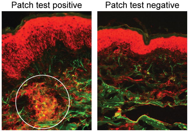
Immunofluorescence of skin biopsy specimens was conducted as previously described to examine colocalization of the CKR CXCR3 (red stain) with T cells (assessed by the T cell specific marker CD3, green stain) in patch-tested skin and matched negative (non-lesional) sites.2 The findings from two representative patients are summarized here.
We report that CXCR3+ T cells are present in the dermis of patch test positive, but not patch-test negative, skin specimens. Immunofluorescence staining revealed co-localization of the chemokine receptor CXCR3 and the T cell marker CD3 in the dermis in patch test positive, but not negative, samples (Figure 1, 20× image). Furthermore, CXCR3+ T cells typically localized in dermal clusters, reminiscent of dermal-dendritic cell-T cell clusters recently described in ACD.2 This corroborates prior work that CXCR3 activating chemokines CXCL9, CXCL10, and CXCL11 are upregulated in allergic contact dermatitis3,4 and that the CXCR3 signaling pathway underlies ACD (Smith et al., in revision). Unlike many other CKRs, the CXCR3 signaling pathway appears relatively selective for allergic, but not irritant, contact dermatitis, making CXCR3 an attractive therapeutic target.5 Targeting CXCR3 holds therapeutic promise for improved treatment of ACD, although more research is needed to follow up on this encouraging preclinical data.
Figure 1.
Acknowledgments
Sources of support
T32GM7171 (JSS), the Duke Medical Scientist Training Program (JSS), and the Duke Pinnell Center for Investigative Dermatology (JSS/ASM/ARA), R21AI28727 (to ASM), K08 AR063729 (to ASM), Duke Physician-Scientist Strong Start Award (to ASM), Dermatology Foundation Research Grant (to ASM).
Footnotes
The authors declare no conflict of interest.
References
- 1.Wilkinson DS, et al. Terminology of contact dermatitis. Acta Derm Venereol. 1970;50:287–292. [PubMed] [Google Scholar]
- 2.Suwanpradid J, et al. Arginase1 Deficiency in Monocytes/Macrophages Upregulates Inducible Nitric Oxide Synthase To Promote Cutaneous Contact Hypersensitivity. J Immunol. 2017;199:1827–1834. doi: 10.4049/jimmunol.1700739. [DOI] [PMC free article] [PubMed] [Google Scholar]
- 3.Flier J, et al. The CXCR3 activating chemokines IP-10, Mig, and IP-9 are expressed in allergic but not in irritant patch test reactions. The Journal of investigative dermatology. 1999;113:574–578. doi: 10.1046/j.1523-1747.1999.00730.x. [DOI] [PubMed] [Google Scholar]
- 4.Goebeler M, et al. Differential and sequential expression of multiple chemokines during elicitation of allergic contact hypersensitivity. Am J Pathol. 2001;158:431–440. doi: 10.1016/s0002-9440(10)63986-7. [DOI] [PMC free article] [PubMed] [Google Scholar]
- 5.Meller S, et al. Chemokine responses distinguish chemical-induced allergic from irritant skin inflammation: memory T cells make the difference. J Allergy Clin Immunol. 2007;119:1470–1480. doi: 10.1016/j.jaci.2006.12.654. [DOI] [PubMed] [Google Scholar]


