Abstract
Wind-up is a frequency-dependent increase in the response of spinal cord neurons, which is thought to underlie temporal summation of nociceptive input. However, whether spinoparabrachial neurons, which likely contribute to the affective component of pain, undergo wind-up was unknown. Here, we addressed this question and investigated the underlying neural circuit. We show that one-fifth of lamina I spinoparabrachial (SPB) neurons undergo wind-up, and provide evidence that wind-up in these cells is mediated in part by a network of spinal excitatory interneurons that show reverberating activity. These findings provide insight into a polysynaptic circuit of sensory augmentation that may contribute to the wind-up of pain’s unpleasantness.
INTRODUCTION
Wind-up is a type of facilitation observed in spinal cord neurons in which the response to repetitive stimulation of peripheral C-fibers increases with each stimulus [18]. This mechanism of amplification was first described over fifty years ago in lamina IV spinocervical neurons, where it was shown to be a frequency-dependent phenomenon [22; 23]. Since then, this form of amplification has been observed in a variety of spinal neurons, including wide dynamic range (WDR) neurons in the deep dorsal horn, unidentified neurons in the superficial dorsal horn, and motor neurons in the ventral horn [32; 44; 45].
It has long been speculated that wind-up could contribute to the sensory experience of temporal summation, the increase in the perception of pain in response to repeated stimulation [2; 8]. Consistent with this possibility, temporal summation in response to electrical, thermal or mechanical stimulation, is frequency-dependent, and requires stimulus intensities capable of activating C-fibers [1; 3; 20; 21; 40]. Both wind-up and temporal summation are physiological mechanisms of amplification that have the potential to contribute to hypersensitivity and allodynia following injury, as suggested by the observations that wind-up is magnified in mice with peripheral inflammation [17; 18; 35; 38], and temporal summation is heightened in pain patients [12; 25; 28; 39]. Thus, understanding the neural circuits of wind-up is important because these circuits may contribute to pathological pain [11; 42].
Although wind-up has been studied extensively, there remain two major unanswered questions. The first pertains to the affective component of pain. Many earlier studies have focused on amplification of reflexes, using wind-up in the ventral horn and/or motor roots as a primary endpoint with which to tease apart mechanisms underlying this phenomenon [27; 32; 44]. Wind-up also occurs in spinal output neurons that project, directly or indirectly to the thalamus, such as WDR spinothalamic neurons [45] and spinocervical neurons [22; 23]. Since somatosensory input to the thalamus contributes to sensory discrimination, wind-up in these cells could account for the psychophysical phenomenon of temporal summation as manifest by reports of increasing pain intensity with repeated application of a noxious stimulus [20]. What remains unclear is whether there is wind-up in sensory affective pathways that mediate the unpleasantness of pain. Spinal output neurons that target the lateral parabrachial nucleus, which convey nociceptive and thermoregulatory information, are thought to contribute to the affective component of pain [5; 14]. However, whether wind-up occurs in spinoparabrachial (SPB) neurons is unknown. Given that pain unpleasantness shows temporal summation [31], we hypothesized that wind-up would occur in SPB neurons.
The second unresolved question is whether synaptic mechanisms alone are sufficient to account for wind-up. The prevailing theory is that wind-up is mediated by NMDA receptors, which are recruited through a relief of magnesium block that occurs upon cumulative depolarization in response to repetitive stimulation [8]. A key role of NMDA receptors in wind-up is supported by the findings that NMDA receptor antagonists inhibit wind-up of WDR neurons [7; 9], ventral horn neurons [36], and motor neurons [43]. Accordingly, NMDA receptor antagonists also reduce temporal summation in healthy humans [2; 29], and abnormal temporal summation in the context of injury-induced pain [13; 41]. However, the block of wind-up by NMDA receptor antagonists is not always complete, raising the possibility that other mechanisms are likely to contribute [7; 9; 36].
One possibility is a circuit-based mechanism in which an excitatory interneuron network in the dorsal horn acts an amplifier. Indeed, such a circuit-based mechanism was originally put forth by Mendell is his seminal study, in which he postulated that wind-up may be due to the reverberatory activity in spinal interneurons that is evoked by afferent C-fiber input, reasoning that “if in this period of time another stimulation arrives to the cord, it sums with the ongoing activity to produce a more intense discharge in the interneurons than the one before it [22].” Consistent with this idea, wind-up is observed in a variety of spinal neurons that do not receive appreciable C-fiber input, indicating that this wind-up must be mediated through a polysynaptic circuit. However, the nature of this circuit, and whether it contributes to (rather than simply propagating) wind-up is not clear.
We recently developed an ex vivo somatosensory preparation that enables the recording from lamina I SPB neurons together with cell-type specific manipulation [16]. We therefore set out to test the hypotheses that wind-up occurs in lamina I SPB neurons, and that a network of excitatory interneurons is involved in mediating this amplification. Here, we report that approximately one-fifth of SPB neurons show wind-up upon repetitive stimulation of the dorsal root. In addition, we provide evidence that optogenetic activation of excitatory interneurons is sufficient for wind-up. This effect is selective for some excitatory networks since it is observed upon activation neurons of the neurotensin lineage (NtsCre) neurons) but not the calretinin lineage (CrCre) neurons. Through optogenetic inhibition, we show that activity in NtsCre neurons is required for dorsal root-evoked wind-up. Finally, our data suggest that this facilitation is mediated, at least in part, by reverberatory activity within an interconnected excitatory network. Together, these studies show the existence of wind-up in lamina I SPB neurons, and provide new insight into the underlying neural circuit basis.
METHODS
Mouse lines
The mouse lines used for this study were all obtained from The Jackson Laboratories and maintained on C57BL/6J background: NtsCre, a non-disruptive IRES-Cre recombinase knock-in at the endogenous Neurotensin locus (Stock number: 017525); CrCre, a non-disruptive IRES-Cre recombinase knock-in at the endogenous Calretinin locus (Stock number: 010774); Ai9, enabling Cre-dependent expression of tdTomato (Gt(ROSA)26Sortm9(CAG-tdTomato), Stock number: 007909); Ai32, enabling Cre-dependent expression of an enhanced channelrhodopsin fusion protein, ChR2(H134)/EYFP (Gt (ROSA)26Sortm32(CAG-COP4*H134R/EYFP), Stock number: 012569); and Ai35, enabling Cre-dependent expression of an Archaerhodopsin fusion protein (Gt(ROSA)26Sortm35.1(CAG-aop3/GFP); Stock number: 012735). Genotyping for these alleles was performed with the following primers: for Ai9, TdTR (GGC ATT AAA GCA GCG TAT CC) and TdTR (CTG TTC CTG TAC GGC ATG G) were used to detect TdTomato (196 bp product); for Ai32, ChR2F (ACA TGG TCC TGC TGG AGT TC) and ChR2R (GGC ATT AAA GCA GCG TAT CC) were used to detect ChR2 (212 bp product); for Ai35, XFPF (GCG AGG GCG AGG GCG ATG) and XFPR (CGA TGT TGT GGC GGA TCT TG) were used to detect EYFP (423 bp product), and for the wild type Rosa allele, RosaWTF (GGA GCG GGA GAA ATG GAT ATG) and RosaWTR (AAA GTC GCT CTG AGT TGT TAT) were used (~550 bp product).
Four- to seven-week-old mice of both sexes were used in this study. Mice were given free access to food and water and housed under standard laboratory conditions. The use of animals was approved by the Institutional Animal Care and Use Committee of the University of Pittsburgh.
Immunohistochemistry
Four NtsCre; Gt(ROSA)26Sortm9(CAG-tdTomato) adult mice were perfused with 4% paraformaldehyde and the lumbar spinal cords from L2 to L3 were dissected and subsequently post-fixed for 4 hours. Transverse 65 micron-thick sections were cut on a vibrating microtome and processed free-floating for immunohistochemistry. Sections were blocked in blocking solution (10% donkey serum, 0.1% triton in phosphate buffered saline) for two hours and incubated in the following primary antibodies for 14 hours overnight at 4 C: NeuN (1:1000 Millipore MAB377) and Pax2 (1:1000, Life Technologies 716000) as well as Biotin-conjugated Isolectin B4 (1:200, Sigma Aldrich L2140). Following three 20-minute washes with wash buffer (0.1% triton, 1% donkey serum, 0.3 M NaCl), sections were incubated with Alexa Fluor-conjugated secondary antibodies (1:500; Life Technologies) and Streptavidin-488 (1:500; Thermo Fisher) and for 2 hours at room temperature. Next, sections were incubated with Hoechst (1:10,000; Thermo Fisher incubation for 1 min to label nuclei. Seven 15-minute washes were performed, and then sections were mounted on slides and coverslipped. The dorsal horns were imaged through a single optical plane using a Nikon A1R confocal microscope with a 20× objective. Only cells with clearly visible nuclei were counted.
Labeling SPB neurons
Four- to six-week-old mice were anesthetized with isoflurane and placed in a stereotaxic frame. A small hole was made on the skull with dental drill. A glass micropipette was used to inject 100 nl of FAST DiI oil (2.5 mg/ml; Invitrogen) into left lateral parabrachial nucleus (relative to lambda: anteroposterior −0.5 mm; lateral 1.3 mm; dorsoventral −2.4 mm). The head wound was closed with stitches. After recovery from the anesthesia, the animals fed and drank normally. The animals were used for electrophysiology 4- to 7-days later.
Whole spinal cord preparation
For electrophysiological recordings, we used a modified semi-intact preparation (Hachisuka et al., 2016). We recorded from neurons in the L2 spinal segment, which are easiest to visualize and record from in this preparation. In brief, five- to seven-week old mice were deeply anesthetized with urethane (1.2 g/kg, I.P.) The animals were perfused transcardially through the left ventricle with ice-cold oxygenated (95% O2 and 5% CO2) sucrose-based artificial cerebrospinal fluid (ACSF) (in mM; 234 sucrose, 2.5 KCl, 0.5 CaCl2, 10 MgSO4, 1.25 NaH2PO4, 26 NaHCO3, 11 Glucose). Immediately following perfusion, the skin was incised along the dorsal midline and the spinal cord was quickly excised and placed it into an ice-cold, sucrose-based Krebs solution. Dura and pia-arachnoid membrane were removed after cutting all of the ventral and dorsal roots except the L2 root on the right. The spinal cord was placed in the recording chamber and pinned into a chamber wall made from Sylguard®. The spinal cord was perfused with Krebs solution saturated with 95% O2, and 5% CO2 at 30–31 °C. The Krebs solution contained (mM): 117 NaCl, 3.6 KCl, 2.5 CaCl2, 1.2 MgCl2, 1.2 NaH2PO4, 25 NaHCO3 and 11 glucose.
Patch clamp recording from dorsal horn neurons
Neurons were visualized using a fixed stage upright microscope (BX51WI Olympus microscope, Tokyo, Japan) equipped with a 40× water immersion objective lens, a CCD camera (ORCA-ER Hamamatsu Photonics) and monitor screen. A narrow beam infrared LED (L850D-06 Marubeni, Tokyo, Japan, emission peak, 850 nm) was positioned outside the solution meniscus, as previously described (Hachisuka et al., 2016; Pinto et al., 2008, 2010; Safronov et al., 2007). To record from NtsCre neurons in optogenetic experiments, mice were generated that harbored both Ai14 and Ai32 alleles, and cells were identified by expression of tdTomato. SPB neurons located within 20 μm from the surface of the dorsal horn were identified by DiI labeling.
Whole-cell patch-clamp recordings were made with an Axopatch 200B amplifier with a Digidata 1322A A/D converter controlled by Clampex software (version 10), all from Molecular Devices. Patch-pipette electrodes had a resistance of 6–12 MΩ when filled with a pipette solution of the following composition (mM); 135 potassium gluconate, 5 KCl, 0.5 CaCl2, 5 EGTA, 5 Hepes, 5 ATP-Mg, pH 7.2. Alexa Fluor 488 was added to aid in visualization. The data were low-pass filtered at 2 kHz and digitized at 10 kHz. The liquid junction potential was not corrected.
Cell recordings were made in voltage-clamp mode at holding potentials of −70 mV to record excitatory postsynaptic currents (EPSCs) and current-clamp mode to record action potentials (APs). Frequency of EPSCs and APs were analyzed using MiniAnalysis (Synaptosoft, Inc.). The L2 dorsal root was stimulated by suction electrode with 100 μs duration. Aδ-fiber evoked responses were considered monosynaptic if the latency remained constant when the root was stimulated at 20 Hz and there was no failure and C-fiber evoked responses were considered monosynaptic if there was no failure at 2 Hz (Nakatsuka et al., 1999). To investigate wind-up, dorsal root stimulation was applied at 2Hz and the number of action potentials (APs) after each stimulation (0.5 s window) was counted. The criteria used to define the presence of wind-up were that the maximum number of APs was at least five and more than twice as many action potentials were evoked in response to subsequent stimuli as were evoked in response to the first stimulus.
Optogenetic activation
During patch clamp recording, photo stimulation was applied to the spinal cord through the objective lens (40x) of the microscope with a Xenon lamp (Lambda DG-4, Sutter Instrument). A Lamdba DG4 (Sutter Inst.) was used for optogenetic stimulation, where switching between filter positions (0.5 ms) was controlled by a TTL pulse from the output of the A/D converter. We used a GFP filter (centered around 485 nm) for activation of ChR2 and a Cy3 filter (centered around 555 nm) for activation of Arch. Light power on the sample was 1.3 mW/mm2. To examine whether the recorded neuron received mono- or polysynaptic input from NtsCre neurons, we applied 0.1 Hz photo stimulation (5 ms). Input was considered monosynaptic if there was no failure and the latency jitter was smaller than 1 ms [16]. For blue light-induced wind-up, we applied 2 Hz photo stimulation (5 ms), using the same wind-up criteria as root-evoked wind-up. To test whether activation of NtsCre primary afferents caused wind-up, blue light pulse of the same light source was applied on the dorsal root, which was approximately 7 mm away from the spinal cord and far enough to prevent blue light-induced ChR2 activation in the spinal cord.
RESULTS
Dorsal root-stimulation induces wind-up in one fifth of lamina I SPB neurons
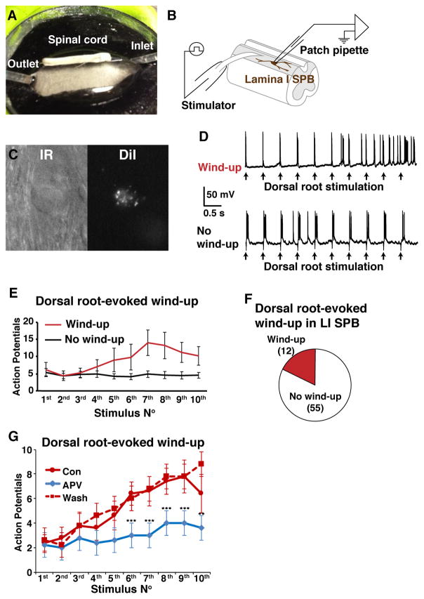
To determine whether wind-up develops in SPB neurons we used an ex vivo spinal cord preparation that preserves intact spinal circuitry and enables whole-cell recordings from SPB neurons in lamina I (Figs. 1A–B), as described previously [16]. Following the injection of DiI into the lateral parabrachial nucleus, retrogradely labeled lamina I SPB neurons were identified for recording with epifluorescence and then visualized in bright field by oblique infrared LED illumination to establish whole-cell patch recording (Fig. 1C) (Hachisuka et al., 2016; Szucs et al., 2009).
Figure 1. Dorsal root-stimulation induces wind-up in 18% of lamina I SPB neurons.
A–B. Photograph (A) and schematic (B) of recording set up in whole spinal cord preparation. Whole-cell patch clamp recording was made from the lamina I SPB neurons. C. Infrared (IR) and fluorescent image of a lamina I SPB neuron that is labeled with DiI. D. Example traces of wind-up and no wind-up in response to 2 Hz dorsal root stimulation. E. Wind-up is observed in a subset of lamina I SPB neurons in response to 2 Hz root stimulation. F. Pie chart illustrating fraction of lamina I SPB neurons that show wind-up. G. Treatment with the NMDA antagonist APV (50 μM) significantly reduced wind-up in lamina I SPB neurons in response to 2 Hz root stimulation, which recovered upon wash. Data are mean ± SEM (n = 5 cells, paired; asterisks indicate significantly different than control, ** p < 0.01, *** p < 0.001, Two-way ANOVA followed by Dunnett’s multiple comparison test).
Electrical stimulation of L2 dorsal root at 0.5 Hz, elicited a stable response to each stimulus in most cases (Figs. 1D, bottom and 1E, black trace). However, at 2 Hz stimulation, there was a progressive increase in AP number across the period of stimulation in 12 of 67 lamina I SBP neurons studied (Figs. 1D, top and 1E, red trace). In these neurons, there was a 299 ± 70% increase in the maximum number of evoked APs by the end of the 10-pulse train relative to the number of APs evoked with the first pulse. This effect was in marked contrast to the absence of a change in the number of evoked action potentials (6 ± 11%) in the remaining neurons (Fig. F). Importantly, and consistent with previous characterizations of wind-up, the increase in the evoked response to dorsal root stimulation significantly reduced by the NMDA receptor antagonist APV (50 μM; Fig. 1G). Thus, wind-up is observed in ~20% of lamina I SPB neurons and, like in other spinal neurons, it is NMDA-receptor dependent.
Neurotensin lineage neurons are sufficient for wind-up in lamina I SPB neurons
To identify spinal neurons potentially underlying the reverberating circuit hypothesized by Mendell [22], we screened for Cre alleles that would enable genetic access to subsets of excitatory neurons. The NtsCre allele was selected because it is relatively specific for excitatory interneurons and it targets a broad population in the dorsal horn. To determine the distribution of these cells, we analyzed L2–L3 spinal dorsal horns from mice harboring both the NtsCre and the Ai14 tdTomato reporter alleles, which were co-stained for NeuN, a marker of neurons, Pax2, a marker of inhibitory neurons, and IB4, a marker of non-peptidergic afferents that was used to help delineate boundaries within the dorsal horn. Using the ventral aspect of IB4 as a boundary that corresponds approximately to the border between high threshold C-fiber and low threshold A-fiber inputs, we found that NtsCre neurons represent 13 ± 1% of neurons within the superficial dorsal horn and 32 ± 2% of neurons within the intermediate dorsal horn (n = 4 mice). Almost all (98 ± 1%) of the NtsCre neurons were deemed to be excitatory as evidenced by the absence of Pax2 staining (Fig. 2A) [6; 30]. Since the genetic population defined by NtsCre-mediated recombination is somewhat broader than that defined by neurotensin protein expression in adult mice [15], we refer to the NtsCre population as neurotensin-lineage neurons.
Figure 2. Activation of excitatory neurons can induce wind-up in lamina I.
A. Spinal cord sections (L2–L3) from adult NtCre; ROSALSL-tdT mice were immunostained to reveal the inhibitory marker Pax2 (green). The vast majority of tdTomato-labeled NtsCre neurons are Pax2-negative. A single confocal optical section of the dorsal horn is shown. For quantification, the ventral border of the IB4 binding (not shown) was used to demarcate the lower boundary of the superficial dorsal horn (SDH); a second boundary, 85 microns below the first, was used to demarcate the intermediate dorsal horn (IDH). B. Schematic of optogenetic stimulation and whole-cell patch-clamp recording from the NtsCre; ROSALSL-ChR2 neuron. C. Optogenetic stimulation (5 ms) evoked APs in an NtsCre neuron, which could follow at 2 Hz. D. Schematic of optogenetic stimulation and whole-cell patch-clamp recording from an unidentified lamina I neuron from NtsCre; ROSA26LSL-ChR2 mice. E – F. Optogenetic stimulation of NtsCre neurons at 2 Hz caused wind-up in six out of nine lamina I neurons; example trace (E) and summary (F). Data are mean ± SEM. G. Schematic of optogenetic stimulation and whole-cell patch-clamp recording from an unidentified lamina I neuron from CrCre; ROSA26LSL-ChR2 mice. H–I. Optogenetic stimulation of CrCre neurons at 2 Hz causes APs in lamina I neurons, but no wind-up (n = 4 cells); example trace (H) and summary (I). Data are mean ± SEM.
Because we wanted to know whether stimulation of NtsCre neurons alone was sufficient to drive wind-up in lamina I SPB neurons, it was first necessary to confirm that we could consistently activate NtsCre neurons expressing ChR2 with blue light (Fig. 2B). We found that brief (5 ms) blue light exposure typically induced one AP in ChR2-expressing NtsCre neurons, which could follow at 2 Hz (Fig. 2C) with a failure rate of less than 5% (4 ± 3%, n = 16 cells).
Having confirmed that it was possible to selectively activate NtsCre neurons, we next sought to determine whether selective activation of these neurons was sufficient to drive wind-up in lamina I (Fig. 2D). For the sake of ease in this preliminary screen, we analyzed unidentified lamina I neurons (rather than retrogradely labeled SPB neurons). Optogenetic stimulation at 2 Hz caused a robust wind-up in six of nine neurons (Figs. 2E–F), indicating that NtsCre interneuron activity is sufficient for wind-up. To determine whether optogenetically-induced wind-up by excitatory neurons in the dorsal horn occurs in response to the stimulation of any excitatory neuron subpopulation, we assessed the impact of optogenetic stimulation of CrCre neurons (Fig. 2G). Calretinin is expressed in ~30% of neurons in the superficial dorsal horn, of which 85% are excitatory [33; 34]. Although optogenetically-evoked action potentials were observed in lamina I neurons upon application of blue light, wind-up was not detected upon activation of CrCre neurons (Figs. 2H–I). Thus, the selective activation of some, but not all, excitatory neurons are sufficient to induce wind-up in lamina I neurons.
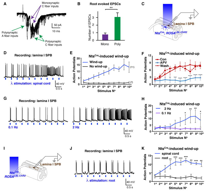
Next, we turned to the analysis of lamina I SPB neurons, since these output neurons may be involved in the affective component of pain. Notably, although some of the input from primary afferents onto lamina I SPB neurons is direct, the majority occurs through polysynaptic connections (Figs. 3A–B), raising the possibility that an excitatory network could contribute to wind-up in lamina I SPB neurons. To test this possibility, we examined the effect of optogenetic simulation of NtsCre neurons (Fig. 3C). Blue light stimulation of NtsCre neurons at 2 Hz caused wind-up in 11 of 18 (61%) lamina I SPB neurons (Figs. 3D–E). Thus, optogenetically-induced wind-up was observed in a significantly larger population of lamina I SPB neurons than dorsal root-evoked wind-up (p < 0.01, Fisher Exact test). In some neurons, the peak response to the wind-up protocol was achieved by the 6th or 7th stimulus in the train, with a decrease from this peak response observed to the remaining stimuli (e.g., Fig. 3D). We suggest that this decrease may be due to a depolarization-induced inactivation of voltage-gated sodium channels. Like dorsal root-evoked wind-up, the NtsCre mediated wind-up of lamina I SPB neurons was blocked by APV (Fig. 3F) and showed frequency dependence (Figs. 3G–H).
Figure 3. Activation of NtsCre neurons induced wind-up in lamina I SPB neurons.
A. Example traces of dorsal root-evoked EPSCs by 2 Hz dorsal root stimulation. Purple arrows indicate likely monosynaptic EPSCs, which have no failure and small jitter. Green arrows indicate polysynaptic EPSCs that have failures or large latency jitter. B. Number of likely monosynaptic EPSCs and polysynaptic EPSCs observed upon dorsal root stimulation. Data are mean ± SEM (n = 13 cells, ** p < 0.01, paired t-test). C. Schematic of optogenetic stimulation and whole-cell patch-clamp recording from a lamina I SPB neuron from NtsCre; ROSA26LSL-ChR2 mice. D – E. Optogenetic stimulation of NtsCre neurons at 2 Hz induced wind-up in 11 of 18 lamina I SPB neurons; example trace (D) and summary (E). Data are mean ± SEM. F. Treatment with APV (50 μM) significantly reduced wind-up by 2 Hz stimulation of NtsCre neurons, which recovered upon wash. Data are mean ± SEM (n = 5 cells, paired; asterisks indicate significantly different than control, ** p < 0.001, *** p < 0.001, **** p < 0.0001, Two-way ANOVA followed by Dunnett’s multiple comparison test). G – H. Optogenetically induced wind-up in lamina I SPB neurons occurs upon stimulation at 2 Hz, but not 0.1 Hz; example trace (G) and summary (H). Data are mean ± SEM (n = 3 cells, paired; * p < 0.01, ** p < 0.01, two-way ANOVA followed by Bonferroni post hoc test). I. Schematic of optogenetic stimulation of the dorsal root and whole-cell patch clamp recording from a lamina I SPB neuron. J. Example trace illustrating that optogenetic stimulation of the dorsal root at 2 Hz does not evoke wind-up in a lamina I SPB neurons. K. Quantification of APs following 2 Hz optogenetic stimulation over the spinal cord (blue) or the dorsal root (black). Data are mean ± SEM (n = 3 cells, paired; ** p < 0.01, *** p < 0.001, Two-way ANOVA followed by Bonferroni post hoc test).
An important consideration is that, in addition to excitatory spinal interneurons, the NtsCre allele causes recombination in approximately 10% of primary afferents, which are mainly small diameter cells, and include both peptidergic and non-peptidergic subtypes (data not shown). To address the potential role of NtsCre primary afferents in optogenetically-induced wind-up, we compared the responses of a given lamina I SPB neuron to optogenetic stimulation over the cord (Fig. 3C) to that observed over the root (Fig. 3I). As before, optogenetic stimulation over the cord induced wind-up (Fig. 3K). In contrast, optogenetic stimulation of the NtsCre afferent input was not sufficient for wind-up in lamina I SPB neurons, even though in all cases it was sufficient to drive action potentials in these cells (Figs. 3J–K). Thus, although we cannot rule out a possible contribution from primary afferents, wind-up in lamina I SPB neurons by optogenetic activation of the NtsCre population likely requires activity in neurotensin lineage neurons in the dorsal horn.
Neurotensin lineage neurons are required for wind-up in lamina I SPB neurons
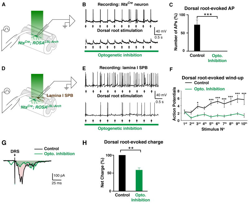
While activation of the local NtsCre network is sufficient for wind-up in lamina I SPB neurons, whether the NtsCre network normally mediates wind-up that is observed upon electrical stimulation of C-fibers remained unclear. To address this question, we examined whether dorsal root-evoked wind-up is abolished upon optogenetic inhibition of neurotensin lineage neurons with Archaerhodopsin (Arch), a light-driven proton pump. To determine whether green light-induced activation of Arch was sufficient to inhibit NtsCre neurons, we recorded from these neurons in voltage- and current-clamp (Fig. 4A). Green light hyperpolarized Arch-expressing NtsCre neurons (10.3 ± 1.4 mV, n = 13 cells) and blocked dorsal root-evoked action potentials in these cells (Figs. 4B–C), thereby confirming the efficacy of this optogenetic strategy. Next, we addressed whether optogenetic inhibition of NtsCre neurons blocked root-evoked wind-up in lamina I SPB neurons. Towards this end, we identified lamina I SPB neurons that showed wind-up upon electrical stimulation of the dorsal root and then we repeated the stimulation in the presence of green light to inhibit NtsCre neurons (Fig 4D). We found that optogenetic inhibition of NtsCre neurons significantly reduced root-evoked wind-up (Figs. 4E–F). To gain insight into the underlying mechanism, we performed recordings in voltage-clamp to analyze the effect of green light on the input received by a lamina I SPB neurons. This analysis revealed that optogenetic inhibition of NtsCre neurons significantly reduced the excitatory input that is observed upon dorsal-root stimulation, as measured by the net influx of charge (Figs. 4G–H. Taken together, these findings suggest that activity in NtsCre neurons is required for dorsal root-evoked wind-up.
Figure 4. Inhibition of NtsCre neurons blocks dorsal root-evoked wind-up.
A. Schematic illustrating optogenetic inhibition and whole-cell patch clamp recording from the NtsCre; ROSA26LSL-Arch neuron. B – C. Optogenetic inhibition of NtsCre neurons blocks root-evoked action potentials in these cells, confirming effectiveness of optogenetic strategy; example traces (B) and quantification (C) Data are mean ± SEM (n = 5 cells, *** p < 0.001, paired t-test) D. Diagram illustrating optogenetic inhibition and whole cell patch clamp recording of a lamina I SPB neuron from NtsCre; ROSA26LSL-Arch mice. E – F. Wind-up in lamina I SPB neurons elicited by dorsal root stimulation was blocked upon optogenetic inhibition of NtsCre neurons; example traces (E) and summary (F). Data are mean ± SEM (n = 5 cells, paired, * p < 0.05, ** p < 0.01, Two-way ANOVA followed by Bonferroni post hoc test). G. Example trace of evoked EPSCs following dorsal root stimulation (DRS) in the absence (Control) or in the presence (Opto. Inhibition) of green light to inhibit NtsCre neurons. Shaded area represents under the curve represents charge. H. Dorsal root-evoked charge, as measured by area under the curve, is significantly reduced upon optogenetic inhibition of NtsCre neurons. Data are mean ± SEM, normalized to control (n = 5 cells, paired; ** p < 0.01; student’s t-test)
NtsCre neurons form an extensive excitatory network
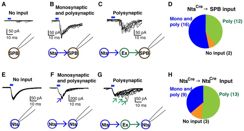
Our results suggested that NtsCre neurons are necessary and sufficient for wind-up, but the underlying circuitry remained unclear. To address this, we analyzed ChR2-evoked currents in voltage-clamp mode. We found that, although a small proportion (2 of 30) of lamina I SPB neurons did not receive any input from NtsCre neurons (Fig. 5A), most showed either monosynaptic and polysynaptic EPSCs (16 of 30; Fig. 5B) or polysynaptic EPSCs (12 of 30; Fig. 5C). Thus, the majority of lamina I SPB neurons receive direct or indirect input from NtsCre neurons (Fig. 5D). Next, we examined the connectivity among NtsCre neurons, recording from NtsCre neurons while activating this population optogenetically. In three of 25 NtsCre neurons, optogenetic stimulation evoked only a single, large amplitude ChR2-evoked current with very short latency (~1 ms) due to the opening of ChR2 in the recorded cell (Fig. 5E). However, in most NtsCre neurons, this large amplitude ChR2-evoked current was accompanied by monosynaptic and polysynaptic EPSCs (Fig. 5F), or polysynaptic EPSCs (Fig. 5G). Together, these findings suggest that NtsCre neurons form an excitatory network.
Figure 5. NtsCre neurons form an extensive excitatory network.
A – C. Whole-cell patch clamp recording from lamina I SPB neurons upon optogenetic stimulation of NtsCre neurons (0.1 Hz ×10, 5 ms duration). Example traces from recorded cells that received no input (A), monosynaptic and polysynaptic input (B), or polysynaptic input alone (C) from NtsCre neurons. D. Summary data; n = 30 cells. E – G. Whole-cell patch clamp recording from NtsCre neurons upon optogenetic stimulation of NtsCre neurons (0.1 Hz ×10, 5 ms duration). Example traces from recorded cells that received no input (E) (inward current is due to opening of ChR2 in the recorded cell), monosynaptic and polysynaptic input, as indicated by the blue arrow (F), or polysynaptic input alone, as indicated by the green arrows (G) from NtsCre neurons. H. Summary data; n = 25 cells.
Activation of NtsCre neurons causes reverberatory activity which correlates with wind-up
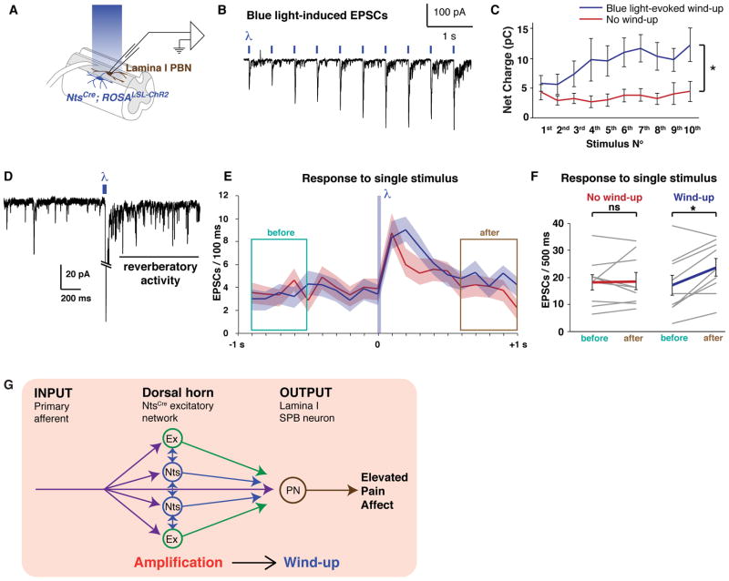
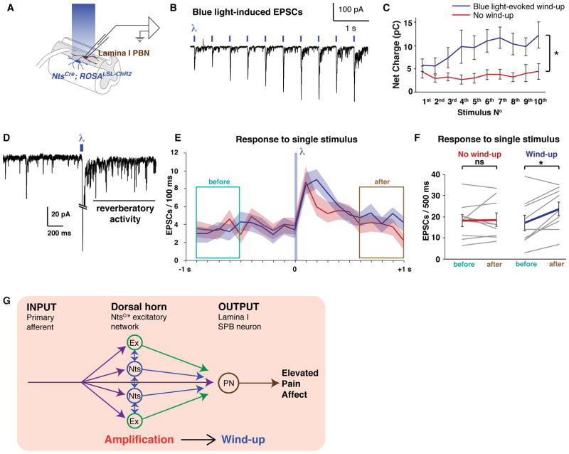
The extensive interconnectivity that we observed within the NtsCre network raised the possibility that a polysynaptic circuit also contributes to the wind-up of in lamina I SPB neurons. If so, we reasoned that we ought to see not only wind-up of the light-evoked EPSCs in lamina I SPB neurons, but also an increase in the subsequent polysynaptic input onto these cells. To investigate this possibility, we recorded from lamina I SPB neurons in voltage-clamp mode in response to optogenetic activation of NtsCre neurons (Fig. 6A). Blue light stimulation at 2 Hz resulted in an increase in both the blue light-evoked EPSC and smaller EPSCs that followed, which appeared to be due to ongoing synaptic input onto the SPB neuron (Fig 6B). To quantify this effect, we measured the net charge influx between stimuli. This analysis revealed that the net charge increase observed upon repeated stimulation was significantly greater in cells that showed wind-up compared to cells that did not. (Fig 6C).
Figure 6. Activation of NtsCre neurons causes reverberatory activity which correlates with wind-up.
A. Diagram illustrating blue light stimulation of NtsCre; ROSA26LSL-ChR2 neurons during whole-cell patch clamp recording from the lamina I SPB neurons. B. Blue light stimulation at 2 Hz resulted in an increase of EPSCs in a SPB neuron which showed wind-up. C. In SPB neurons that show wind-up, there is a frequency dependent increase in net charge onto the recorded cell (i.e., area under the curve) of each stimulation, which is significantly different than that observed in lamina I SPB neurons that do not undergo wind-up. Data are mean ± SEM (n = 11 and 7 cells for blue light evoked wind-up and no wind-up, respectively; * p < 0.05, Two-way ANOVA). D. Single blue light stimulation (5 ms) of NtsCre neurons evoked persistent EPSCs in a lamina I PBN, indicative of reverberatory activity. E. EPSCs in lamina I PBN neurons observed before (green box) or after (brown box) a single stimulation (5 ms) of NtsCre neurons with blue light. Lamina I SPB neurons that subsequently showed wind-up are shown in blue (n = 9); those that showed no wind-up are in red (n = 9). Data are mean ± SEM. F. A comparison of the number of EPSCs 1000 – 500 ms before optogenetic stimulation (green box in E) to that 500 – 1000 ms after optogenetic stimulation (brown box in E). In lamina I SPB neurons that do not show wind-up, there is no significant change in the number of EPSCs before and after a single optogenetic stimulus (red), whereas in lamina I neurons that do show wind up, there the number of EPSCs after optogenetic stimulation is significantly greater than before (blue). Data from individual neurons as well as mean ± SEM are shown (n = 9; * p < 0.05, student’s t-test). G. Model: During wind-up, primary afferent input is amplified by a network of excitatory NtsCre interneurons that synapse onto each other as well and other excitatory interneurons (Ex), thereby generating reverberatory activity, which contributes to wind-up in lamina I SPB neurons.
One of the factors that could lead to increased net charge influx is reverberatory activity within the network. We found that brief (5 ms) blue light stimulation induced EPSCs (Figs. 6D) that continued for prolonged period of time (e.g., > 1 s). Given the extended time course of this reverberatory activity, we reasoned that a second stimulus during this time could sum with previous activity, thereby contributing to wind-up as previously suggested [22]. To test this idea, we compared the reverberating activity evoked by a single stimulus between lamina I SPB neurons that subsequently showed wind-up to those that did not. In this retrospective analysis, we found that in lamina I SPB neurons where no wind-up was observed, there was no difference in the EPSC frequency during the 1000 – 500 ms before optogenetic stimulation compared to the EPSC frequency during the 500 – 1000 ms after optogenetic stimulation. In contrast, in lamina I SPB neurons where wind-up was observed, there was a significant increase in the EPSC frequency observed after optogenetic stimulation compared to before (Figs. 6E–F). Together, these findings suggest that wind-up in lamina I SPB neurons is mediated, at least in part, by a polysynaptic circuit that shows reverberatory activity (Fig. 6G).
DISCUSSION
Wind-up is a mechanism of facilitation that is thought to account for the intensification of pain observed with repeated or prolonged application of noxious stimuli, a psychophysical phenomenon referred to as temporal summation. Importantly, both the sensory/discriminative (intensity) and the emotional/affective (unpleasantness) components of pain are subject to temporal summation. Spinothalamic neurons, which are thought to contribute to intensity coding, undergo wind-up, but whether spinoparabrachial neurons, which are thought to be contribute to unpleasantness coding, are subject to wind-up, was less clear. Our study reveals that lamina I SPB neurons show wind-up in response to repetitive stimulation of the dorsal root. In addition, we provide evidence that this wind-up is mediated, at least in part, by NtsCre neurons, since selective activation of these cells was sufficient to evoke wind-up in lamina I SPB neurons, whereas optogenetic inhibition of these cells inhibited dorsal root-evoked wind-up. Finally, our data suggest that the duration of reverberatory input onto lamina I SPB neurons contributes to the manifestation of wind-up. These findings provide new insight into the circuit mechanisms that could underlie the temporal summation of pain’s unpleasantness, and uncover a long-speculated contribution of an excitatory network in this sensory augmentation (Figure 6E).
Our study reveals that wind-up in lamina I SPB neurons upon optogenetic stimulation of NtsCre neurons was more frequent in occurrence than that observed upon electrical stimulation of the dorsal root. Although the reason for this difference is not known, we suggest the key role of inhibitory neurons within nociceptive circuits of the dorsal horn are likely to account for the difference. That is, primary afferent input normally recruits activity in inhibitory interneurons to effect feed-forward, feedback and lateral inhibition. In contrast, our optogenetic stimulation of NtsCre excitatory interneurons may have bypassed this inhibition, thereby shifting the balance of activity in favor of excitation, leading to an increase in the prevalence of wind-up.
Our data suggest that reverberatory activity within a neural circuit contributes to wind-up lamina I SPB neurons. It has long been speculated that interconnectivity among excitatory interneurons may provide a substrate for reverberatory activity, allowing for summation [22]. Under physiological conditions, this summation is limited by activity in inhibitory interneurons, as evidenced by the observation that disinhibition by GABA and glycine antagonists causes long-lasting EPSCs by dorsal root stimulation [4; 10; 26; 37]. Thus, inhibition may normally limit wind-up. Since a decrease in inhibitory tone is frequently seen upon injury [19; 24], this loss of inhibition may contribute to abnormal reverberatory activity, giving rise to both heightened wind-up and abnormally elevated wind-up that are observed in pathological conditions [18].
Acknowledgments
Research reported in this publication was supported by the National Institute of Arthritis and Musculoskeletal and Skin Diseases of the National Institutes of Health under Award Number R01 AR063772 and R21 AR064445 to SER; the National Institute of Neurological Disorder and Stroke under Award Number NS023725 and NS096705 to HRK, NS083347 to MSG, and NS096860 to MMC; and the National Institutes of Diabetes and Digestive and Kidney Diseases under Award Number DK107966 to MSG. Part of this work was supported by a grant from the Rita Allen Foundation to SER who is a Rita Allen Foundation Pain Scholar. The authors declare no conflict of interest.
References
- 1.Andrew D, Greenspan JD. Peripheral coding of tonic mechanical cutaneous pain: comparison of nociceptor activity in rat and human psychophysics. J Neurophysiol. 1999;82(5):2641–2648. doi: 10.1152/jn.1999.82.5.2641. [DOI] [PubMed] [Google Scholar]
- 2.Arendt-Nielsen L, Petersen-Felix S. Wind-up and neuroplasticity: is there a correlation to clinical pain? Eur J Anaesthesiol Suppl. 1995;10:1–7. [PubMed] [Google Scholar]
- 3.Arendt-Nielsen L, Petersen-Felix S, Fischer M, Bak P, Bjerring P, Zbinden AM. The effect of N-methyl-D-aspartate antagonist (ketamine) on single and repeated nociceptive stimuli: a placebo-controlled experimental human study. Anesth Analg. 1995;81(1):63–68. doi: 10.1097/00000539-199507000-00013. [DOI] [PubMed] [Google Scholar]
- 4.Baba H, Ji RR, Kohno T, Moore KA, Ataka T, Wakai A, Okamoto M, Woolf CJ. Removal of GABAergic inhibition facilitates polysynaptic A fiber-mediated excitatory transmission to the superficial spinal dorsal horn. Mol Cell Neurosci. 2003;24(3):818–830. doi: 10.1016/s1044-7431(03)00236-7. [DOI] [PubMed] [Google Scholar]
- 5.Bourgeais L, Gauriau C, Monconduit L, Villanueva L, Bernard JF. Dendritic domains of nociceptive-responsive parabrachial neurons match terminal fields of lamina I neurons in the rat. J Comp Neurol. 2003;464(2):238–256. doi: 10.1002/cne.10793. [DOI] [PubMed] [Google Scholar]
- 6.Cheng L, Arata A, Mizuguchi R, Qian Y, Karunaratne A, Gray PA, Arata S, Shirasawa S, Bouchard M, Luo P, Chen CL, Busslinger M, Goulding M, Onimaru H, Ma Q. Tlx3 and Tlx1 are post-mitotic selector genes determining glutamatergic over GABAergic cell fates. Nat Neurosci. 2004;7(5):510–517. doi: 10.1038/nn1221. [DOI] [PubMed] [Google Scholar]
- 7.Davies SN, Lodge D. Evidence for involvement of N-methylaspartate receptors in ‘wind-up’ of class 2 neurones in the dorsal horn of the rat. Brain Res. 1987;424(2):402–406. doi: 10.1016/0006-8993(87)91487-9. [DOI] [PubMed] [Google Scholar]
- 8.Dickenson AH. A cure for wind up: NMDA receptor antagonists as potential analgesics. Trends Pharmacol Sci. 1990;11(8):307–309. doi: 10.1016/0165-6147(90)90228-z. [DOI] [PubMed] [Google Scholar]
- 9.Dickenson AH, Sullivan AF. Evidence for a role of the NMDA receptor in the frequency dependent potentiation of deep rat dorsal horn nociceptive neurones following C fibre stimulation. Neuropharmacology. 1987;26(8):1235–1238. doi: 10.1016/0028-3908(87)90275-9. [DOI] [PubMed] [Google Scholar]
- 10.Duan B, Cheng L, Bourane S, Britz O, Padilla C, Garcia-Campmany L, Krashes M, Knowlton W, Velasquez T, Ren X, Ross S, Lowell BB, Wang Y, Goulding M, Ma Q. Identification of spinal circuits transmitting and gating mechanical pain. Cell. 2014;159(6):1417–1432. doi: 10.1016/j.cell.2014.11.003. [DOI] [PMC free article] [PubMed] [Google Scholar]
- 11.Eide PK. Wind-up and the NMDA receptor complex from a clinical perspective. Eur J Pain. 2000;4(1):5–15. doi: 10.1053/eujp.1999.0154. [DOI] [PubMed] [Google Scholar]
- 12.Eide PK, Jorum E, Stenehjem AE. Somatosensory findings in patients with spinal cord injury and central dysaesthesia pain. J Neurol Neurosurg Psychiatry. 1996;60(4):411–415. doi: 10.1136/jnnp.60.4.411. [DOI] [PMC free article] [PubMed] [Google Scholar]
- 13.Eide PK, Jorum E, Stubhaug A, Bremnes J, Breivik H. Relief of post-herpetic neuralgia with the N-methyl-D-aspartic acid receptor antagonist ketamine: a double-blind, cross-over comparison with morphine and placebo. Pain. 1994;58(3):347–354. doi: 10.1016/0304-3959(94)90129-5. [DOI] [PubMed] [Google Scholar]
- 14.Gauriau C, Bernard JF. Pain pathways and parabrachial circuits in the rat. Exp Physiol. 2002;87(2):251–258. doi: 10.1113/eph8702357. [DOI] [PubMed] [Google Scholar]
- 15.Gutierrez-Mecinas M, Furuta T, Watanabe M, Todd AJ. A quantitative study of neurochemically defined excitatory interneuron populations in laminae I–III of the mouse spinal cord. Mol Pain. 2016:12. doi: 10.1177/1744806916629065. [DOI] [PMC free article] [PubMed] [Google Scholar]
- 16.Hachisuka J, Baumbauer KM, Omori Y, Snyder LM, Koerber HR, Ross SE. Semi-intact ex vivo approach to investigate spinal somatosensory circuits. Elife. 2016:5. doi: 10.7554/eLife.22866. [DOI] [PMC free article] [PubMed] [Google Scholar]
- 17.Hedo G, Laird JM, Lopez-Garcia JA. Time-course of spinal sensitization following carrageenan-induced inflammation in the young rat: a comparative electrophysiological and behavioural study in vitro and in vivo. Neuroscience. 1999;92(1):309–318. doi: 10.1016/s0306-4522(98)00734-9. [DOI] [PubMed] [Google Scholar]
- 18.Herrero JF, Laird JM, Lopez-Garcia JA. Wind-up of spinal cord neurones and pain sensation: much ado about something? Prog Neurobiol. 2000;61(2):169–203. doi: 10.1016/s0301-0082(99)00051-9. [DOI] [PubMed] [Google Scholar]
- 19.Koerber HR, Brown PB. Quantitative analysis of dorsal horn cell receptive fields following limited deafferentation. J Neurophysiol. 1995;74(5):2065–2076. doi: 10.1152/jn.1995.74.5.2065. [DOI] [PubMed] [Google Scholar]
- 20.Koltzenburg M, Handwerker HO. Differential ability of human cutaneous nociceptors to signal mechanical pain and to produce vasodilatation. J Neurosci. 1994;14(3 Pt 2):1756–1765. doi: 10.1523/JNEUROSCI.14-03-01756.1994. [DOI] [PMC free article] [PubMed] [Google Scholar]
- 21.Lundberg LE, Jorum E, Holm E, Torebjork HE. Intra-neural electrical stimulation of cutaneous nociceptive fibres in humans: effects of different pulse patterns on magnitude of pain. Acta Physiol Scand. 1992;146(1):41–48. doi: 10.1111/j.1748-1716.1992.tb09391.x. [DOI] [PubMed] [Google Scholar]
- 22.Mendell LM. Physiological properties of unmyelinated fiber projection to the spinal cord. Exp Neurol. 1966;16(3):316–332. doi: 10.1016/0014-4886(66)90068-9. [DOI] [PubMed] [Google Scholar]
- 23.Mendell LM, Wall PD. Responses of Single Dorsal Cord Cells to Peripheral Cutaneous Unmyelinated Fibres. Nature. 1965;206:97–99. doi: 10.1038/206097a0. [DOI] [PubMed] [Google Scholar]
- 24.Moore KA, Kohno T, Karchewski LA, Scholz J, Baba H, Woolf CJ. Partial peripheral nerve injury promotes a selective loss of GABAergic inhibition in the superficial dorsal horn of the spinal cord. J Neurosci. 2002;22(15):6724–6731. doi: 10.1523/JNEUROSCI.22-15-06724.2002. [DOI] [PMC free article] [PubMed] [Google Scholar]
- 25.Nikolajsen L, Hansen CL, Nielsen J, Keller J, Arendt-Nielsen L, Jensen TS. The effect of ketamine on phantom pain: a central neuropathic disorder maintained by peripheral input. Pain. 1996;67(1):69–77. doi: 10.1016/0304-3959(96)03080-1. [DOI] [PubMed] [Google Scholar]
- 26.Peirs C, Williams SP, Zhao X, Walsh CE, Gedeon JY, Cagle NE, Goldring AC, Hioki H, Liu Z, Marell PS, Seal RP. Dorsal Horn Circuits for Persistent Mechanical Pain. Neuron. 2015;87(4):797–812. doi: 10.1016/j.neuron.2015.07.029. [DOI] [PMC free article] [PubMed] [Google Scholar]
- 27.Price DD. Characteristics of second pain and flexion reflexes indicative of prolonged central summation. Exp Neurol. 1972;37(2):371–387. doi: 10.1016/0014-4886(72)90081-7. [DOI] [PubMed] [Google Scholar]
- 28.Price DD, Long S, Huitt C. Sensory testing of pathophysiological mechanisms of pain in patients with reflex sympathetic dystrophy. Pain. 1992;49(2):163–173. doi: 10.1016/0304-3959(92)90139-3. [DOI] [PubMed] [Google Scholar]
- 29.Price DD, Mao J, Frenk H, Mayer DJ. The N-methyl-D-aspartate receptor antagonist dextromethorphan selectively reduces temporal summation of second pain in man. Pain. 1994;59(2):165–174. doi: 10.1016/0304-3959(94)90069-8. [DOI] [PubMed] [Google Scholar]
- 30.Punnakkal P, von Schoultz C, Haenraets K, Wildner H, Zeilhofer HU. Morphological, biophysical and synaptic properties of glutamatergic neurons of the mouse spinal dorsal horn. J Physiol. 2014;592(4):759–776. doi: 10.1113/jphysiol.2013.264937. [DOI] [PMC free article] [PubMed] [Google Scholar]
- 31.Sarlani E, Grace EG, Reynolds MA, Greenspan JD. Sex differences in temporal summation of pain and aftersensations following repetitive noxious mechanical stimulation. Pain. 2004;109(1–2):115–123. doi: 10.1016/j.pain.2004.01.019. [DOI] [PubMed] [Google Scholar]
- 32.Schouenborg J, Sjolund BH. Activity evoked by A- and C-afferent fibers in rat dorsal horn neurons and its relation to a flexion reflex. J Neurophysiol. 1983;50(5):1108–1121. doi: 10.1152/jn.1983.50.5.1108. [DOI] [PubMed] [Google Scholar]
- 33.Smith KM, Boyle KA, Madden JF, Dickinson SA, Jobling P, Callister RJ, Hughes DI, Graham BA. Functional heterogeneity of calretinin-expressing neurons in the mouse superficial dorsal horn: implications for spinal pain processing. J Physiol. 2015;593(19):4319–4339. doi: 10.1113/JP270855. [DOI] [PMC free article] [PubMed] [Google Scholar]
- 34.Smith KM, Boyle KA, Mustapa M, Jobling P, Callister RJ, Hughes DI, Graham BA. Distinct forms of synaptic inhibition and neuromodulation regulate calretinin-positive neuron excitability in the spinal cord dorsal horn. Neuroscience. 2016;326:10–21. doi: 10.1016/j.neuroscience.2016.03.058. [DOI] [PMC free article] [PubMed] [Google Scholar]
- 35.Thompson SW, Dray A, McCarson KE, Krause JE, Urban L. Nerve growth factor induces mechanical allodynia associated with novel A fibre-evoked spinal reflex activity and enhanced neurokinin-1 receptor activation in the rat. Pain. 1995;62(2):219–231. doi: 10.1016/0304-3959(94)00271-F. [DOI] [PubMed] [Google Scholar]
- 36.Thompson SW, King AE, Woolf CJ. Activity-Dependent Changes in Rat Ventral Horn Neurons in vitro; Summation of Prolonged Afferent Evoked Postsynaptic Depolarizations Produce a d-2-Amino-5-Phosphonovaleric Acid Sensitive Windup. Eur J Neurosci. 1990;2(7):638–649. doi: 10.1111/j.1460-9568.1990.tb00453.x. [DOI] [PubMed] [Google Scholar]
- 37.Torsney C, MacDermott AB. Disinhibition opens the gate to pathological pain signaling in superficial neurokinin 1 receptor-expressing neurons in rat spinal cord. J Neurosci. 2006;26(6):1833–1843. doi: 10.1523/JNEUROSCI.4584-05.2006. [DOI] [PMC free article] [PubMed] [Google Scholar]
- 38.Traub RJ. Spinal modulation of the induction of central sensitization. Brain Res. 1997;778(1):34–42. doi: 10.1016/s0006-8993(97)00946-3. [DOI] [PubMed] [Google Scholar]
- 39.Vase L, Svensson P, Nikolajsen L, Arendt-Nielsen L, Jensen TS. The effects of menthol on cold allodynia and wind-up-like pain in upper limb amputees with different levels of phantom limb pain. Neurosci Lett. 2013;534:52–57. doi: 10.1016/j.neulet.2012.11.005. [DOI] [PubMed] [Google Scholar]
- 40.Vierck CJ, Jr, Cannon RL, Fry G, Maixner W, Whitsel BL. Characteristics of temporal summation of second pain sensations elicited by brief contact of glabrous skin by a preheated thermode. J Neurophysiol. 1997;78(2):992–1002. doi: 10.1152/jn.1997.78.2.992. [DOI] [PubMed] [Google Scholar]
- 41.Warncke T, Stubhaug A, Jorum E. Ketamine, an NMDA receptor antagonist, suppresses spatial and temporal properties of burn-induced secondary hyperalgesia in man: a double-blind, cross-over comparison with morphine and placebo. Pain. 1997;72(1–2):99–106. doi: 10.1016/s0304-3959(97)00006-7. [DOI] [PubMed] [Google Scholar]
- 42.Woolf CJ. Windup and central sensitization are not equivalent. Pain. 1996;66(2–3):105–108. [PubMed] [Google Scholar]
- 43.Woolf CJ, Thompson SW. The induction and maintenance of central sensitization is dependent on N-methyl-D-aspartic acid receptor activation; implications for the treatment of post-injury pain hypersensitivity states. Pain. 1991;44(3):293–299. doi: 10.1016/0304-3959(91)90100-C. [DOI] [PubMed] [Google Scholar]
- 44.Woolf CJ, Wall PD. Relative effectiveness of C primary afferent fibers of different origins in evoking a prolonged facilitation of the flexor reflex in the rat. J Neurosci. 1986;6(5):1433–1442. doi: 10.1523/JNEUROSCI.06-05-01433.1986. [DOI] [PMC free article] [PubMed] [Google Scholar]
- 45.Zhang DX, Owens CM, Willis WD. Intracellular study of electrophysiological features of primate spinothalamic tract neurons and their responses to afferent inputs. J Neurophysiol. 1991;65(6):1554–1566. doi: 10.1152/jn.1991.65.6.1554. [DOI] [PubMed] [Google Scholar]


