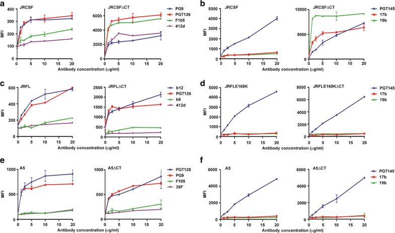
Fig. 6.
CT-deleted JRCSF but not JRFL and A5, have altered cell surface antigenicity/conformation. a, c, e FACS based cell surface staining assays of JRCSF, JRCSFΔCT with bNAbs, PGT126, PG9 and non-NAbs, F105, 412d; JRFL, JRFLΔCT with bNAbs, b12, PGT126 and non-NAbs, b6 and 412d; A5, A5ΔCT with bNAbs, PGT128 and PG9 and non-NAbs, F105, 39F over a range of antibody concentrations. b, d, f FACS based cell surface staining assays of JRFLE168K, JRFLE168KΔCT, A5, A5ΔCT, JRCSF, JRCSFΔCT with trimer-selective, cleavage-specific bNAb, PGT145 and non-NAbs, 17b (CD4i) and 19b (V3) over a range of antibody concentrations

