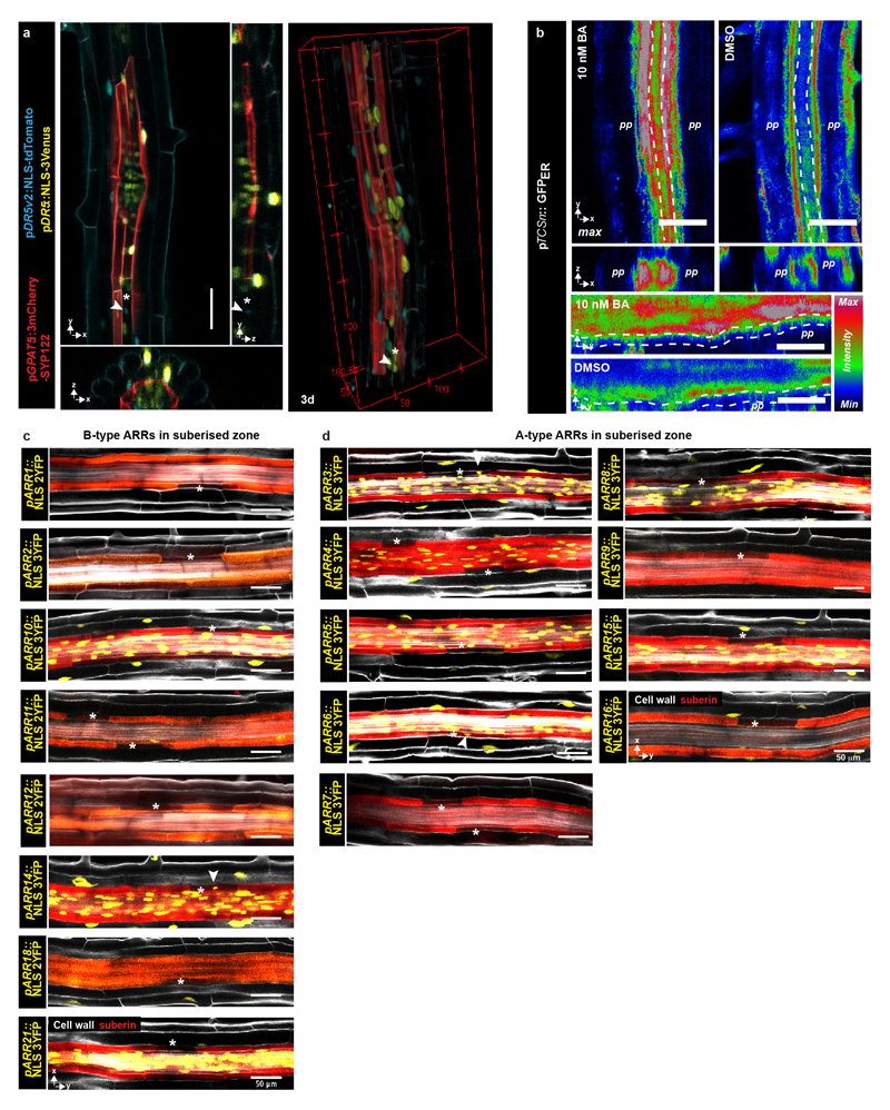
Extended Data Figure 2. Auxin and cytokinin signaling in the suberised root zone.
a) Activity of the auxin-signaling reporter DR5 (yellow) as well as the highly sensitive DR5v2 (blue) in an area of the suberised zone of a 5-day-old root with an emerging lateral root. Suberised cells were visualized based on the suberin biosynthetic gene GPAT5 driving expression of a plasma membrane localized 3mCherry-based reporter b) Expression of ER-localized GFP driven by the cytokinin signaling reporter TCSn in the phloem and xylem poles of 5-day-old roots in the suberized zone. Plants were either grown on plates containing 5 nM cytokinin (BA) or a mock treatment (DMSO). Punctured lines indicate the endodermis. c) Expression of B-type ARRs in the suberised endodermis of 5-day-old roots, suberin and cellwalls are stained using a Clearsee protocol in combination with Nile Red and Calcoflour White, respectively33 d) Expression of A-type ARRs in the suberised endodermis of 5-day-old roots, suberin and cell walls as in d). All stainings were repeated 3 times. For a) and b) the image is representative of 8 independent lines. White arrowheads depict passage cell nuclei. Asterisk marks passage cells. BA; Benzyladenine, Co; Cortex, En; Endodermis. Scale bars represent 50 μm.

