Abstract
Abortion in sheep and goats causes enormous economic losses. This study revealed the epidemiology of abortion caused by Brucella melitensis, Coxiella burnetii and Salmonella abortusovis in Baluchi sheep in Sistan region. In the autumn of 2015 and winter of 2016, a total of 78 aborted sheep fetuses were collected from all over the Sistan region. Risk factors, including location of livestock, history of abortion, gender of fetus, age of fetus, age of ewe and parity were obtained using a questionnaire. The results showed that 27 fetuses (35%) were infected with these organisms. Infection with B. melitensis, C. burnetii and S. abortusovis were identified respectively in 15 (19.2%), 13 (16.6%) and 1 (1.3%) fetus. Logistic regression analysis showed that infection with B. melitensis in male fetuses is higher than females (OR=3.73, P=0.040), also infection with C. burnetii in ≤2 years’ ewes (OR=0.047, P=0.009) and 2-5 years’ ewes (OR=0.197, P=0.069) is lower than ≥5 years’ ewes.
Key Words: Abortion, Brucella melitensis, Coxiella burnetii, Salmonella abortusovis, Sheep
Introduction
Abortion is a condition in which the pregnancy terminates before the fetus is viable (Boden and Andrews, 2015 ▶). Infectious and non-infectious multiple factors can cause spontaneous abortions. Infective abortions are caused by bacteria, fungi, protozoan and viruses. Non-infective factors are stress, housing conditions, transport, toxemia, metabolic disorders, inappropriate nutrition, hereditary factors, physical factors, etc (Beuzon et al., 1997 ▶; Vidić et al., 2007 ▶).
In the Sistan region, many people breed sheep and goats, much of the profit from sheep breeding belongs to lambing. Therefore, any disruption in this process can cause enormous economic losses. Importation of livestock from Pakistan, slaughtering of animals outside slaughterhouses and incomplete coverage of vaccination programs can cause spread of infectious disease among livestock in this region.
Brucella melitensis, Campylobacter fetus, Salmonella abortusovis, and Chlamidophila abortus are the most important pathogens involved in the abortion of ewes in Iran (Atyabi et al., 2012 ▶). Some of the abortive microorganisms such as Brucella and Coxiella have been reported in the Sistan region (Esmaeili et al., 2016 ▶). Thus, this study was conducted to investigate the prevalence of B. abortus, C. brunetti and S. abortusovis among aborted fetuses in the Sistan region.
Materials and Methods
Sampling methods
During the autumn of 2015 and winter of 2016, a total of 78 aborted Baluchi sheep fetuses were collected from all over the Sistan region. Risk factors including the location of livestock, history of abortion, gender of fetus, age of fetus, age of ewe and parity were obtained using a questionnaire. Age of the fetuses was estimated based on the crown rump length (Noakes et al., 2001 ▶). Location of livestock was classified as eastern part of Sistan (Zahak and Hirmand counties), central part of Sistan (Zabol county) and western part of Sistan (Nimrooz and Hamoon counties).
Sheep owners were asked to transfer their aborted fetus to a nearby veterinary clinic. Biosecurity principles were taught to the sheep owners. Fetuses were placed in foam container on ice. Aborted fetuses were transferred to laboratory of anatomy of veterinary faculty within 24 h after abortion. After the autopsy, contents of abomasum and tissue of spleen were placed in sterile Eppendorf. Samples were stored at -20°C until use for DNA extraction.
The polymerase chain reaction (PCR)
Spleen and abomasum samples was used following the manufacturer’s instructions of a commercial DNA extraction kit (DNPTM yield DNA purification Kit, Cinnagen Biotech Co.©, Karaj, Iran). The DNA extracts were used as a template for PCR. Primer design was done as using prior studies (Table 1). The PCR solutions were placed in the thermocycler (Eppendorf, Germany). Predenaturation was performed at 94°C for 4 min, following that, 30 thermal cycle was set as follows: 45 s of denaturation at 94°C, 1 min of primer annealing at 54°C, 59°C and 64°C, respectively for B. melitensis, C. brunetti and S. abortusovis, 1 min of primer extension at 72°C, after completing these cycles a final extension was performed at 72°C for 10 min. Positive control (B. melitensis ATCC 23457, C. brunetti ATCC VR-615, and S. abortusovis ATCC 31684) and negative control (sample without genomic DNA) were used in all reactions. PCR products were used for electrophoresis in a 2% buffer TAE (Tris Acetic Acid EDTA, MERCK, Germany).
Table 1.
Characterization of primers using in this study
| Gene | Primers | Length | Bacteria | Source |
|---|---|---|---|---|
| IS711 | 5´-AAA TCG CGT CCT TGC TGG TCT GA-3´ | 731 bp | B. melitensis | Unver et al., (2006) |
| 5´-TGC CGA TCA CTT AAG GGC CTT CAT-3´ | ||||
| IS1111 | 5´-TAT GTA TCC ACC GTA GCC AGT-3´ | 687 bp | C. burnetii | Parisi et al., (2006) |
| 5´-CCC AAC AAC ACC TCC TTA TTC-3´ | ||||
| IS200 | 5´-CGA TGA AAG CGT AAA TAA GG-3´ | 900 bp | S. abortusovis | Belloy et al., (2009) |
| 5´-TTC TCT TGT CAG TCT CAA AC-3´ |
Statistical method
In this study, when one or both the spleen and abomasum of a fetus was positive in PCR test, that fetus was considered as positive case. Since the dependent variable was a dichotomous variable, the multivariate logistic regression method was used for statistical analysis of the data. Before applying logistics regression, the relation between each independent variable with infection was analyzed using Chi-square test. When p-value in Chi-square test was less than 0.25, that variable was considered in the regression model.
Also, contamination of spleen and abomasum were compared using McNemar test. 95% confidence interval for prevalence of bacterial infection was calculated using binomial distribution. SPSS version 23 was used for statistical analysis. The significant level was considered P<0.05.
Results
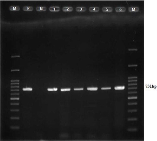
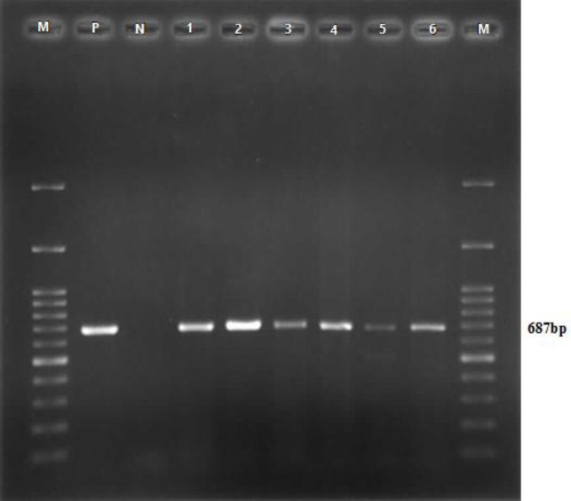
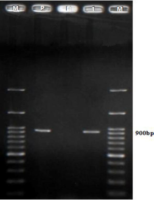
Among 78 aborted fetuses, 27 cases (35%) (95% CI: 24%-46%) were identified positive by PCR method. Infection with B. melitensis, C. burnetii and S. abortusovis was observed in 15 (19.2%), 13 (16.7%), and 1 (1.3%) fetuses, respectively (Figs. 1, 2 and 3). Among these fetuses, 2 cases (2.6%) were infected with both B. melitensis and C. burnetii.
Fig. 1.
PCR reactions for detection of B. melitensis. M: The 100 bp marker, P: The positive control, and N: The negative control. Positive samples (Lanes 1 to 6) characterized by identifying a gene fragment of 731 bp
Fig. 2.
PCR reactions or detection of C. burnetii. M: The 100 bp marker, P: The positive control, and N: The negative control. Positive samples (Lanes 1 to 6) characterized by identifying a gene fragment of 687 bp
Fig. 3.
PCR reactions for detection of S. abortusovis. M: The 100 bp marker, P: The positive control, and N: negative control. Positive sample (Lane 1) identifying characteristic gene fragment length of 900 bp
The prevalence of each bacterium was calculated based on independent variables (Table 2). Because of the collinearity between parity and age of ewe, the parity was not considered in multivariate logistic regression model. The results showed that relationship between gender of fetus and infection with B. melitensis was significant, also the relationship between age of ewe and infection with C. burnetii was significant (Table 3). There was no statistically significant relationship between other independent variables and infection of fetuses.
Table 2.
Prevalence of infection with B. melitensis, C. burnetii and S. abortusovis by independent variables in 87 aborted fetuses of Baluchi sheep in Sistan region
| Category | Levels | No. of tested fetus |
B. melitensis
|
C. burnetii
|
S. abortusovis
|
|---|---|---|---|---|---|
| No. (%) of infected fetus | No. (%) of infected fetus | No. (%) of infected fetus | |||
| Location of livestock | Eastern part | 25 | 5 (20) | 5 (20) | 0 (0) |
| Central part | 38 | 6 (16) | 5 (13) | 1 (3) | |
| Western part | 15 | 4 (27) | 3 (20) | 0 (0) | |
| History of abortion | Yes | 3 | 1 (33) | 1 (33) | 0 (0) |
| No | 75 | 14 (19) | 12 (16) | 1(1) | |
| Gender of fetus | Male | 38 | 11 (29)a | 7 (18) | 0 (0) |
| Female | 40 | 4 (10) | 6 (15) | 1 (2) | |
| Age of fetus | ≤3 month | 12 | 2 (17) | 2 (17)a | 0 (0) |
| 4 month | 30 | 7 (23) | 8 (27) | 0 (0) | |
| 5 month | 36 | 6 (17) | 3 (8) | 1(3) | |
| Age of ewe | ≤2 years | 26 | 4 (15) | 2 (8)a | 0 (0) |
| 2-5 years | 41 | 9 (22) | 7 (17) | 1 (2) | |
| ≥5 years | 11 | 2 (18) | 4(36) | 0 (0) | |
| parity | First | 25 | 5 (20) | 4 (16) | 0 (0) |
| Second | 23 | 2 (9) | 4 (17) | 0 (0) | |
| Third | 18 | 5 (28) | 2 (11) | 1 (6) | |
| Forth ≥ | 12 | 3 (25) | 3 (25) | 0 (0) |
Significant variables based on P<0.25 that were considered in the regression model
Table 3.
Multivariable logistic regression model for variables associated with fetuses’ contamination in 87 aborted fetuses of Baluchi sheep in Sistan region
| Infection | Independent variable | Levels | Odds Ratio | 95% CI for OR | P-value |
|---|---|---|---|---|---|
| B. melitensis | Gender of fetus | Male | 3.73 | 1.06-13.15 | 0.040 |
| Female | Reference category | ||||
| C. burnetii | Age of ewe | ≤2 years | 0.047 | 0.005-0472 | 0.009 |
| 2-5 years | 0.197 | 0.028-1.143 | 0.069 | ||
| ≥5 years | Reference category | - | |||
Among a total of 78 aborted fetuses, 7 spleens (9%) and 9 abomasa (12%) were infected with B. melitensis and 4 spleens (5%) and 11 abomasa (14%) were infected with C. burnetii. McNemar test showed that there was not a statistical difference between the detection rate of B. melitensis in spleen and in abomasum (P=0.791) also there was not a statistical difference between the detection rate of C. burnetii in spleen and in abomasum (P=0.065).
Discussion
In the present study, B. melitensis was the most prevalent bacteria among three bacterial agents which were investigated in the present study. In a study in Shiraz, 198 aborted fetuses of sheep were collected and bacterial culture used for detection of Brucella in which 22 fetuses (11%) were infected with Brucella (Firouzi, 2006 ▶). In another study conducted in Kalaleh and Gonbad Kavus (Golestan province), a total of 57 aborted sheep fetuses were collected and their abomasum contents were tested by PCR (Mohammadi, 2016 ▶). It was concluded that 10 fetuses (17.5%) were infected with B. melitensis. In another study among aborted sheep fetuses referred to Hamedan Provincial Veterinary Service, it was found that 5.3% of fetuses were infected with Brucella (Gharahkhani et al., 2011 ▶).
Because of the abundance of rural and nomadic populations and consumption of non-pasteurized dairy products, there is a high prevalence of brucellosis in the Sistan region (Sharifi Mood et al., 2007 ▶; Khammar et al., 2015 ▶). In another study from Sistan, 100 samples of cow’s milk were tested by PCR, 7 samples of which were infected with Brucella (Noori Jangi, 2016 ▶).
According to the results, prevalence of B. melitensis in male fetuses was more than female ones. More research in this field is necessary to find out the cause of it.
In a study conducted in Sistan and Baluchestan province in 2011, 190 sera were collected from butchers and slaughterhouse workers and it was established that the seroprevalence of phase I and II of Q fever were18.1% and 14.4%, respectively (Esmaeili et al., 2016 ▶). Although C. burnetti has previously been reported from the human population of Sistan and Baluchestan province, the present study, is the first report of C. burnetti from the livestock population in the Sistan region. In the present study 17% of aborted fetuses were infected with C. burnetii. Another study was conducted in sheep herds with a history of abortion, in different regions of Iran (Asadi et al., 2013 ▶), and it was found that 19.5% of sheep have antibodies against C. burnetii. In a study on aborted fetuses in Italy (Masala et al., 2007 ▶), 18.1% of fetuses were infected with C. burnetii. But in a similar study conducted in Hungary (Szeredi et al., 2006 ▶), only 2% of aborted fetuses were infected with C. burnetii.
In the present study, the prevalence of C. burnetii was increased with increasing age of ewes. Such a trend has also been reported in other studies (García-Pérez et al., 2009 ▶; Knobel et al., 2013 ▶), it may be due to cumulative exposure to the pathogen from a young age (Knobel et al., 2013 ▶).
In the present study, S. abortusovis was detected from abomasum of one fetus. Other researchers have reported that S. abortusovis is one of the major causes of abortion in sheep. A research that was carried out in Charmahal va Bakhtyari province showed that 30 fetuses out of 54 aborted sheep fetuses (55%) were infected with S. abortusovis (Sharifzadeh et al., 2007 ▶). Belloy’s study in Switzerland showed that 20 fetuses out of 24 aborted sheep fetuses (83%) were infected with S. aborusovis (Belloy et al., 2009 ▶). Other Salmonella serotypes may be more prevalent than S. abortusovis in the Sistan region.
The present study showed that there is a high prevalence of B. melitensis and C. burnetti in aborted fetuses in the Sistan region. These bacteria caused not only economic losses due to abortion in livestock, but also the health of consumers of livestock products to be compromised.
Acknowledgements
The present study was financially supported by Iran’s Student Basij Organization. The authors would like to thank Dr. Hashem Zadeh, Dr. Mish Mast, Dr. Bakhshi, Dr. Zamanian and Dr. Bameri, veterinary clinician in Sistan for collaboration in collecting samples. Also, Mr. Shahriari, laboratory staff, for his assistance in performing PCR procedure.
References
- Asadi, J , Kafi, M , Khalili, M Seroprevalence of Q fever in sheep and goat flocks with a history of abortion in Iran between 2011 and 2012. Vet. Ital. 2013;49:163–168. [PubMed] [Google Scholar]
- Atyabi, N , Youssefi, R , Javdani, G , Tavasoli, A , Vojgani, M , Gharegozloo, F Isolation of Erysipelothrix rhusiopathiae from aborted lambs in Iran: a case report. Iran. J. Vet. Med. 2012;6:129–132. [Google Scholar]
- Belloy, L , Decrausaz, L , Boujon, P , Hächler, H , Waldvogel, AS Diagnosis by culture and PCR of Salmonella abortusovis infection under clinical conditions in aborting sheep in Switzerland. Vet. Microbiol. 2009;138:373–377. doi: 10.1016/j.vetmic.2009.03.026. [DOI] [PubMed] [Google Scholar]
- Beuzon, CR , Schiaffino, A , Leori, G , Cappuccinelli, P , Rubino, S , Casadesús, J Identification of Salmonella abortusovis by PCR amplification of a serovar-specific IS200 element. Appl. Environ. Microbiol. 1997;63:2082–2085. doi: 10.1128/aem.63.5.2082-2085.1997. [DOI] [PMC free article] [PubMed] [Google Scholar]
- Boden, E , Andrews, A . Black’s veterinary dictionary. 22th Edn. UK: Bloomsbury Publishing; 2015. p. 1. [Google Scholar]
- Esmaeili, S , Naddaf, SR , Pourhossein, B , Shahraki, AH , Amiri, FB , Gouya, MM , Mostafavi, E Seroprevalence of brucellosis, leptospirosis, and Q fever among butchers and slaughterhouse workers in south-eastern Iran. PloS one. 2016;10:1–12. doi: 10.1371/journal.pone.0144953. [DOI] [PMC free article] [PubMed] [Google Scholar]
- Firouzi, R Bacteriological study of abortion in ewes of Shiraz area. J. Vet. Res. 2006;61:15–17. [Google Scholar]
- García Perez, AL , Astobiza, I , Barandika, JF , Atxaerandio, R , Hurtado, A , Juste, RA Short communication: investigation of Coxiella burnetii occurrence in dairy sheep flocks by bulk-tank milk analysis and antibody level determination. J. Dairy Sci. 2009;92:1581–1584. doi: 10.3168/jds.2008-1672. [DOI] [PubMed] [Google Scholar]
- Gharahkhani, J , Karimi Makhsus, A , Sadeghi, B Study of bacterial agents of ovine abortion in the Hamedan province. J. Large Anim. Clin. Sci. Res. 2011;5:9–14. [Google Scholar]
- Khammar, F, Layegh Rafaat, P, Hosseini, M. The most important reasons for the high prevalence of brucellosis in Sistan region. In: International Conference on Sustainable Development With a Focus on Agriculture, Environment and Tourism; Permanent Secretariat of the Conference; Sept.16.2015; Tabriz, Iran. 2015. [Google Scholar]
- Knobel, DL , Maina, AN , Cutler, SJ , Ogola, E , Feikin, DR , Junghae, M , Halliday, JE , Richards, AL , Breiman, RF , Cleaveland, S Coxiella burnetii in humans, domestic ruminants, and ticks in rural western Kenya. Am. J. Trop. Med. Hyg. 2013;88:513–518. doi: 10.4269/ajtmh.12-0169. [DOI] [PMC free article] [PubMed] [Google Scholar]
- Masala, G , Porcu, R , Daga, C , Denti, S , Canu, G , Patta, C , Tola, S Detection of pathogens in ovine and caprine abortion samples from Sardinia, Italy, by PCR. J. Vet. Diagn. Invest. 2007;19:96–98. doi: 10.1177/104063870701900116. [DOI] [PubMed] [Google Scholar]
- Mohammadi, N. A survey of the prevalence of Brucella melitensis, Campylobacter fetus and Chlamydophila abortus induced abortion in sheep flocks in Kalaleh and Gonbad Kavus by PCR. DVM Thesis. University of Zabol; 2016. pp. 36–37. [Google Scholar]
- Noakes, DE , Parkinson, TJ , England, GCW . Arthur’s veterinary reproduction and obstetrics. 8th Edn., (reprinted 2008) Philadelphia, USA: Saunders Ltd; 2001. p. 68. [Google Scholar]
- Noori Jangi, A. Prevalence and diagnosis of Brucella abortus and Brucella mellitensis in cow’s milk in Zabol city with using the polymerase chain reaction. DVM Thesis. University of Zabol; 2016. pp. 75–76. [Google Scholar]
- Sharifi Mood, B , Metanat, M , Moradiyan, A , Alimardani, M Inappropriate nutritional behaviors and incorrect therapeutic beliefs: important risk factors associated with brucellosis in Sistan and Baluchestan province. Iran. J. Infect. Dis. Trop. Med. 2007;38:59–62. [Google Scholar]
- Sharifzadeh, A , Doosti, A , Khaksar, Kh A multiplex PCR for the detection of Brucella spp and Salmonella abortusovis from aborted ovine fetus in Charmahal va Bakhtyari province. Iran J. Comp. Pathol. 2007;4:247–250. [Google Scholar]
- Szeredi, L , Jánosi, S , Tenk, M , Tekes, L , Bozsó, M , Deim, Z , Molnár, T Epidemiological and pathological study on the causes of abortion in sheep and goats in Hungary (1998-2005) Acta Vet. Hung. 2006;54:503–515. doi: 10.1556/AVet.54.2006.4.8. [DOI] [PubMed] [Google Scholar]
- Vidić, B , Savić-Jevđenić, S , Grgić, Ž , Bugarski, D , Maljković, M Infectious abortion in sheep. Biotechnol. Anim. Husb. 2007;23:383–389. [Google Scholar]


