
Figure 3.
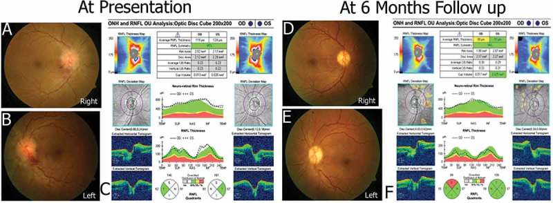
Showing regression of changes in retinal nerve fibre layer thickness in coherence with papilledemain a prototype patient. Figure 3C shows retinal nerve fibre layer thickness at presentation of prototype patient. The findings were consistent with papilledema both eyes(Figure 3A/B) at presentation. Normalisation of retinal nerve fibre layer thickness is seen over 6 months(Figure 3F), consistent with resolution of papilledema(Figure 3D/E).