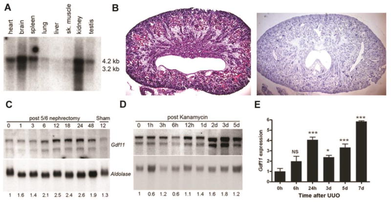
Figure 1. Expression of Gdf11 in normal adult kidney, in tubular epithelial cells and after kidney injury.
(A) Northern blot analysis of 2 µg twice poly(A)-selected mRNA shows two transcripts hybridizing to a cDNA probe corresponding to the C-terminal domain of Gdf11 in various adult mouse tissues. (B) H&E staining (left) and in situ hybridization for Gdf11 (right) in neonatal kidney shows expression in distal tubules, cortical collecting ducts, and medullary rays. (C) Northern blot analysis of Gdf11 expression in 10 µg total RNA from the remnant kidney at the number of hours indicated after removal of one kidney and resection of 2/3 of the other, and from (D) kidneys after kanamycin treatment at the indicated hours (h) or days (d) after injury. Gdf11 was normalized to aldolase expression as a loading control. (E) Gdf11 levels in total RNA isolated from ligated kidneys after unilateral ureteric obstruction (UUO) at the indicated hours (h) or days (d) after injury were measured by quantitative real-time RT-PCR (qRT-PCR) and normalized to Gapdh in this and all subsequent figures. Gdf11 levels are plotted relative to sham-operated kidney at time 0 (n = 3–5 mice each). *P < 0.05, **P < 0.01, ***P < 0.001

