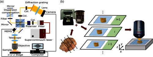
Fig. 1.
The visible-light OCM and the tissue processing pipeline. (a) The visible light OCM system (Col. = Collimator). (b) The extracted brain tissue is embedded in 5% agarose. A vibratome is used to section the brain-agarose block in slices. These sections are glued onto glass slides and are then optically cleared for 15 min following the SWITCH protocol.24 The cleared sections are imaged with the OCM setup. An automated stage is integrated in the sample arm to acquire images with a large field-of-view.

