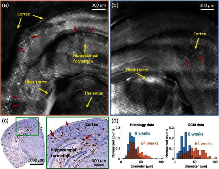
Fig. 5.
Large field-of-view images of brain tissue of an (a) 64-week- and a (b) 8-week-old AD mouse, acquired with the water immersion objective. (c) Immunohistochemically and hematoxylin stained histological section of the AD mouse shown in (a). (d) Histograms showing the plaque diameter distribution in the 8- and the 64-weeks-old mice in histology and OCM images. Amyloid-beta plaques in all panels are indicated by red arrows and anatomical features such as white matter tracts are indicated in yellow.

