Abstract
Background
Circular RNAs (circRNAs) are a type of covalently closed loop structure of endogenous RNAs. Recent studies have shown that circular RNAs may play an important role in human cancer. However, there is limited information on the function of circRNA in oral squamous cell carcinoma (OSCC).
Methods
Hsa_circ_001242 expression levels in 40 paired OSCC tissues and four OSCC cell lines were selected using real-time quantitative reverse transcription polymerase chain reaction (qRT-PCR). A receiver operating characteristic (ROC) curve was used to evaluate the diagnostic value of hsa_circ_001242 in OSCC.
Results
Hsa_circ_001242 was significantly downregulated in OSCC tissues compared to paired adjacent normal tissues (P < 0.001). Hsa_circ_001242 expression levels were significantly downregulated in four OSCC cell lines (SCC-9, SCC-15, SCC25, and CAL-27) than in human normal oral keratinocyte (HOK) cell lines. Moreover, the expression level of hsa_circ_001242 was negatively correlated with tumor size and T stage (P < 0.05). The area under the ROC curve was 0.784.
Conclusion
This study showed that hsa_circ_001242 was significantly downregulated in OSCC and may act as a potential novel biomarker for the diagnosis and treatment of OSCC.
1. Introduction
Oral squamous cell carcinoma (OSCC) is the third most common cancer in developing countries and ranks sixth among systemic cancers worldwide [1, 2]. Although there have been some improvements in the diagnosis and clinical treatment of OSCC, the 5-year overall survival rate of OSCC patients has not improved and remains <50% over the last three decades [3, 4]. In addition, many patients have a poor response to therapy and high recurrence rates [5]. Therefore, it is important to determine effective molecular diagnostic markers and therapeutic targets of OSCC.
Circular RNAs (circRNAs) are a type of covalently closed loop structure of endogenous RNAs, which are characterized by linking the 3′ and 5′ ends generated by backsplicing. Unlike linear RNAs, circRNAs require high conservation, have high stability and tissue specificity, and are not easily degraded by enonuclease [6, 7]. In the 1970s, circRNAs were first discovered in Sendai viruses and then were clearly observed in eukaryotes [8, 9]. CircRNAs were initially deemed to the products of aberrant RNA splicing, without attracting attention [10]. With the development of RNA high-throughput sequencing technology and bioinformatics, thousands of circRNAs have been discovered in the human genome [11]. Numerous researches have shown that circRNAs can be used as new biomarkers for clinical diagnosis and treatment of cancer [12–14]. However, there is little known about the role of circRNA in OSCC.
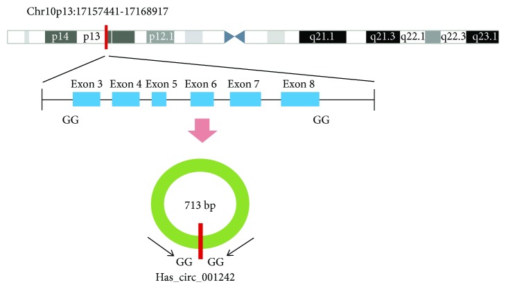
Searching OSCC associated circRNAs from circBase [15]. We first confirmed that hsa_circ_001242, which is located at chr10: 17157441–17168917 (Figure 1), was significantly downregulated in OSCC cell lines and OSCC tissues. Hsa_circ_001242, with a spliced sequence length of 713 bp, associated gene symbol is TRDMT1 (tRNA aspartic acid methyltransferase 1) and composed of six exons from exon 3 to exon 8. Its expression levels in tissues at various stages of OSCC were then explored, and the potential relationship between hsa_circ_001242 expression levels and patients' clinicopathological factors were investigated. A receiver operating characteristic (ROC) curve was constructed for evaluating the diagnostic value of hsa_circ_001242. Our data indicate that hsa_circ_001242 may be a potential biomarker for the diagnosis of OSCC.
Figure 1.
Hsa_circ_001242 is encoded from chromosomal region 10p13. Six exons of them form hsa_circ_001242 from exon 3 to exon 8.
2. Materials and Methods
2.1. Patients and Specimens
Forty oral cancer tissue samples and paired adjacent normal tissues were collected from the Department of Oral and Maxillofacial Surgery, Shenzhen Hospital, Peking University (Shenzhen, China), from December 2016 to May 2017. All samples were verified by histopathology. No patient previously underwent OSCC surgery, chemotherapy, or radiotherapy. Tissue samples were stored at −80°C before use. This study was approved by Ethics Committee of Peking University Health Science Center (IRB00001053-08043).
2.2. RNA Extraction
Total RNA was extracted from OSCC tissues and paired adjacent normal tissues using the TRIzol reagent (Invitrogen, Carlsbad, CA, USA) according to the manufacturer's protocol. Total RNA from each specimen was quantified, and quality assurance was conducted using a NanoDrop ND2000 spectrophotometer (NanoDrop, Wilmington, DE, USA). Reaction mixture (20 μl) containing 1 μg total RNA was reverse transcribed to cDNA using PrimeScript RT-polymerase (Takara, Dalian, China).
The RNA sample was dissolved in RNase-free water. The yield and purity were measured using a NanoDrop 2000 instrument (Thermo Fisher Scientific, Waltham, MA, USA). The integrity of the RNA was determined using 1% formaldehyde denaturing gel electrophoresis. A PrimeScript RT Reagent Kit (Takara Bio, Nojihigashi, Kusatsu, Japan) was used for the production of complementary DNA (cDNA) by reverse transcription, according to the manufacturer's instructions. qRT-PCR was performed using SYBR-Green Premix Ex Taq (Takara Bio, Nojihigashi, Kusatsu, Japan) and was monitored using the ABI PRISM 7500 Sequence Detection System (Applied Biosystems, Life Technologies, Waltham, MA, USA). The relative expression levels of circRNAs were determined by qRT-PCR. The sequences of the primers used in the qRT-PCR assay are shown in Supplementary Table 1.
The sequences of the hsa_circ_001242 primers were as follow: 5′-GCCCACTTGTAGAAGGTCCG-3′ (forward primer) and reverse 5′-CTGGCAGGGAGGGCTCATTA-3′ (reverse primer). The primer sequences for β-actin were 5′-AAACTGGAACGTTGAGAGTG-3′ (forward primer) and 5′-AGTGGTCTGGCTTTTAGGT-3′ (reverse primer). The reaction conditions were as follows: 95°C at 5 min for a preincubation and 40 cycles of 95°C for 5 s, annealing temperature of 60°C for primer pairs for 30 s, and 72°C for 20 s. RNA levels were normalized using β-actin as the internal control.
2.3. Cell Culture
The human OSCC cell lines, SCC9, SCC15, SCC25, and CAL27, were obtained from the College of Stomatology, Wuhan University (Wuhan, China). Human oral keratinocytes (HOK) cells were obtained from the cell bank of the Chinese Academy of Sciences (Shanghai, China). SCC9 cells were cultured in DMED/F12 medium supplemented with 1% penicillin/streptomycin. SCC15, SCC25, CAL27, and HOK cells were cultured in Dulbecco's Modified Eagle Medium (DMED, GIBCO, China) supplemented with 1% penicillin/streptomycin (Life Technologies Inc., USA). All cells were cultured at 37°C under 5% CO2.
2.4. Statistical Analysis
GraphPad Prism 5.0 Software (GraphPad Software, La Jolla, CA, USA) was used to analyze the obtained data. Results of hsa_circ_001242 expression for OSCC tissues and paired adjacent normal tissues or between OSCC cell lines and HOK cell lines were compared using a paired t-test. A nonpaired t-test was used to analyze the relationship between hsa_circ_001242 expression level and clinicopathological factors in OSCC patients. The ROC curve was constructed to evaluate the diagnostic values. ∗ P < 0.05, ∗∗ P < 0.01, and ∗∗∗ P < 0.001 were considered statistically significant.
3. Results
3.1. Expression of hsa_circ_001242 in OSCC Tissues
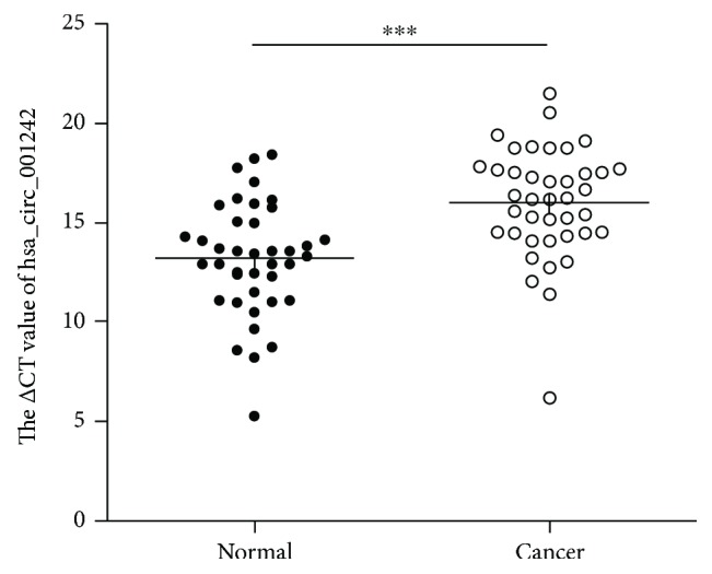
In this study, we first observed that the expression levels of hsa_circ_001242 were evidently downregulated in 40 OSCC tissue samples compared to that in paired adjacent normal tissues by qRT-PCR (n = 40, P < 0.001) (Figure 2).
Figure 2.

Hsa_circ_001242 expression levels in 40 pairs of OSCC tissues compared to that in paired adjacent normal tissues. Higher △Ct value indicates lower expression. Data are expressed as mean ± SD; ∗∗∗ P < 0.001.
3.2. Hsa_circ_001242 Expression Levels in OSCC Cell Lines
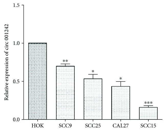
Furthermore, we explored the expression levels of hsa_circ_001242 in the HOK cell lines and four human OSCC cell lines, SCC-9, SCC-15, SCC-25, and CAL27. Expression levels in the OSCC cell lines were significantly lower than those in the HOK cell lines (Figure 3).
Figure 3.

Hsa_circ_001242 expression in different OSCC cell lines. Hsa_circ_001242 expression levels in four OSCC cell lines (SCC-9, SCC-15, SCC-25, and CAL-27), and normal human oral keratinocyte cell lines were assessed by qRT-PCR. Data are presented as mean ± SD; ∗ P < 0.05, ∗∗ P < 0.01, and ∗∗∗ P < 0.001.
3.3. Relationship between hsa_circ_001242 Levels and Clinicopathological Factors
Clinicopathological analysis revealed that hsa_circ_001242 expression level was significantly associated with clinicopathological factors of OSCC patients. As presented in Table 1, hsa_circ_001242 expression levels were negatively related to tumor size (P = 0.0125) and T stage (P = 0.0434) but were not associated with other clinicopathological features, such as age, gender, tumor differentiation, TNM stage, or lymphatic metastasis (P > 0.05).
Table 1.
Primer sequences.
| Primer set | Forward primer | Reverse primer |
|---|---|---|
| hsa_circ_001242 | GCCCACTTGTAGAAGGTCCG | CTGGCAGGGAGGGCTCATTA |
| β-Actin | AAACTGGAACGTTGAGAGTG | AGTGGTCTGGCTTTTAGGT |
3.4. The Diagnostic Value of hsa_circ_001242 in OSCC
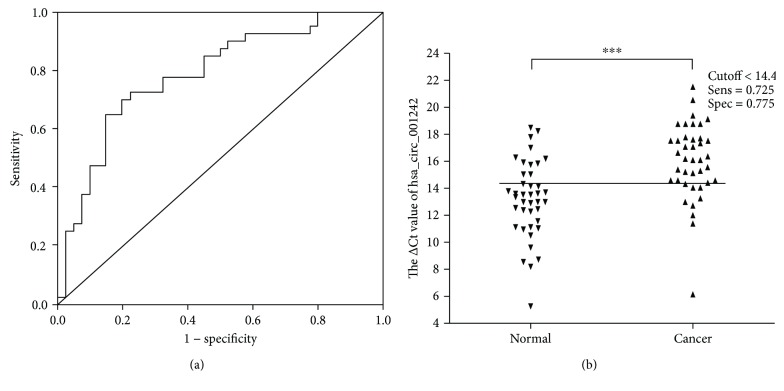
To estimate the diagnostic value of hsa_circ_001242 in OSCC, an ROC curve was constructed for differentiating OSCC tissues from paired adjacent normal tissues. The area under the ROC curve (AUC) was 0.784 (95% confidence interval (CI) = 0.717–0.867; P < 0.001; Figure 4(a)). The cutoff value was 14.4 (Figure 4(b)), and the corresponding sensitivity and specificity of hsa_circ_001242 were 72.5% and 77.5%, respectively. When the area under the curve is larger, the diagnostic value of the variable is higher. Values below the cutoff value are negative, while those exceeding the cutoff value are positive. Therefore, hsa_circ_001242 may act as a potential biomarker for the diagnosis of OSCC.
Figure 4.
The diagnostic value of hsa_circ_001242 in OSCC. (a) The area under the ROC curve (AUC) was 0.784 (95% CI = 0.683–0.885, P < 0.001), (b) and the cutoff of hsa_circ_001242 was 14.4. The sensitivity and specificity were 0.725 and 0.775, respectively. Data are presented as mean ± SD; ∗∗∗ P < 0.001.
4. Discussion
CircRNAs are a class of noncoding RNAs (ncRNAs) that have been neglected as transcriptional noise in eukaryotes for the past 30 years [10, 16, 17]. Recent studies have found that some circRNAs serve as an important regulatory role and do not splice noise as previously thought [18]. Compelling evidence has manifested that circRNA can regulate gene expression through multiple mechanisms. CircRNAs act as a sponge of miRNA molecules and competitively bind miRNAs to regulate gene expression [19, 20]. Exon-intron circRNAs (EIciRNAs) can regulate transcription by RNA-RNA interactions in the nucleus [21, 22]. Some reports showed that circRNAs have the function of being translated into proteins [23, 24]. These studies indicate that the circular RNA plays an important role in transcriptional and posttranscriptional levels and can serve as an ideal marker for disease diagnosis. Previous studies have suggested that circRNAs may play an important role in the development of cancer and act as potential biomarkers for cancer [25, 26]. For example, Li Wan et al. [27] have revealed that circ-ITCH is overexpressed in lung cancer tissues and inhibits the Wnt/β-catenin signaling pathway by acting as a sponge for miR-7 and miR-214. Zhu et al. [28] have reported that circ-BANP expression in colorectal cancerous tissues is significantly upregulated than those in adjacent normal tissues. Huang et al. [29] have observed that hsa_circ_0000745 is downregulated in gastric cancer tissues as well as in the plasma samples from patients with gastric cancer. Their study revealed that the circular RNA hsa_circ_0000745 may serve as a diagnostic marker for gastric cancer. Yao et al. [30] have confirmed that circZKSCAN1 is downregulated in hepatocellular carcinoma and may act as a diagnostic biomarker. Thus, we believe that circRNAs may serve as novel molecular markers for cancer.
In this study, we first observed that hsa_circ_001242 was downregulated in both OSCC tissues and OSCC cell lines. Furthermore, the study on the clinical features and the expression of hsa_circ_001242 indicated that hsa_circ_001242 expression level was negatively relevant to tumor size and T stage (Table 2). These data suggest that circRNAs may play a role in carcinogenesis and progress of OSCC. The ROC curve was constructed for differentiating OSCC tissues from the controls. Our results demonstrated that AUC was 0.784. The sensitivity and specificity of hsa_circ_001242 were 0.725 and 0.775, respectively. Furthermore, we observed that the cutoff value of hsa_circ_001242 was 14.4. To further understand the biological role of circRNAs in OSCC, both in vitro and in vivo assays should be performed in the future after successfully engineering of circRNAs.
Table 2.
Correlation between clinicopathological factors and hsa_circ_001242 expression levels (ΔCt) in oral squamous cell carcinoma patients.
| Characteristics | Number of patients | Mean ± SD | P value |
|---|---|---|---|
| Age (year) | |||
| ≥60 | 14 | 16.88 ± 0.644 | 0.145 |
| <60 | 26 | 15.52 ± 0.572 | |
| Gender | |||
| Male | 28 | 16.05 ± 0.537 | 0.871 |
| Female | 12 | 15.89 ± 0.813 | |
| Tumor size(cm) | |||
| ≥5 | 6 | 13.43 ± 0.543 | 0.0125∗ |
| <5 | 34 | 16.45 ± 0.408 | |
| Differentiation | |||
| Well and moderate | 34 | 16.23 ± 0.424 | 0.221 |
| Poor | 6 | 14.70 ± 0.747 | |
| T stage | |||
| T1–2 | 29 | 16.58 ± 0.45 | 0.0317∗ |
| T3–4 | 11 | 14.47 ± 0.99 | |
| TNM | |||
| I and II | 19 | 16.76 ± 0.61 | 0.103 |
| III and IV | 21 | 15.31 ± 0.61 | |
| Lymphatic metastasis | |||
| N0 | 24 | 16.50 ± 0.516 | 0.170 |
| N1–3 | 16 | 15.25 ± 0.773 | |
∗ indicated statistical significance.
In conclusion, our study manifested that hsa_circ_001242 was significantly downregulated in OSCC tissues and OSCC cell lines. In addition, the expression of hsa_circ_001242 expression is negatively correlated with tumor size and T stage. Thus, hsa_circ_001242 may serve as a potential diagnostic biomarker for OSCC.
Acknowledgments
This study was supported by the National Natural Science Foundation of China (Grant no. 81572654), the Basic Research Program of Shenzhen Innovation Council of China (Grant nos. JCYJ20150403091443303, JCYJ20150403091443286, JCYJ201604287 3933559, and SZBC2017023), and Sanming Project of Medicine in Shenzhen (SZSM 201512036, Oral and Maxillofacial Surgery Team, Professor Yu Guangyan, Stomatology Hospital Peking University).
Contributor Information
Wei Zhang, Email: zhangweispace@yeah.net.
Hongyu Yang, Email: hyyang192@hotmail.com.
Data Availability
The data used to support the findings of this study are available from the corresponding author upon request.
Conflicts of Interest
The authors declare that there is no conflict of interest regarding the publication of this article.
Authors' Contributions
Shuai Sun and Bowen Li contributed equally to this work.
Supplementary Materials
Supplementary Information Table 1. Detailed information on clinical and oral squamous cell carcinoma characteristics of patients and the information of metastasis.
References
- 1.Rosebush M. S., Rao S. K., Samant S., et al. Oral cancer: enduring characteristics and emerging trends. The Journal of the Tennessee Dental Association. 2012;91(2):24–27. [PubMed] [Google Scholar]
- 2.Casiglia J., Woo S. B. A comprehensive review of oral cancer. General Dentistry. 2001;49(1):72–82. [PubMed] [Google Scholar]
- 3.Brocklehurst P. R., Baker S. R., M Speight P. Oral cancer screening: what have we learnt and what is there still to achieve? Future Oncology. 2010;6(2):299–304. doi: 10.2217/fon.09.163. [DOI] [PubMed] [Google Scholar]
- 4.Siegel R., Ma J., Zou Z., Jemal A. Cancer statistics, 2014. CA: a Cancer Journal for Clinicians. 2014;64(1):9–29. doi: 10.3322/caac.21208. [DOI] [PubMed] [Google Scholar]
- 5.Wangsa D., Ryott M., Åvall-Lundqvist E., et al. Ki-67 expression predicts locoregional recurrence in stage I oral tongue carcinoma. British Journal of Cancer. 2008;99(7):1121–1128. doi: 10.1038/sj.bjc.6604633. [DOI] [PMC free article] [PubMed] [Google Scholar]
- 6.Chen L.-L., Yang L. Regulation of circRNA biogenesis. RNA Biology. 2015;12(4):381–388. doi: 10.1080/15476286.2015.1020271. [DOI] [PMC free article] [PubMed] [Google Scholar]
- 7.Suzuki H., Zuo Y., Wang J., Zhang M. Q., Malhotra A., Mayeda A. Characterization of RNase R-digested cellular RNA source that consists of lariat and circular RNAs from pre-mRNA splicing. Nucleic Acids Research. 2006;34(8, article e63) doi: 10.1093/nar/gkl151. [DOI] [PMC free article] [PubMed] [Google Scholar]
- 8.Kolakofsky D. Isolation and characterization of Sendai virus DI-RNAs. Cell. 1976;8(4):547–555. doi: 10.1016/0092-8674(76)90223-3. [DOI] [PubMed] [Google Scholar]
- 9.Hsu M.-T., Coca-Prados M. Electron microscopic evidence for the circular form of RNA in the cytoplasm of eukaryotic cells. Nature. 1979;280(5720):339–340. doi: 10.1038/280339a0. [DOI] [PubMed] [Google Scholar]
- 10.Cocquerelle C., Mascrez B., Hétuin D., Bailleul B. Mis-splicing yields circular RNA molecules. The FASEB Journal. 1993;7(1):155–160. doi: 10.1096/fasebj.7.1.7678559. [DOI] [PubMed] [Google Scholar]
- 11.Xia S., Feng J., Lei L., et al. Comprehensive characterization of tissue-specific circular RNAs in the human and mouse genomes. Briefings in Bioinformatics. 2016;18(6):984–992. doi: 10.1093/bib/bbw081. [DOI] [PubMed] [Google Scholar]
- 12.Li P., Chen S., Chen H., et al. Using circular RNA as a novel type of biomarker in the screening of gastric cancer. Clinica Chimica Acta. 2015;444:132–136. doi: 10.1016/j.cca.2015.02.018. [DOI] [PubMed] [Google Scholar]
- 13.Szabo L., Salzman J. Detecting circular RNAs: bioinformatic and experimental challenges. Nature Reviews Genetics. 2016;17(11):679–692. doi: 10.1038/nrg.2016.114. [DOI] [PMC free article] [PubMed] [Google Scholar]
- 14.Rong D., Tang W., Li Z., et al. Novel insights into circular RNAs in clinical application of carcinomas. OncoTargets and Therapy. 2017;10:2183–2188. doi: 10.2147/OTT.S134403. [DOI] [PMC free article] [PubMed] [Google Scholar]
- 15.Circbase. http://circbase.org/cgi-bin/simplesearch.cgi.
- 16.Kos A., Dijkema R., Arnberg A. C., Van der Meide P. H., Schellekens H. The hepatitis delta (δ) virus possesses a circular RNA. Nature. 1986;323(6088):558–560. doi: 10.1038/323558a0. [DOI] [PubMed] [Google Scholar]
- 17.Zaphiropoulos P. G. Circular RNAs from transcripts of the rat cytochrome P450 2C24 gene: correlation with exon skipping. Proceedings of the National Academy of Sciences of the United States of America. 1996;93(13):6536–6541. doi: 10.1073/pnas.93.13.6536. [DOI] [PMC free article] [PubMed] [Google Scholar]
- 18.Chen L., Huang C., Wang X., Shan G. Circular RNAs in eukaryotic cells. Current Genomics. 2015;16(5):312–318. doi: 10.2174/1389202916666150707161554. [DOI] [PMC free article] [PubMed] [Google Scholar]
- 19.Karreth F. A., Tay Y., Perna D., et al. In vivo identification of tumor- suppressive PTEN ceRNAs in an oncogenic BRAF-induced mouse model of melanoma. Cell. 2011;147(2):382–395. doi: 10.1016/j.cell.2011.09.032. [DOI] [PMC free article] [PubMed] [Google Scholar]
- 20.Hansen T. B., Kjems J., Damgaard C. K. Circular RNA and miR-7 in cancer. Cancer Research. 2013;73(18):5609–5612. doi: 10.1158/0008-5472.CAN-13-1568. [DOI] [PubMed] [Google Scholar]
- 21.Li Z. Y., Huang C., Bao C., et al. Exon-intron circular RNAs regulate transcription in the nucleus. Nature Structural & Molecular Biology. 2015;22(3):256–264. doi: 10.1038/nsmb.2959. [DOI] [PubMed] [Google Scholar]
- 22.Huang C., Shan G. What happens at or after transcription: insights into circRNA biogenesis and function. Transcription. 2015;6(4):61–64. doi: 10.1080/21541264.2015.1071301. [DOI] [PMC free article] [PubMed] [Google Scholar]
- 23.Abe N., Matsumoto K., Nishihara M., et al. Rolling circle translation of circular RNA in living human cells. Scientific Reports. 2015;5(1, article 16435) doi: 10.1038/srep16435. [DOI] [PMC free article] [PubMed] [Google Scholar]
- 24.Wang Y., Wang Z. Efficient backsplicing produces translatable circular mRNAs. RNA. 2015;21(2):172–179. doi: 10.1261/rna.048272.114. [DOI] [PMC free article] [PubMed] [Google Scholar]
- 25.Chen Y., Li C., Tan C., Liu X. Circular RNAs: a new frontier in the study of human diseases. Journal of Medical Genetics. 2016;53(6):359–365. doi: 10.1136/jmedgenet-2016-103758. [DOI] [PubMed] [Google Scholar]
- 26.Meng S., Zhou H., Feng Z., et al. CircRNA: functions and properties of a novel potential biomarker for cancer. Molecular Cancer. 2017;16(1):p. 94. doi: 10.1186/s12943-017-0663-2. [DOI] [PMC free article] [PubMed] [Google Scholar]
- 27.Wan L., Zhang L., Fan K., Cheng Z. X., Sun Q. C., Wang J. J. Circular RNA-ITCH suppresses lung cancer proliferation via inhibiting the Wnt/β-catenin pathway. BioMed Research International. 2016;2016:11. doi: 10.1155/2016/1579490.1579490 [DOI] [PMC free article] [PubMed] [Google Scholar]
- 28.Zhu M., Xu Y., Chen Y., Yan F. Circular BANP, an upregulated circular RNA that modulates cell proliferation in colorectal cancer. Biomedicine & Pharmacotherapy. 2017;88:138–144. doi: 10.1016/j.biopha.2016.12.097. [DOI] [PubMed] [Google Scholar]
- 29.Huang M., He Y.-R., Liang L.-C., Huang Q., Zhu Z.-Q. Circular RNA hsa_circ_0000745 may serve as a diagnostic marker for gastric cancer. World Journal of Gastroenterology. 2017;23(34):6330–6338. doi: 10.3748/wjg.v23.i34.6330. [DOI] [PMC free article] [PubMed] [Google Scholar]
- 30.Yao Z., Luo J., Hu K., et al. ZKSCAN1 gene and its related circular RNA (circZKSCAN1) both inhibit hepatocellular carcinoma cell growth, migration, and invasion but through different signaling pathways. Molecular Oncology. 2017;11(4):422–437. doi: 10.1002/1878-0261.12045. [DOI] [PMC free article] [PubMed] [Google Scholar]
Associated Data
This section collects any data citations, data availability statements, or supplementary materials included in this article.
Supplementary Materials
Supplementary Information Table 1. Detailed information on clinical and oral squamous cell carcinoma characteristics of patients and the information of metastasis.
Data Availability Statement
The data used to support the findings of this study are available from the corresponding author upon request.


