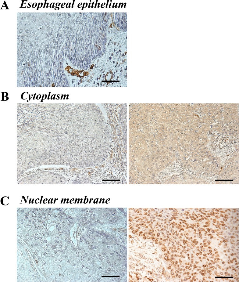
Figure 1. AQP1 protein expression in human ESCC.
(A) Immunohistochemical staining of human esophageal epithelia using an anti-AQP1 antibody. AQP1 was not expressed in noncancerous esophageal squamous epithelium. Magnification: ×400. Bar 50 μm. (B) Immunohistochemical staining of the cytoplasm in primary human ESCC samples using an anti-AQP1 antibody. Photomicrographs are shown with the examples of negative cells (left), positive cells (right). Magnification: ×200. Bar 100 μm. (C) Immunohistochemical staining of the nuclear membrane in primary human ESCC samples with an anti-AQP1 antibody. Photomicrographs are shown with the examples of the low AQP1 expression in the nuclear membrane (left) and of the high AQP1 expression in the nuclear membrane (right). Magnification: ×400. Bar 50 μm.

