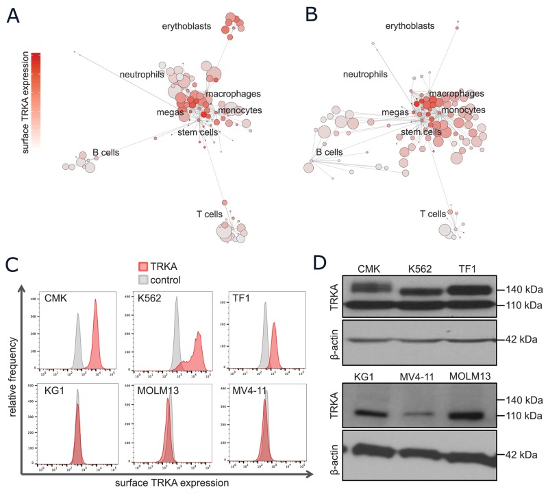
Figure 2. A subset of normal and leukemic myeloid cells express surface TRKA protein.
(A-B) Scaffold visual summary of single cell mass cytometry data depicting total surface TRKA expression in the context of normal hematopoiesis (A) and an example AML sample (B). (C) Cells were stained with anti-TRKA-phycoerythrin (PE) (red) and an isotype control mAb (grey). The cell lines that expressed evident surface TRKA, CMK, K562, and TF-1, are displayed along with 3 cell lines that express NTRK1 mRNA but no surface TRKA protein. (D) A western blot analysis of TRKA protein expression was performed on cell lysates prepared from the same cells as the previous flow experiment using a rabbit anti-TRKA antibody with a β-actin loading control. The anti-TRKA antibody identifies both the immature (110 kDa) and mature glycosylated (typically 140 kDa) form of the TRKA protein.

