Abstract
A 74-year-old man presented with a progressive decrease in visual acuity and foreign body sensation in his right eye 8 days post uncomplicated phacoemulsification cataract surgery and intraocular lens insertion. The patient had been placed on a perioperative cataract regimen which consisted of G. Maxitrol (dexamethasone, polymyxin B sulfate, neomycin sulfate) four times a day and G. Yellox twice daily (bromfenac, a non-steroidal anti-inflammatory) for 2 weeks. On examination, he had a corneal ulcer and stromal thinning in his right eye which progressed to a full thickness perforation 12 hours later. The patient required a full thickness tectonic corneal transplant. Direct questioning revealed that this patient had both dry mouth and eyes. Serology revealed that the patient was positive for rheumatoid factor and for anti-Ro and anti-La antibodies. A parotid gland biopsy revealed significant lymphocytic infiltrate consistent with Sjögren’s syndrome.
Keywords: eye, ophthalmology, anterior chamber, sjogren’s syndrome
Background
Corneal melt or perforation has been described in the literature in association with topical non-steroidal anti-inflammatory drug (NSAID) use1 2; however, it is extremely rare for it to occur in such a short period without a coexisting epithelial insult.3 Topical NSAIDs have been used to both prevent and treat postoperative cystoid macular oedema (CMO) and to date the published data suggest that bromfenac is safe and effective when used to treat or prevent postoperative CMO4 although there are rare reported cases of corneal melt.3 5 Our patient had no notable previous medical history and preoperative ophthalmic examination was unremarkable. Although he was subsequently diagnosed with Sjögren’s syndrome, ocular manifestations were absent or minimal. This case highlights the risk of using topical NSAID and/or preservative containing drops in patients with potential ocular surface disease to prevent ocular surface complications post routine cataract extraction.
Case presentation
A 74-year-old man presented to the Eye Emergency Department of Galway University Hospital complaining of a gradual decrease in visual acuity and foreign body sensation in his right eye 8 days post uncomplicated phacoemulsification cataract surgery and intraocular lens insertion. His preoperative assessment was routine with no specific preoperative concerns and he had a normal Schirmer test and normal corneal sensation. He had no relevant medical or surgical history and was not currently taking any medication. Postoperatively, the patient was placed on a regimen which consisted of G. Maxitrol (dexamethasone, polymyxin B sulfate, neomycin sulfate and benzalkonium chloride), one drop four times a day for 2 weeks post procedure and G. Yellox (bromfenac) one drop two times a day for 3 days before and 11 days post procedure. The patient stated that his vision had improved initially in the postoperative period but began to gradually deteriorate from day 4 post procedure. He denied any ocular pain but complained of a persistent ‘gritty’ sensation in his eye. His visual acuity on presentation to the emergency department was 6/60 in his right eye and 6/6 in his left eye (left eye examination was unremarkable). There was a 5.1 mm × 6.6 mm infratemporal epithelial defect in his right cornea. The patient had a superior on-axis incision for his cataract surgery and so the defect was not related to the primary incision. Stromal thinning was present to approximately 50% of total corneal thickness. The cause of the defect at the time was unknown. An initial assumption was made that he may have inadvertently caused a corneal abrasion while instilling his mediations. The drops were stopped, and chloramphenicol ointment was applied to the eye, which was padded overnight.
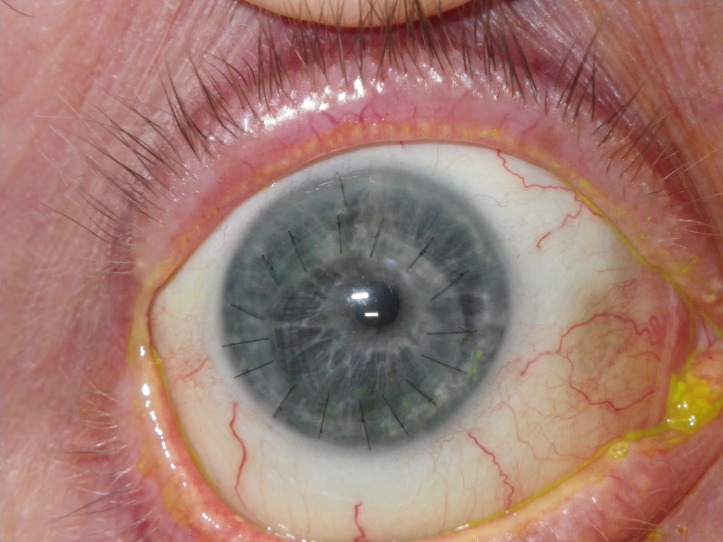
On review 12 hours later, the stromal defect had progressed to a localised full-thickness perforation of the inferior cornea and the anterior chamber was shallow. As a temporary measure, he underwent sealing of the corneal perforation with cyanoacrylate glue (Histoacryl). A bandage contact lens was inserted and the patient was commenced on preservative-free prednisolone 0.5% and preservative-free chloramphenicol 0.5% drops, both four times a day. Day 1 post glue application, the anterior chamber was fully reformed and the cornea was Seidel negative. Over the course of the following week, the defect began to leak and the anterior chamber depth fluctuated. Secondary to the fact that it was a non-healing defect in the inferior cornea, a decision was made that the patient undergo a corneal graft. He subsequently had an uneventful slightly eccentric full-thickness tectonic corneal transplant due to the inferior site of the corneal melt and perforation. The patient was placed on 2 hourly preservative-free prednisolone 0.5% and 4 hourly preservative-free chloramphenicol 0.5% drops. His vision improved to 6/36 4 days post transplant. Re-epithelisation was slow but was complete 21 days post operation (figure 1).
Figure 1.
25 days post penetrating keratoplasty. Graft completely re-epithelialised.
Direct questioning revealed that this patient had mild dry mouth and eye symptoms. It was noted on gross examination that he had mildly enlarged parotid glands. Serology showed that the patient was positive for rheumatoid factor and for anti-Ro and anti-La antibodies. CT revealed multiple lesions in his parotid gland. A parotid gland biopsy was taken which revealed significant lymphocytic infiltrate consistent with Sjögren’s syndrome.
He was placed on g. Hylo Forte (0.2% sodium hyaluronate, preservative free) to be used hourly as a substitute for his reduced tear flow and lacrimal punctal plugs were inserted.
Investigations
Serology was positive for anti-Ro, anti-La and rheumatoid factor.
CT revealed multiple lesions in his parotid gland and parotid gland biopsy was positive for a lymphocytic infiltrate.
Differential diagnosis
Chronic epitheliopathy.
Perforation secondary to keratoconjunctivitis sicca related to Sjögren’s syndrome.
Toxicity to preservative in topical medication.
Treatment
Patient underwent a penetrating keratoplasty in his right eye due to full-thickness corneal melt and perforation.
Outcome and follow-up
The patient is currently being followed up in outpatients. His graft remains clear but due to its eccentric position the patient has substantial astigmatism and his best corrected visual acuity remains at 6/36.
Discussion
This is an unusual case of corneal perforation secondary to topical medication use post routine phacoemulification procedure. It is likely that there are several factors that predisposed this patient to a corneal perforation: his undiagnosed Sjögren’s syndrome the use of the topical NSAID therapy, the use of topical steroids and use of preservative-containing drops. There are reports of corneal melt with the use of steroids alone,6 as an initial presentation of Sjögren’s syndrome7 and therefore it is highly probable that it is a combination of the above factors that subjected this patient to his subsequent melt.
Sjögren’s syndrome is an autoimmune disorder characterised by a variable degree of sicca symptoms including keratoconjunctivitis sicca and xerostomia.8 In the eye, it is considered predominately as an aqueous tear deficiency with an additional insufficient lipid layer component. Most patients with this syndrome are stable from an ophthalmic point of view with ocular lubricants. Severe dry eyes can be associated with keratitis, stromal scarring and ulceration.9 There is some evidence that the use of NSAIDs in Sjögren’s syndrome can improve symptoms of ocular discomfort but should be used with caution.10 In the setting of cataract surgery, in addition to their anti-inflammatory effect NSAIDS may also act as mild anaesthetics, decrease corneal sensation and may impede corneal healing. Patients with symptomatic Sjögren’s syndrome should employ frequent use of preservative-free tear substitutes with ocular lubricating ointments reserved for nocturnal use.11 Controlled trials support the use of topical 0.05% cyclosporine two times a day for patients with moderate-to-severe dry eye disease.12 Patients who have dry eye secondary to a rheumatological condition are at an increased risk of corneal melt with the use of preservative containing drops.13 In this case, there was no known history of Sjögren’s syndrome or dry eyes preoperatively. There is limited literature on protocols for postoperative drops after cataract surgery in patients with Sjögren’s syndrome. However, standard preoperative and postoperative treatment regimens need to be tailored for those with compromised ocular surfaces, perhaps even withholding NSAIDs completely. Avoidance of preservative in drops and cautious use of topical NSAIDs should be considered in patients prone to corneal epithelial breakdown.
A history of diabetes is an independent risk factor for the development of postoperative CMO14 and recent literature has demonstrated that patients with diabetes would benefit from a course of G. Nepafenac 0.1% eye drops (NSAID) 1 day before to 3 months post cataract extraction to lower the risk of postoperative CMO from 16.7% to 3.2%.15 Use of NSAIDs in diabetics post cataract surgery is becoming more routine, but these patients have compromised ocular surfaces and thus are more prone to corneal complications post surgery. This highlights the need of judicious prescribing of NSAID and preservative-containing drops in patients for the prevention of postoperative CMO.
Patient’s perspective.
‘I believed that I was going in for a standard cataract operation. A lot of my friends had gotten it done and they had great outcomes. After the surgery my eye didn’t feel right but I thought it would improve. It was a shock to me when I heard that the skin at the front of me eye had broken down and that I would need a graft. My vision isn’t bad at the moment and I am happy with the outcome overall but there were many clinic appointments and lots of drops to be put in all the time!’
Learning points.
Preoperative assessment in cataract surgery is essential and patients should be questioned as regards any rheumatological conditions which may predispose to corneal problems post surgery.
Patients with Sjögren’s syndrome and aqueous deficiency dry eye require an appropriate postoperative regimen of ocular surface lubrication with preservative free lubricants.
Recent studies support the use of perioperative non-steroidal anti-inflammatory drug (NSAIDs) as a prophylactic treatment for postoperative cystoid macular oedema especially in patients with diabetes. We advise judicious use particularly in those with impaired ocular surface. Avoidance of preservative in drops and cautious use of topical NSAIDs should be considered in patients prone to corneal epithelial breakdown.
Footnotes
Contributors: RC initially saw the patient and underwent emergency treatment. He was referred to GF for further intervention. RC suggested to PM that the case should be written up and PM wrote the case report under the guidance of both GF and RC. GF and RC reviewed numerous drafts until they deemed it fit for submission.
Funding: The authors have not declared a specific grant for this research from any funding agency in the public, commercial or not-for-profit sectors.
Competing interests: None declared.
Patient consent: Obtained.
Provenance and peer review: Not commissioned; externally peer reviewed.
References
- 1.Lin JC, Rapuano CJ, Laibson PR, et al. Corneal melting associated with use of topical nonsteroidal anti-inflammatory drugs after ocular surgery. Arch Ophthalmol 2000;118:1129–32. [PubMed] [Google Scholar]
- 2.Congdon NG, Schein OD, von Kulajta P, et al. Corneal complications associated with topical ophthalmic use of nonsteroidal antiinflammatory drugs. J Cataract Refract Surg 2001;27:622–31. 10.1016/S0886-3350(01)00801-X [DOI] [PubMed] [Google Scholar]
- 3.Asai T, Nakagami T, Mochizuki M, et al. Three cases of corneal melting after instillation of a new nonsteroidal anti-inflammatory drug. Cornea 2006;25:224–7. 10.1097/01.ico.0000177835.93130.d4 [DOI] [PubMed] [Google Scholar]
- 4.Sheppard JD. Topical bromfenac for prevention and treatment of cystoid macular edema following cataract surgery: a review. Clin Ophthalmol 2016;10:2099–111. 10.2147/OPTH.S86971 [DOI] [PMC free article] [PubMed] [Google Scholar]
- 5.Isawi H, Dhaliwal DK. Corneal melting and perforation in Stevens Johnson syndrome following topical bromfenac use. J Cataract Refract Surg 2007;33:1644–6. 10.1016/j.jcrs.2007.04.041 [DOI] [PubMed] [Google Scholar]
- 6.Praidou A, Brazitikos P, Dastiridou A, et al. Severe unilateral corneal melting after uneventful phacoemulsification cataract surgery. Clin Exp Optom 2013;96:109–11. 10.1111/j.1444-0938.2012.00750.x [DOI] [PubMed] [Google Scholar]
- 7.Vivino FB, Minerva P, Huang CH, et al. Corneal melt as the initial presentation of primary Sjögren’s syndrome. J Rheumatol 2001;28:379–82. [PubMed] [Google Scholar]
- 8.Brito-Zerón P, Theander E, Baldini C, et al. Early diagnosis of primary Sjögren’s syndrome: EULAR-SS task force clinical recommendations. Expert Rev Clin Immunol 2016;12:137–56. 10.1586/1744666X.2016.1109449 [DOI] [PubMed] [Google Scholar]
- 9.Hyon JY, Lee YJ, Yun PY. Management of ocular surface inflammation in Sjögren syndrome. Cornea 2007;26(9 Suppl 1):S13–15. 10.1097/ICO.0b013e31812f6782 [DOI] [PubMed] [Google Scholar]
- 10.Aragona P, Stilo A, Ferreri F, et al. Effects of the topical treatment with NSAIDs on corneal sensitivity and ocular surface of Sjögren’s syndrome patients. Eye 2005;19:535–9. 10.1038/sj.eye.6701537 [DOI] [PubMed] [Google Scholar]
- 11.Kassan SS, Moutsopoulos HM. Clinical manifestations and early diagnosis of Sjögren syndrome. Arch Intern Med 2004;164:1275–84. 10.1001/archinte.164.12.1275 [DOI] [PubMed] [Google Scholar]
- 12.Sall K, Stevenson OD, Mundorf TK, et al. Two multicenter, randomized studies of the efficacy and safety of cyclosporine ophthalmic emulsion in moderate to severe dry eye disease. CsA Phase 3 Study Group. Ophthalmology 2000;107:631–9. [DOI] [PubMed] [Google Scholar]
- 13.Ram J, Sharma A, Pandav SS, et al. Cataract surgery in patients with dry eyes. J Cataract Refract Surg 1998;24:1119–24. 10.1016/S0886-3350(98)80107-7 [DOI] [PubMed] [Google Scholar]
- 14.Oyewole K, Tsogkas F, Westcott M, et al. Benchmarking cataract surgery outcomes in an ethnically diverse and diabetic population: final post-operative visual acuity and rates of post-operative cystoid macular oedema. Eye 2017;31:1672–7. 10.1038/eye.2017.96 [DOI] [PMC free article] [PubMed] [Google Scholar]
- 15.Singh R, Alpern L, Jaffe GJ, et al. Evaluation of nepafenac in prevention of macular edema following cataract surgery in patients with diabetic retinopathy. Clin Ophthalmol 2012;6:1259–69. 10.2147/OPTH.S31902 [DOI] [PMC free article] [PubMed] [Google Scholar]


