Abstract
The early diagnosis of dental caries has an important role in pregnancy, as it allows establishing preventive measures. Besides the clinical examination, there are modern preclinical ways of detecting odontal lesions such as electrical conductivity (EC) and quantitative Light-Induced Fluorescence (QLF). Dental radiography and three-dimensional (3D) orthopantomography, although useful, are forbidden during pregnancy (6). Bacteriological evaluation and early detection of demineralized areas allow preventive measures aimed at stopping the destructive process and permit measures for the restoration of the damaged dental structures.
Regarding the treatment of caries, superficial coronal odontal lesions in enamel can be treated noninvasively by remineralization. Reconstruction, obturation or inscruction therapy involves loss of dental material, sometimes even healthy one; they are also expensive and stressful for the patient and therefore, remineralization and sealing of dental retention areas is the treatment of choice for children and pregnant women (8). For the restoration of the damaged dental structure, fluoride topics, laques or fluoride gels are applied locally (3).
An adequate diet during pregnancy plays an important role in maintaining the general and oral health; it must be high in calories, proteins, vitamins and minerals, and it must have a balanced proportion of salts, carbohydrates and lipids. As with the rest of the population, proper dental brushing at least twice a day in the morning and evening as well as the use of yarn thread are effective ways of oral hygiene, which also prevent the appearance and evolution of dental caries (1).
Keywords:electrical conductivity, fluorescence of dental hard tissues, 3D radiography, sealing, fluoride topics
Early diagnosis of dental caries in pregnant women is an essential factor in prophylactic and curative therapy in puerperality. Early detection and treatment of dental caries in pregnant women are all the more important if we think that restorative therapy is more expensive and sometimes involves certain risks. The purpose of contemporary dental medicine is to offer practitioners advanced diagnosis techniques in order to objectify dental caries with more than just the help of a dental probe (2).
Clinical examination is beneficial and mandatory only if it is done with great care, thoroughly with non-traumatic fine instrumentation after removing the dental plaque, cleansing, isolating and drying the dental surfaces (4).
The medical algorithm for early detection of carious lesions requires two basic directions that will allow the correct approach to dental caries therapy:
- evaluation of risk factors with real potential in the appearance of tooth caries, but which did not cause any actual injury;
- early detection of demineralized areas prior to their objectification by clinical and paraclinical examination (5).
The quantitative detection of dental caries is based on a mandatory clinical examination, along with two other distinct techniques: electrical conductivity and quantitative Light-Induced Fluorescence. More recently, dental radiography and orthopantomography with three-dimensional visualization is considered to be the most effective method of early detection of dental lesions. These methods are able to identify a lesion that has invaded at least half of the thickness of the dental enamel (4), but unfortunately, they are forbidden during pregnancy.
The main advantage of these approaches of identifying primary decalcification lesions – chalky spots – is that these can be treated by remineralization. Electrical conductivity studies are based on the increase of electrical current transmission due to the presence of oral fluid, a good electrical conductor, infiltrated into the porosity of the demineralized or carious enamel. Technically, these measurements can be established by attaching a reference microelectrode, simultaneously with the palpation of the dental surfaces with an intraoral microcamera (2).
Fluorescent light causes enamel variations that are proportional to mineral deficiency at the dental level, thus facilitating the quantitative assessment of demineralization. The dental surfaces are explored using a device that includes a video camera and the images are obtained, processed and stored by a computer (6). Three-dimensional image radiography reveals the difference in brightness between normal mineralization areas and those with deficient mineralization or demineralized. By comparing results successively, an assessment of a carious lesion evolution can be accomplished by assessing the immediate study of demineralization, stationary activity or remineralization after therapy. There is a consensus on the clinical principles of differential diagnosis between active, inactive or stationary dental caries (4).
Determination of oral microbial flora with a cariogenic potential is useful, but not defining for the risk of dental caries. Bacteriological evaluation refers to the presence of lactobacilli and Streptococcus mutans in the bacterial plaque, which are especially found in retention areas (approximal surfaces, fissures and fossas).
Methods for dental caries prophylaxis include a series of measures such as use of fluoride topics, fluoride lacquer and gels, methods which are only applicable for accessible areas. These are especially recommended for children and pregnant women (7).
Dental caries, being a multifactorial pathology, also involve complex methods of prophylactic and curative treatment such as optimal diet, fighting rickets, correcting dento-maxillary abnormalities and also local prophylaxis.
Pregnancy diet is of great importance in both maintaining general and oral health and fetal development. It has to be calorically efficient, rich in protein, vitamins and mineral salts, and balanced in carbohydrates and lipids (8).
To reduce the harmful effect of carbohydrates, three rules must be followed:
- carbohydrates are not to be consumed between meals;
- meals are not to be ended with refined carbohydrates;
- teeth hygiene is to be maintained by brushing after each carbohydrate ingestion.
It is advisable to avoid a series of eating habits such as snacking (biscuits, crackers, candies) or consumption of sweet refreshing drinks, because they can generate harmful effects by the carbohydrate composition and by not stimulating the salivary secretion, known to have a buffering capacity beneficial for orodental health (5).
Saliva plays an indispensable role in protecting against caries and in maintaining oral health. Its constituents maintain and help the repairing of dental enamel, inhibit the growth and multiplication of bacteria and help eliminate food debris (7).
Far from being an inactive structure in the oral environment, dental enamel undergoes a continuous reshuffle by abrasion, demineralization, restoration and remineralization. Minerals in saliva, especially calcium, and salivary pH are part of the enamel repairing process. Its remodeling may even reduce or remove the chalky white dots in the teeth, which are signs of an incipient cavity (8).In addition, saliva contains a series of substances that act as a buffer and neutralizes acidity in the dental surfaces and even the oral cavity. Another critical role of saliva is to remove food debris from the external surfaces of the teeth. Salivary flow varies from person to person based on general and local factors. It influences the contact time of food with dental surfaces, as well as the demineralization and remineralization processes.
Salivary flow is increased in mastication and it is affected by the consistency and chemical composition of food. During sleep, the salivary flow is reduced, while the “clearance” of the oral cavity is minimal. For this reason, it is very important to clean the teeth by brushing before bedtime and to avoid food intake after cleaning (3).
In dental caries prophylaxis in general, and especially in pregnant women, the use of a fluoride- containing mouthwash in the form of sodium or selenium fluoride (sodium or selenium 1-2%) is recommended along with a correct brushing technique. Fluoride-containing chewing gum is a pleasant vector, acting both locally and generally. It stimulates salivary secretion by increasing the interproximal flow. Chewing gum reduces food stores by about 80%, as well as microbial plaque. Varieties containing xylitol reduce the number of Streptococci mutans and also increase the pH of the mouth and bacterial plaque (7). Investigations, followed by appropriate instrumentation methods, are decisive to the preservation of dental structures. Introduction of fluoridated remineralizing substances contributes to the achievement of contemporary dentistry goals (8).
Based on these means, dentists and obstetricians are given the opportunity to adopt a model for the early detection and treatment of dental caries with beneficial effects on the oral and systemic health status of pregnant women (4). The dilemma appears about the decision on how to approach and treat microscopic odontal coronal lesions. Reconstitution therapy is laborious, requires tissue resection imposed by the principles of odontal therapy, is stressful for the pregnant women and is also expensive. In these cases, the remineralization and sealing of fissures and fossets are preferable in pregnancy, being less invasive and much cheaper (8).
During pregnancy, there is a more rapid development of already existing dental caries lesions and thus, a possible aggravation. They often appear in the teeth previously reconstituted by obturations or incrustations, secondary caries and recurrences with brisk evolution and pulp complications. In most cases, the lack or neglect of oral hygiene is at fault (3).
It has been observed that, at a relatively short time after conception, dentinal hyperesthesia occurs, especially in the cervical area of the teeth, at the erosion, wear or abrasion zones, being triggered by temperature change and sapids (sweet, sour). This is particularly noticeable in the first trimester of pregnancy as a result of vomiting and consumption of acidic foods.
For this reason, it is recommended to rinse one’s mouth with mouthwash (Listerine) or 5% bicarbonate solution after each vomit or acidic food ingestion. Special protective shields (bite guard) can also be used along with a gel or fluoridated solutions (1).
Conflict of interests: none declared.
Financial support: none declared.
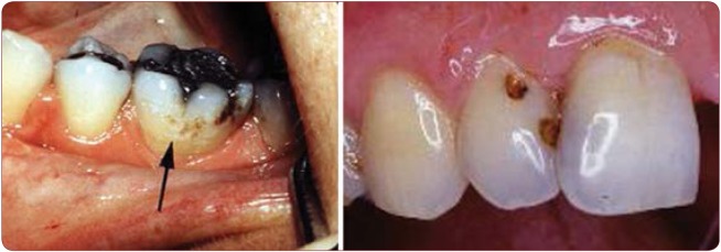
FIGURE 1.
Caries of enamel in pregnant woman
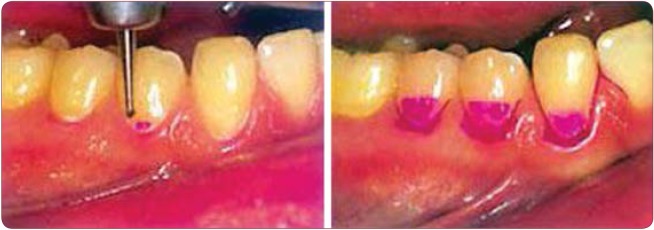
FIGURE 2.
Detection of clinical signs of caries
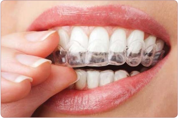
FIGURE 3.
Fluoride topics, fluoride lacquers and gels to a pregnant woman
Contributor Information
Diana POPOVICI, 3rd Clinic of Obstetrics and Gynecology,“Gr. T. Popa” University of Medicine and Pharmacy, Iasi, Romania; Mother and Child Health Department, “Gr. T. Popa” University of Medicine and Pharmacy, Iasi, Romania.
Eduard CRAUCIUC, 3rd Clinic of Obstetrics and Gynecology,“Gr. T. Popa” University of Medicine and Pharmacy, Iasi, Romania; Mother and Child Health Department, “Gr. T. Popa” University of Medicine and Pharmacy, Iasi, Romania.
Razvan SOCOLOV, 3rd Clinic of Obstetrics and Gynecology,“Gr. T. Popa” University of Medicine and Pharmacy, Iasi, Romania; Mother and Child Health Department, “Gr. T. Popa” University of Medicine and Pharmacy, Iasi, Romania.
Raluca BALAN, Morpho-Functional Sciences Department, “Gr. T. Popa” University of Medicine and Pharmacy, Iasi, Romania.
Loredana HURJUI, Morpho-Functional Sciences Department, “Gr. T. Popa” University of Medicine and Pharmacy, Iasi, Romania.
Ioana SCRIPCARIU, Mother and Child Health Department, “Gr. T. Popa” University of Medicine and Pharmacy, Iasi, Romania.
Ioana PAVALEANU, 3rd Clinic of Obstetrics and Gynecology,“Gr. T. Popa” University of Medicine and Pharmacy, Iasi, Romania; Mother and Child Health Department, “Gr. T. Popa” University of Medicine and Pharmacy, Iasi, Romania.
REFERENCES
- Alcouffe F. Oral hygiene behavior, differences between men and pregnant woman. Clinical preventive Dentistry. 1989;3 [PubMed] [Google Scholar]
- American Dental Association, Council on Access Prevention and Interprofessional Relations. Caries diagnosis and risk assessment review of preventive strategies and management. Journal American Dental Assoc. 1995;%nbsp; [PubMed] [Google Scholar]
- Axelson P, Lindhe J, Nystrom B. On the prevention of caries and periodontal disease. Results of a 15-year longitudinal study in adults. Journal of Clinical Periodontology. 1991;13:182–189. doi: 10.1111/j.1600-051x.1991.tb01131.x. [DOI] [PubMed] [Google Scholar]
- Ismail Al. Clinical diagnosis of precavitaded carious lesions. Community Dentistry and Oral Epidemiology. 1997;1:13–23. doi: 10.1111/j.1600-0528.1997.tb00895.x. [DOI] [PubMed] [Google Scholar]
- Lacatusu S. Caria dentara exploziva. Ed. Cronica, Iasi. 1996; [Google Scholar]
- Lussi A. Impact of including of excluding methods for the diagnosis of occlusal caries. Caries Research. 1996;30:389–393. doi: 10.1159/000262349. [DOI] [PubMed] [Google Scholar]
- Mandel ID. Caries prevention: current strategies new directions. JADA. 1996;10:1477–1488. doi: 10.14219/jada.archive.1996.0057. [DOI] [PubMed] [Google Scholar]
- Silverstone LM. Remineralization and enamel caries: new concepts. Dental Update. 1983;4:261–273. [PubMed] [Google Scholar]


